Introduction
The increasing need to cryopreserve supernumerary embryos following IVF has led to the development of efficient cryopreservation techniques (Larman & Gardner Reference Larman and Gardner2014; Liebermann, Reference Liebermann2015). Vitrification is a relatively simple and quick method, which has yielded higher survival rates compared with slow freezing in human (Wikland et al., Reference Wikland, Hardarson, Hillensjö, Westin, Westlander, Wood and Wennerholm2010; Zhu et al., Reference Zhu, Zhang, Cao, Zhang, Heng, Huang, Ling, Duan and Tong2011; Vanderzwalmen et al., Reference Vanderzwalmen, Connan, Grobet, Wirleitner, Remy, Vanderzwalmen, Zech and Ectors2013; Levron et al., Reference Levron, Leibovitz, Brengauz, Gitman, Yerushalmi, Katorza, Gat and Elizur2014; Xue et al., Reference Xue, Tong, Jiang, Zhu, Yang and Zhang2014, Debrock et al., Reference Debrock, Peeraer, Fernandez Gallardo, De Neubourg, Spiessens and D'Hooghe2015), mice (Huang et al., 2005; Martino et al., Reference Martino, Dell'aquila, Cardone, Somoskoi, Lacalandra and Cseh2013; Kim et al., Reference Kim, Lee, Park, Choi and Yang2015), and bovine embryos (Vajta et al., Reference Vajta, Holm, Kuwayama, Booth, Jacobsen, Greve and Callesen1998). Vitrification has the advantage of preventing ice crystal formation by subjecting embryos to a short exposure of high concentrations of cryoprotectants (Vajta et al., Reference Vajta, Holm, Kuwayama, Booth, Jacobsen, Greve and Callesen1998; Vanderzwalmen et al., Reference Vanderzwalmen, Connan, Grobet, Wirleitner, Remy, Vanderzwalmen, Zech and Ectors2013).
In human, vitrification has been successfully applied to embryos at all stages and clinical trials have shown high pregnancy rates following transfer of thawed embryos (Vanderzwalmen et al., Reference Vanderzwalmen, Bertin, Debauche, Standaert, van Roosendaal, Vandervorst, Bollen, Zech, Mukaida, Takahashi and Schoysman2002, Reference Vanderzwalmen, Ectors, Grobet, Prapas, Panagiotidis, Vanderzwalmen, Stecher, Frias, Liebermann and Zech2009; Liebermann & Tucker, Reference Liebermann and Tucker2006; Mukaida et al., Reference Mukaida, Oka, Goto and Takahashi2006; Mukaida et al., Reference Mukaida, Takahashi, Goto and Oka2008; Hong et al., Reference Hong, Sepilian, Chung and Kim2009; Desai et al., Reference Desai, Abdelhafez, Bedaiwy, Goldberg, Falcone and Goldfarb2010; Wikland et al., Reference Wikland, Hardarson, Hillensjö, Westin, Westlander, Wood and Wennerholm2010; Chen et al., Reference Chen, Zheng, Yan, Qiao and Liu2013; Li et al., Reference Li, Wang, Ledger, Edgar and Sullivan2014; Chi et al., Reference Chi, Luo, Yin, Hong, Ruan, Huang, Duan and Tong2015; Debrock et al., Reference Debrock, Peeraer, Fernandez Gallardo, De Neubourg, Spiessens and D'Hooghe2015; Devine et al., Reference Devine, Connell, Richter, Ramirez, Levens, DeCherney, Stillman and Widra2015; Galliano et al., Reference Galliano, Garrido, Serra-Serra and Pellicer2015; Van Landuyt et al., Reference Van Landuyt, Polyzos, De Munck, Blockeel, Van de Velde and Verheyen2015).
Evaluation of vitrification efficacy has mainly been performed by assessment of embryo survival and cytoskeletal integrity. Most studies that evaluated embryo survival, however, have used light microscopy for this purpose, while no studies have been performed, so far, using fluorescent viability markers.
Conversely, evaluation of vitrification by examining ultrastructure and cytoskeletal alterations with measurements of fluorescence intensities of actin and tubulin has been carried out in ovine and murine embryos (Bettencourt et al., Reference Bettencourt, Bettencourt, Silva, Ferreira, de Matos, Oliveira, Romão, Rocha and Sousa2009; Dalcin et al., Reference Dalcin, Silva, Paulini, Silva, Neves and Lucci2013; Dasiman et al., Reference Dasiman, Rahman, Othman, Mustafa, Yusoff, Jusoff, Rajikin, Froemming and Khan2013), but cytoskeletal integrity by analysing spindle/chromosome configurations has been carried out only in human blastocysts post warming (Chatzimeletiou et al., Reference Chatzimeletiou, Morrison, Panagiotidis, Vanderzwalmen, Prapas, Prapas, Tarlatzis and Handyside2012).
The purpose of the current study was to investigate the effect of vitrification on blastomere/embryo viability, by staining with fluorescent viability markers, and on cytoskeletal integrity, by examining spindle/chromosome configurations in mouse cleavage/morula stage embryos. As it is well known the mouse embryo has been used as a model in numerous publications so far in the literature. Considering the difficulty of finding spare cleavage-stage human embryos to monitor development following vitrification and assess spindle/chromosome configurations, the mouse embryo was used as a model in the current study.
Materials and methods
Culture of mouse embryos
Background CBA/C57BL6J female mice were used, which were superovulated by intraperitoneal administration of 5 IU of pregnant mare's serum (PMS; MSD Animal Health) and 48 h later by administration of a second injection of 5 IU of human chorionic gonadotropin (HCG; Chorulon, MSD Animal Health). The experimental protocols and animal handling procedures were performed with the approval of the European Union and applicable laws for the protection and proper treatment of vertebrates (N.1197/81 Article 4 N.2015/92 PD160/91, PD30/96). The superovulation and embryo isolation procedures took place at the BSRC Alexander Fleming Institute, Greece, by trained personnel of the institute and in accordance with Ittner & Gotz (Reference Lawitts and Biggers2007). In turn, female mice were caged with males (Harlan, UK) and checked daily for vaginal plugs. Pregnant females (n = 15) were sacrificed on day 2 or day 3 (embryo stages: 4- to 8-cell) post coitus. One hundred fifty-five embryos were flushed out of the oviducts in HEPES-buffered M2 medium (Sigma, Dorset, UK) supplemented with 2 mg/ml bovine serum albumen (BSA; ICN Pharmaceuticals, Ltd, Hampshire, UK) (M2 + BSA). Embryos were washed twice in gassed and prewarmed KSOM medium with 2 mg/ml BSA (KSOM-Appendix) (Lawitts & Biggers, 1991) and cultured in 30 μl drops under mineral oil (Sigma, Dorset, UK) in a humidified atmosphere at 37°C in 5% CO2 in air.
Vitrification of embryos and assessment of blastomere viability
Out of the 155 embryos, 95 were allocated randomly at either the fresh (n = 45) or the vitrified group (n = 50). Each group consisted of three subgroups depending on the embryo stage of development. In the fresh group, 15 embryos were allocated at the 4–6-cell stage, 15 embryos at the 8-cell stage and 15 embryos at the compacted morula stage. In the vitrification group, 15 embryos were allocated at the 4–6-cell stage, 18 embryos at the 8-cell stage and 17 embryos at the compacted morula stage.
Embryo vitrification was performed using the Cook vitrification kit according to the manufacturer's instructions, with modifications (Cook Medical, Bloomington, Indiana, USA). In brief, mouse embryos were vitrified on day 2 or 3 under aseptic conditions in accordance with Vanderzwalmen et al. (Reference Vanderzwalmen, Ectors, Grobet, Prapas, Panagiotidis, Vanderzwalmen, Stecher, Frias, Liebermann and Zech2009, 2013). After gradual exposure to dimethylsulphoxide:ethylene glycol (DMSO:EG) (5%:5%, 10%:10% and 20%:20%) for 3 min, 3 min and 60 s respectively, the cleavage and morula stage embryos were loaded into the gutter of a plug (Vitri-Safe, MTG, Germany) before insertion into an outer protective straw (CryoBioSystẻme, France) that was welded and plunged into liquid nitrogen (LN2). The vitrified embryos were warmed by immersing the tip of the straw (Vitri-Safe) in 1, 0.5 or 0.25 M sucrose solutions for 1 min, 1 min and 2 min respectively and were allowed to recover in culture.
Blastomere and embryo viability after vitrification was assessed using two fluorescent markers as previously described by Chatzimeletiou et al. (Reference Chatzimeletiou, Picton and Handyside2001). These markers included: (1) carboxyfluorescein-diacetate succinimidylester (CFSE; Molecular Probes Inc., Cambridge Bioscience, Cambridge, UK), which binds covalently in the cytoplasm of viable cells and stains them green; and (2) propidium iodide (PI) (Sigma, Dorset, UK), which is excluded from viable cells but can enter cells with damaged plasma membranes, binds to double-stranded DNA in the nucleus and labels it red. Fresh and vitrified/warmed embryos were first incubated in CFSE at a concentration of 20 µg/ml in Ca2+/Mg2+-free phosphate-buffered saline (PBS; Gibco; BRL) for 30 min at 37°C and then transferred into a well containing PI: CFSE/PBS (1:10 dilution) for 5 min at room temperature. Each embryo was then washed twice in Ca2+/Mg2+-free PBS for 2 min, transferred to a microscope slide in 10 µl PBS, and examined by fluorescence microscopy using standard fluorescein (CFSE) and rhodamine (PI) filter sets. Images were captured using the Zeiss fluorescence microscope, and ISIS software (Metasystems).
Vitrification of embryos and assessment of spindle chromosome configurations
Out of the 155 embryos, 60 were allocated randomly at either the fresh (n = 20) or the vitrified group (n = 40). In particular, 20 8-cell embryos and 20 compacted morulae were vitrified as described above. Following warming, embryo survival was recorded using visual assessment by light microscopy. The embryos were allowed to recover in culture for 24–48h until they reached the blastocyst stage. Twenty fresh 8-cell embryos were allowed to develop to the blastocyst stage in culture and were used as control.
Immunostaining of cleavage and morula stage embryos was performed according to Chatzimeletiou et al. (Reference Chatzimeletiou, Morrison, Prapas, Prapas and Handyside2005a, Reference Chatzimeletiou, Morrison, Panagiotidis, Prapas, Prapas, Rutherford, Grudzinskas and Handysideb), using a primary monoclonal antibody specific for α-tubulin (Sigma, Dorset, UK) in order to visualize microtubules in combination with 4–6-diamidino-2-phenylindole (DAPI) (Sigma, Dorset, UK) or PI in order to visualize DNA. In brief, all embryos were rapidly fixed in ice-cold methanol, washed in Ca2+/Mg2+-free PBS (Gibco BRL) containing 2% bovine serum albumin (BSA; Sigma, Dorset, UK) and transferred into 10-μl drops of the primary antibody under mineral oil (Sigma) and incubated for 1 h. The embryos were then washed in PBS/BSA and transferred into 10-μl drops of the secondary antibody [highly cross-adsorbed Alexa Fluor 488 (Molecular Probes)]. Following incubation of 1 h in the secondary antibody, embryos were washed in PBS/BSA and mounted on slides (BDH) in Vectashield antifade medium with DAPI or PI (Vector Laboratories, CA, USA) under a coverslip. The coverslips were then sealed with nail varnish. Cleavage and morula stage embryos were examined using the Zeiss fluorescence microscope and the Leica TCS-SP laser scanning confocal microscope (Leica Microsystems, Cambridge, UK). Standard fluorescence images were captured using the ISIS software (Metasystems) and confocal image analysis was typically accomplished by capturing a z-series stack of 1-mm thick sections encompassing the entire embryo.
Classification of spindle abnormalities
The criteria used for classifying spindle abnormalities were previously described by Chatzimeletiou et al. (Reference Chatzimeletiou, Morrison, Prapas, Prapas and Handyside2005a, Reference Chatzimeletiou, Morrison, Panagiotidis, Vanderzwalmen, Prapas, Prapas, Tarlatzis and Handyside2012). A spindle with astral-shaped or fusiform poles and with chromosomes aligned at the equator was classified as normal. A spindle with one or two poorly defined or apparently absent poles, generally with misaligned chromosomes, was classified as having an abnormal shape. Spindles with more than two clearly defined astral poles and the characteristic ‘Y’ or ‘X’ shaped arrangement of chromosomes were classified as multipolar.
Statistical analysis
Statistical analysis was carried out with Stata 13 (Stata Corporation, 4905 Lakeway Drive, College Station, Texas 77845, USA). Fisher's exact test was used to compare nominal or binary variables. Logistic regression was used to analyse the effect of vitrification and that of stage of embryo development, at which vitrification occurred, on embryo viability. Generalized estimating equations were used to analyse the effect of vitrification on spindle morphology. Continuous variables are expressed as mean ± standard error (SE), while binary or nominal variables are expressed as proportion (95% CI). Statistical significance was set at the level of P < 0.05.
Results
Viability of fresh and vitrified embryos
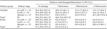
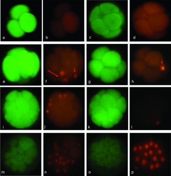
All vitrified embryos (n = 50) were processed for viability staining immediately after warming, as there was no initial loss. Viability of vitrified embryos immediately after warming as compared with fresh embryos at various stages of development is shown in Table 1. A significantly lower proportion of embryos with non-damaged blastomeres, identified by CFSE/PI staining, was observed in the vitrified/warmed embryos as compared with fresh embryos (54.0% vs. 91.1%; difference −37.1, 95% CI: −51.7 to −19.5) (Fig. 1).
Viability of fresh and vitrified mouse embryos following labelling with carboxyfluorescein-diacetate succinimidylester (CFSE) and propidium iodide (PI)

a P ≤ 0.01 (comparison of the proportion of embryos with non-damaged blastomeres between the DMSO and the fresh group).
b P = 0.986 (comparison of the proportion of embryos with non-damaged blastomeres amongst different stages of embryo development in the vitrified group).
c P = 0.302 (comparison of the proportion of embryos with non-damaged blastomeres amongst different stages of embryo development in the fresh group).
CI, confidence interval.
Effects of vitrification on embryo viability assessed by CFSE (green: viable) and PI (red: damaged) labelling. (a–d) Fresh mouse embryos at the 4-cell (a, b) and the morula stage (c, d) showing no damaged blastomeres. (e–p) vitrified mouse embryos at cleavage and morula stages showing: (e, f) a 6-cell embryo with the majority of its blastomeres viable (green) (e) and a binucleate cell damaged (red) (f; long arrow). Also note the damaged polar body in (f) (short arrow). (g, h) 8-cell embryo with all its blastomeres viable (green). Note the damaged polar body in (h) (short arrow). (i, j). A morula stage embryo with five cells damaged (red) (j) and all remaining cells viable (green) (i). (k, l) A morula stage embryo with one cell damaged (red) (l) and all remaining cells viable (green) (k). (m, n) A morula stage embryo with the majority of its cells damaged (red) (n) and a minority of viable cells (green) (m). (o, p) A morula stage embryo with extensive cell damage.

No significant difference was observed in the proportion of embryos with non-damaged blastomeres amongst different stages of development in both the vitrified and the fresh embryos (Table 1). In particular, no damage was observed in fresh embryos at cleavage stages, while at the morula stage, only 3/15 (20%) embryos showed damage in 15–18% of their cells. In contrast, in the vitrified group, the highest proportion of damaged cells was observed at the morula stage, where 8/17 (47.1%) embryos showed damage in 17–56% of their cells.
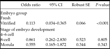
Logistic regression with dependent variable: viability (presence of damaged versus presence of exclusively non-damaged blastomeres in each embryo) and independent variables: stage of embryo development and embryo group (vitrified versus fresh) showed that embryo viability was significantly decreased in vitrified/warmed embryos as compared with fresh embryos, regardless of the stage of embryo development (Table 2).
Logistic regression with dependent variable: viability (presence of damaged versus presence of exclusively non-damaged blastomeres) and independent variables: stage of embryo development and embryo group (fresh versus vitrified)
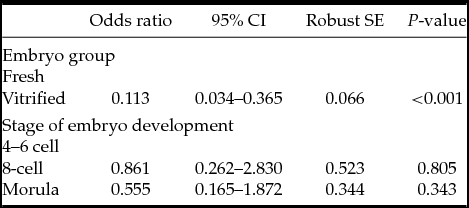
Model P< 0.001. CI, confidence interval; SE, standard error.
Development of fresh and vitrified embryos
No significant difference was observed regarding the timing of cavitation and blastocyst formation between vitrified and fresh embryos. All 20 fresh embryos reached the blastocyst stage (95% CI: 83.2–100, 20/20), while this occurred in 95% of the vitrified at the compacted morula stage (95% CI: 75.1–99.9, 19/20) and in 90% of the 8-cell vitrified embryos (95% CI: 68.3–98.7, 18/20).
Spindle abnormalities in fresh blastocysts
Out of the 20 fresh blastocysts, which were processed for cytoskeletal analysis, 17 (85%, 95% CI: 62.1–96.8) had cells with mitotic spindles (range of mitotic spindles per fresh blastocyst: 1–3). The mean ± SE cell/nuclei number per blastocyst analysed was 44.2±1.0.
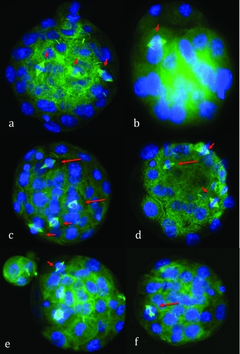
Cytoskeletal analysis showed that 58.8% (95% CI: 32.9–81.6, 10/17) of the fresh blastocysts had exclusively normal spindles, while 41.2 % (95% CI: 18.4–67.1, 7/17) had a mixture of normal and multipolar or abnormally shaped spindles (Fig. 2).
Fluorescence photomicrographs showing (a) fresh blastocyst with normal spindle chromosome configurations (short arrows), (b) vitrified embryo at the 8-cell stage that reached the blastocyst stage 24 h post-warming with a normal bipolar spindle (short arrow), (c–f) vitrified morulae that reached the blastocyst stage 24 h post warming with (c) two normal (short arrows) and two abnormally shaped spindles (long arrows), (d) two normal (short arrows) and one abnormally shaped spindle (short arrow), (e) one normal spindle (short arrow) and (f) one multipolar spindle (long arrow).

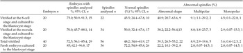
In total, 36 spindles were analysed, of which 72.2% were normal, 22.2% were abnormally shaped, 2.8% were multipolar and 2.8% were monopolar (Table 3).
Cytoskeletal analysis in fresh and vitrified mouse embryos

CI, confidence interval.
Post-warming survival and spindle abnormalities in vitrified embryos
8-cell stage
Out of the 20 vitrified 8-cell embryos, 18 (90% 95% CI: 68.3–98.7) survived post-warming.
Cytoskeletal analysis showed that 83.3% (95% CI: 58.6–96.4, 15/18) had cells with mitotic spindles. The range of mitotic spindles was 1–3 per 8-cell vitrified embryo. The mean cell number of the embryos analysed 24–36 h post warming was 34.2 ± 0.7.
Cytoskeletal analysis showed that 40% (95% CI: 16.3–67.7, 6/15) of the vitrified/warmed 8-cell embryos had only normal spindles, 40% (95% CI: 16.3–67.7, 6/15) had only abnormal spindles and 20% (95% CI: 4.3–48.1, 3/15) had a mixture of normal and abnormal spindles, including abnormally shaped, monopolar and multipolar spindles. In total, 22 spindles were analysed, of which 45.5% were normal, 41% were abnormally shaped (all of which had abnormalities in both spindle morphology and chromosome alignment), 9% were multipolar and 4.5% was monopolar (Table 3), (Fig. 2).
Morula stage
Out of the 20 vitrified morulae, 19 (95 % 95% CI: 75.1–99.9) survived post-warming. Cytoskeletal analysis showed that 73.7% of the warmed morulae had cells with mitotic spindles (range of mitotic spindles per warmed morula developed to blastocyst: 1–5). The mean ± SE cell/nuclei number per warmed morula analysed 24 h post warming was 41.8 ± 2.4.
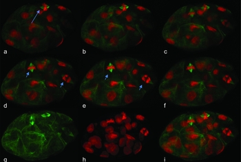
Cytoskeletal analysis showed that 28.6% (95% CI: 8.4–58.1, 4/14) of the vitrified/warmed morulae which survived to the blastocyst stage, had exclusively normal spindles, 28.6% (95% CI: 8.4–58.1, 4/14) had only abnormal spindles, while 42.9% (95% CI: 17.7–71.1, 6/14) had a mixture of normal and abnormal spindles, including abnormally shaped, monopolar and multipolar spindles. In total, 34 spindles were analysed, of which 50% were normal, 38.2% were abnormally shaped of which eight (61.5%) had abnormalities in both spindle morphology and chromosome alignment, two (15.4%) had normal spindle morphology but misaligned chromosomes and three (23.1%) were elongated), 8.8% were multipolar, and 2.9% were monopolar (Table 3), (Figs 2 and 3).
Confocal sections (a–f) and projections (g–i) of an embryo that was vitrified at the morula stage and developed to the blastocyst stage 24 h after warming. Note the normal metaphase and anaphase spindles (short arrows) and the abnormally shaped metaphase spindle (long arrow). Spindles are stained with α-tubulin in green and DNA with PI in red.

Spindle abnormalities in fresh versus vitrified embryos
Generalized estimating equations with dependent variable: spindle morphology assessed at the blastocyst stage (normal vs. abnormal) and independent variable: embryo group (fresh vs. vitrified) showed that vitrification was associated with a significantly lower proportion of normal spindles as compared with fresh embryos (Table 4).
Generalized estimating equations with dependent variable: spindle morphology (normal vs. abnormal) and independent variable: embryo group (fresh vs. vitrified)

CI, confidence interval; SE, standard error.
Discussion
To the best of our knowledge, this study is the first to examine in mice the immediate effects of vitrification on blastomere viability, using fluorescent markers. In addition, it is the first study to evaluate the type and incidence of cytoskeletal abnormalities in vitrified cleavage and morula stage embryos as compared with fresh embryos.
The current study demonstrated that vitrified embryos showed high survival rates following warming but analysis with the sensitive viability markers CFSE/PI revealed that vitrified/warmed embryos had a higher incidence of damaged blastomeres (PI-stained nuclei) compared with fresh embryos, regardless of the stage of embryo development. Most mitotic spindles examined in all groups were normal, but multivariable analysis revealed that the proportion of abnormal spindles was significantly higher in vitrified/warmed embryos as compared with fresh embryos (P ≤ 0.05). Conversely, the overall high survival rates following warming and the ability of the vast majority of embryos to develop to the blastocyst stage, suggest that embryos may compensate for a limited number of damaged cells and continue their development (Van Landuyt et al., 2013).
These results are in agreement with previous studies recording high survival rates following vitrification in animal models (Linlin et al., Reference Linlin, Bei, Guijin and Cuilian2013; Fang et al., Reference Fang, Zeng, Fu, Jia, Li, An, Chen and Zhu2014; Varago et al., Reference Varago, Moutacas, Carvalho, Serapião, Vieira, Chiarini-Garcia, Brandão, Camargo, Henry and Lagares2014; Mito et al., Reference Mito, Yoshioka, Noguchi, Yamashita, Misumi, Hoshi and Hoshi2015) and clinical trials in human that have shown high survival, implantation and live birth rates following vitrification and warming at all stages (Liebermann & Tucker, Reference Liebermann and Tucker2006; Mukaida et al., Reference Mukaida, Oka, Goto and Takahashi2006, Reference Mukaida, Takahashi, Goto and Oka2008; Hong et al., Reference Hong, Sepilian, Chung and Kim2009; Vanderzwalmen et al., Reference Vanderzwalmen, Bertin, Debauche, Standaert, Bollen, van Roosendaal, Vandervorst, Schoysman and Zech2003, Reference Vanderzwalmen, Ectors, Grobet, Prapas, Panagiotidis, Vanderzwalmen, Stecher, Frias, Liebermann and Zech2009; Desai et al., Reference Desai, Abdelhafez, Bedaiwy, Goldberg, Falcone and Goldfarb2010; Wikland et al., Reference Wikland, Hardarson, Hillensjö, Westin, Westlander, Wood and Wennerholm2010; Shi et al., Reference Shi, Xue, Zhang, Zhao, Liu, Zhou, Wang and Shi2012; Liu et al., Reference Liu, Teng, Fu, Li, Zheng and Sun2013; Debrock et al., Reference Debrock, Peeraer, Fernandez Gallardo, De Neubourg, Spiessens and D'Hooghe2015; Devine et al., Reference Devine, Connell, Richter, Ramirez, Levens, DeCherney, Stillman and Widra2015; Lopes et al., Reference Lopes, Frederickx, Van Kerkhoven, Campo, Puttemans and Gordts2015; Van Landuyt et al., Reference Van Landuyt, Polyzos, De Munck, Blockeel, Van de Velde and Verheyen2015).
Based on the results of the current study, it appears that although vitrification may result in damage of a proportion of blastomeres, this is not dependent on the stage that vitrification is performed. Damage of blastomeres may play an important role for embryo implantation since embryos with many damaged blastomeres may have a reduced developmental potential and be unable to implant. Moreover, cells with damaged membranes may be incapable to complete normal mitosis, leading to subsequent chromosomally abnormal cellular divisions. Failure of cytoplasmic divisions in the presence of karyokinesis will lead to the formation of binucleate cells, which in turn may progress through multipolar spindles into mitosis. As there is no checkpoint control to monitor excess spindle poles, these multipolar spindles are likely to give rise to cells with chaotic chromosomal constitutions, as the segregating chromosomes would be pulled to three or four directions respectively (Chatzimeletiou et al., Reference Chatzimeletiou, Morrison, Prapas, Prapas and Handyside2005a, 2012).
Most mitotic spindles examined in all groups in the current study were normal (bipolar with normal configuration). However, the proportion of abnormal spindles (multipolar/abnormally shaped) was significantly higher in vitrified/warmed embryos as compared with fresh embryos, probably due to mechanical stress following exposure to high concentrations of cryoprotectants and subsequent dehydration (Vanderzwalmen et al., Reference Vanderzwalmen, Zech, Lejeune, Wirtleitner, Zech and Ectors2010; Chatzimeletiou et al., Reference Chatzimeletiou, Morrison, Panagiotidis, Vanderzwalmen, Prapas, Prapas, Tarlatzis and Handyside2012). It should also be noted that intracellullar concentration of cryoprotectants during vitrification was not higher than in slow freezing (Vanderzwalmen et al., 2013). These results are in line with previously published data in human embryos (Chatzimeletiou et al., Reference Chatzimeletiou, Morrison, Panagiotidis, Vanderzwalmen, Prapas, Prapas, Tarlatzis and Handyside2012) and constitute mechanisms that may lead to postzygotic chromosomal abnormalities in early development (Chatzimeletiou et al., Reference Chatzimeletiou, Morrison, Prapas, Prapas and Handyside2005a, Reference Chatzimeletiou, Morrison, Panagiotidis, Prapas, Prapas, Rutherford, Grudzinskas and Handysideb, Reference Chatzimeletiou, Morrison, Prapas, Prapas and Handyside2008). Further investigation is necessary to establish if the morphological changes in the structure of the spindles, have a functional effect on the way spindles progress into mitosis, the fate of the abnormal spindles and the potential effects these may have on the chromosomal constitution of the developing embryos.
In conclusion, embryo viability, as assessed by the presence of damaged versus exclusively non-damaged blastomeres, was significantly decreased in vitrified/warmed embryos as compared with fresh embryos, regardless of the stage of embryo development. In addition, vitrified embryos had a significantly higher incidence of abnormal spindles compared with fresh embryos. Further research is necessary to assess whether the effects of vitrification on viability and cytoskeletal integrity observed in the present study in mice are also evident in human cleavage and morula stage embryos. Whether or not or to what extent vitrification may have further effects on methylation and at the transcriptomic and proteomic level remains to be established (Zhao et al., Reference Zhao, Du, Hao, Wang, Qin, Liu and Zhu2012; Saenz-de-Juano et al., Reference Saenz-de-Juano, Marco-Jimenez, Schmaltz-Panneau, Jimenez-Trigos, Viudes-de-Castro, Peñaranda, Jouneau, Lecardonnel, Lavara, Naturil-Alfonso, Duranthon and Vicente2014; Kopeika et al., Reference Kopeika, Thornhill and Khalaf2015).
Financial support
No external funding was raised for this study.
Statement of interest
All authors state that they have no conflicts of interest to disclose.