Introduction
There are over 60 species of digenean trematodes within the family Echinostomatidae (Sorensen et al., Reference Sorensen, Curtis and Minchella1998). The definitive hosts of echinostomes include aquatic birds, poultry, rodents and mammals, including humans (Belding, Reference Belding1964; Huffman & Fried, Reference Huffman and Fried1990, Fried et al., Reference Fried, Graczyk and Tamang2004). Human echinostomiasis is caused by at least 19 species of echinostomes (from eight genera), and has been reported in China, India, Indonesia, Japan, Korea, Malaysia, the Philippines, Russia, Taiwan and Thailand (Graczyk & Fried, Reference Graczyk and Fried1998; Miliotis & Bier, Reference Miliotis and Bier2003; Fried et al., Reference Fried, Graczyk and Tamang2004). For example, in Thailand, five species of echinostome have been reported to infect humans (Joe et al., Reference Joe, Nasemary and Impand1973).
Echinostomiasis is also a very important disease in domestic animals and wildlife. Morbidity and mortality due to echinostomiasis are related to parasite load (Huffman & Fried, Reference Huffman and Fried1990; Chai et al., Reference Chai, Hong, Lee, Lee and Min1994). Anaemia, headache, dizziness, stomach ache, gastric pain, and loose stools have most often been reported for light-to-moderate infections (Belding, Reference Belding1964; Graczyk & Fried, Reference Graczyk and Fried1998). Heavy infections are associated with eosinophilia, abdominal pain, profuse watery diarrhoea, anaemia, oedema and anorexia (Belding, Reference Belding1964; Carney, Reference Carney1991; Graczyk & Fried, Reference Graczyk and Fried1998). Pathologically, echinostomes damage the intestinal mucosa and cause extensive intestinal and duodenal erosions (Chai et al., Reference Chai, Hong, Lee, Lee and Min1994) and catarrhal inflammation (Belding, Reference Belding1964).
A number of species, including Echinostoma revolutum, E. miyagawai, E. gotoi and E. koidzumii were reported to infect poultry (Eom et al., Reference Eom, Rim and Jang1984). In Thailand, the two principal species that infect domestic ducks are E. revolutum and Hypodereaum conoideum (Pethleart et al., Reference Pethleart, Nateeworanart and Kaewsook2005). The adults of E. revolutum and H. conoideum can be distinguished from one another based on several morphological characters, such as the number of collar spines, the size of circumoral disc, and the shape of the testes. Echinostoma revolutum has 37 collar spines, a large circumoral disc and oval or lobed testes, whereas H. conoideum possesses 45–53 collar spines, a small circumoral disc and the testes are sausage-shaped (Miliotis & Bier, Reference Miliotis and Bier2003; Radomyos et al., Reference Radomyos, Tangtrongchitr, Krudsood, Wilairatana and Looareesuwan2003). However, it has been noted that the E. revolutum adults infecting humans have differently shaped testes compared to specimens of the same species in ducks, i.e. adult E. revolutum in humans have oval-shaped testes (Radomyos et al., Reference Radomyos, Bunnag and Harinasuta1982) whereas those in ducks have both oval and multi-lobed testes (Pethleart et al., Reference Pethleart, Nateeworanart and Kaewsook2005).
It is often difficult to distinguish morphologically among immature echinostomes, and no morphological characters are available to distinguish the eggs of different echinostome species. Therefore, molecular markers have been developed to identify and differentiate between species of echinostome and to examine their genetic relationships (Voltz et al., Reference Voltz, Richard and Pesson1987; Morgan & Blair, Reference Morgan and Blair1995, Reference Morgan and Blair1998a, Reference Morgan and Blairb; Sloss et al., Reference Sloss, Meece, Romano and Nollen1995; Petrie et al., Reference Petrie, Burg and Cain1996; Sorensen et al., Reference Sorensen, Curtis and Minchella1998). Multilocus enzyme electrophoresis (MEE) has been used effectively to investigate genetic differentiation in parasitic trematodes, including Opisthorchis viverrini (Saijuntha et al., Reference Saijuntha, Sithithaworn, Wongkham, Laha, Pipitgool, Tesana, Chilton, Petney and Andrews2007), Clornorchis sinensis (Park et al., Reference Park, Yong, Im and Lee2000) and species of Fasciola (Agatsuma et al., Reference Agatsuma, Terasaki, Yang and Blair1994), Paragonimus (Agatsuma & Habe, Reference Agatsuma and Habe1986) and Echinostoma (Voltz et al., Reference Voltz, Richard and Pesson1987; Sloss et al., Reference Sloss, Meece, Romano and Nollen1995). For example, Sloss et al. (Reference Sloss, Meece, Romano and Nollen1995) established six enzyme loci to differentiate genetically among three species of echinostomes (i.e. E. caproni, E. paraensei and E. trivolvis).
The aim of this study was to use MEE to compare genetically between two medically important species of echinostomes, E. revolutum and H. conoideum collected from domestic ducks in Thailand, to determine the extent of the genetic differences between them and to establish genetic markers for their identification. In addition, the polymorphic loci detected within each species were also established for subsequent studies of population genetics.
Materials and methods
Sample collection
Adult worms of E. revolutum and H. conoideum were collected from intestines of domestic ducks from a slaughterhouse in Khon Kaen Province during April and June of 2008. The 76 adults of H. conoideum were from 11 ducks with between 3 and 20 worms/duck, whereas the 39 E. revolutum adults were from another 12 ducks with 1–7 worms/duck. Adult trematodes were examined using a light microscope and identified to species level according to size of the circumoral disc, morphology of testes and number of collar spines (Gower, Reference Gower1939; Yamaguti, Reference Yamaguti1971; Miliotis & Bier, Reference Miliotis and Bier2003). Several individual worms of each species were selected randomly for carmine staining to confirm their species identity by morphology (Miliotis & Bier, Reference Miliotis and Bier2003; Radomyos et al., Reference Radomyos, Tangtrongchitr, Krudsood, Wilairatana and Looareesuwan2003). Other individuals were washed extensively in normal saline, then frozen for subsequent electrophoretic analyses.
Sample preparation for electrophoresis
Individual worms were placed in a microcentrifuge tube to which 10–30 μl of lysing solution (100 ml distilled water, 100 μl β-mercaptoethanol, 10 mg NADP) was added. Samples were homogenized by hand using a glass rod. The homogenates were then centrifuged at 10,000 rpm for 10 min while maintained at a temperature of 4°C. Supernatants were stored in capillary tubes as 5 μl aliquots at − 20°C until required.
Multilocus enzyme electrophoresis
MEE was performed using cellulose acetate gels (Cellogel, Milan, Italy) as the support medium, with 0.02 m phosphate (pH 8.0) used as the running buffer. MEE gels were run for 120–150 min at a constant voltage (200 V) and at a constant temperature (4°C). The histochemical staining methods used follow those described by Richardson et al. (Reference Richardson, Baverstock and Adams1986).
Parasite samples were compared using 22 enzymes as follows (abbreviation, enzyme commission no. in parentheses): aldolase (Ald, 4.1.2.13), adenylate kinase (Ak, 2.7.4.3), enolase (Enol, 4.2.1.11), fructose-1,6-diphosphatase (Fdp, 3.1.3.11), fumarate hydratase (Fum, 4.2.1.2), glyceraldehyde-3-phosphate dehydrogenase (Gapd, 1.2.1.12), glucose-6-phosphate dehydrogenase (G6pd, 1.1.1.49), aspartate amino transferase (Got, 2.6.1.1), glucose-phosphate isomerase (Gpi, 5.3.1.9), hexokinase (Hk, 2.7.1.1), isocitrate dehydrogenase (Idh, 1.1.1.42), malate dehydrogenase (Mdh, 1.1.1.37), malic enzyme (Me, 1.1.1.40), nucleotide diphosphate kinase (Ndpk, 2.7.4.6), peptidase valine–leucine (PepA, 3.4.13.11), peptidase leucine–glycine–glycine (PepB, 3.4.11.4), peptidase phenylalanine-proline (PepD, 3.4.13.9), phosphoglycerate mutase (Pgam, 2.7.5.3), phosphoglucomutase (Pgm, 2.7.5.1), pyruvate kinase (Pk, 2.7.1.40), triose phosphate isomerase (Tpi, 5.3.1.1), and uridine monophosphate kinase (Umpk, 2.7.1.48).
For each enzyme, bands detected in each sample were scored alphabetically, the allozyme with the least electrophoretic mobility from the cathode designated as allele a. The multiple banding patterns of individual worms at a particular locus were consistent with the expectations of heterozygous individuals for enzymes with a quaternary structure (e.g. two-banded and triple-banded patterns for heterozygous individuals for monomeric and dimeric enzymes, respectively) (Richardson et al., Reference Richardson, Baverstock and Adams1986). The allelic profiles of specimens were compared and the fixed allelic (hence genetic) differences among samples representing the two echinostome species were recorded. A fixed genetic difference occurs when two groups of samples do not have any alleles in common at an enzyme locus (Richardson et al., Reference Richardson, Baverstock and Adams1986; Andrews & Chilton, Reference Andrews and Chilton1999).
Results
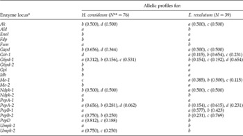
The present study represents the first time that allozyme (genetic) markers have been established to differentiate between E. revolutum and H. conoideum, two species that infect domestic ducks in Thailand. Thirty-nine E. revolutum adults and 76 H. conoideum adults were compared genetically by MEE at 22 enzymes encoding 30 presumptive enzyme loci (table 1). Genetic variation among individual trematodes was detected at 22 loci. The eight remaining loci were invariant. Fixed genetic differences between H. conoideum and E. revolutum were detected at 17 (57%) of the 30 enzyme loci (i.e. at Ak, Ald, Enol, Fdp, Fum, Gapd, Got-1, G6pd-2, Gpi, Idh, Me-2, Ndpk-1, Ndpk-2, PepA-1, PepD, Umpk-1 and Umpk-2) (fig. 1 and table 1). Genetic variation among specimens of H. conoideum were detected at eight loci (Ak, Gapd, G6pd-1, Ndpk-1, PepA-2, PepB-2, PepD and Umpk-2), whereas nine loci were polymorphic for E. revolutum (i.e. Ak, Gapd, Got-1, G6pd-1, Me-1, Ndpk-1, PepA-2, PepB-1 and PepB-2) (fig. 2 and table 1).
Allelic profiles of H. conoideum and E. revolutum at 30 enzyme loci, with allele frequency indicated in parentheses.

* Excludes monomorphic loci (Got-2, Hk, Mdh, Pgm, Pgam-1, Pgam-2, Pk and Tpi).
** Sample size.
The electrophoretic banding patterns of individual specimens of H. conoideum and E. revolutum for Idh.

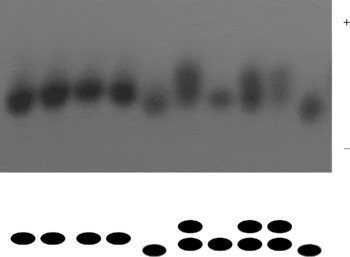
Example of electrophoretic patterns of a polymorphic locus, Me-1, comparing ten individuals of E. revolutum.

Allele frequency of the polymorphic loci is indicated in table 1. The PepA-2, PepB-2 and G6pd-1 showed higher frequencies in unique alleles than in common shared alleles. For example, alleles b and d of PepA-2 were common alleles between H. conoideum and E. revolutum, which showed freqencies of 0.062 to 0.281, whereas allele a was a unique allele for H. conoideum and showed a frequency of 0.656, and allele c, a unique allele for E. revolutum, showed a frequency of 0.615 (table 1).
Discussion
At least 17 of the 30 enzyme loci tested can be used to distinguish between these two species of echinostome. This indicates the high levels of genetic diversity of echinostomes in Thailand (Sloss et al., Reference Sloss, Meece, Romano and Nollen1995). Moreover, intraspecific variation was also detected by using allozyme markers, as the result showed that up to eight loci showed fixed differences among individual worms within each species. This number of enzyme loci provides the opportunity for comprehensive analyses of population genetics in these parasites, as achieved in a previous study on Opisthorchis viverrini sensu lato (Saijuntha et al., Reference Saijuntha, Sithithaworn, Wongkham, Laha, Satrawaha, Chilton, Petney and Andrews2008).
As suggested by Andrews & Chilton (Reference Andrews and Chilton1999), the percentage fixed difference should be greater than 15% between different species. In this study, 57% of fixed differences were detected between E. revolutum and H. conoideum. Moreover, the number of enzyme loci also affect percentage fixed differences – more than 15 loci should be examined for reliable interpretation. In this study, we examined up to 22 loci which, compared to previous studies (Voltz et al., Reference Voltz, Richard and Pesson1987; Sloss et al., Reference Sloss, Meece, Romano and Nollen1995), is the largest number of loci of allozyme markers studied in echinostomes.
An application of enzyme markers to the investigation of genetic relationships or co-evolution between hosts and parasitic trematodes has been reported (e.g. Saijuntha et al., Reference Saijuntha, Sithithaworn, Wongkham, Laha, Pipitgool, Tesana, Chilton, Petney and Andrews2007). Various freshwater snails, fish, crustaceans and amphibians have have been commonly found as first and second intermediate hosts of echinostomes (Graczyk & Fried, Reference Graczyk and Fried1998). Morever, several kinds of mammals and poultry, e.g. rat, dog, human, water bird, chicken and duck, have been reported as final hosts of echinostomes. Thus, the enzyme markers from this study may be used for further investigation of co-evolution of echinostomes and their hosts. These enzyme markers could also be used for further genetic identification in other echinostomes of other host, e.g. E. malayanum in mammals. In addition, some loci can be used as markers for further population genetic analyses, e.g. G6pd-1 for both E. revolutum and H. conoideum or Got-1, Me-2 and PepB-1 for E. revolutum.
Acknowledgements
This research was supported by the Thailand Research Fund and the Commission on Higher Education (grant no. MRG5180102). The locations and facilities were provided by Mahasarakham University and Department of Parasitology, Liver Fluke and Cholangiocarcinoma Research Center, Faculty of Medicine, Khon Kaen University. Many thanks to Dr Neil Chilton for his kind proofreading of this manuscript and Ms Nadda Kiatsopit and Ms Kunyarat Duenngai for helping with sample collection.