DNA-based scaffolds for the spatial confinement of enzymes
The highly complex network of chemical reactions within the cell is mainly controlled through the confinement of enzymes into restricted spatial environments, such as on filaments, on biological membranes, and within organelles and other nanoscale compartments. Reference Kuchler, Yoshimoto, Luginbuhl, Mavelli and Walde1,Reference Chen and Silver2 In this way, high local concentrations of specific enzymes and signaling molecules in prescribed stoichiometric ratios can be realized to enhance reaction kinetics, substrates can be channeled through specific series of enzyme cascades (i.e., a sequence of successive activation reactions involving enzymes), and intermediates can be sequestered, thereby minimizing toxicity and competing cross reactions.
In the effort to mimic such features, in vitro systems have been developed to immobilize enzymes with control over their spatial coordinates and dynamic temporal actuation (see Schulman and Simmel in this issue). Reference Simmel and Schulman3 A promising approach toward this goal uses DNA to construct ordered molecular assemblies, up to few hundreds of nanometers in size, with unique spatial addressability. Reference Seeman4,Reference Rothemund5 This, together with the development of different chemical conjugation strategies to couple proteins to nucleic acids (see Gothelf et al. in this issue), Reference Gothelf6 has now enabled the programmable organization of a distinct number of enzymes into linear arrays or small tile-tethered constructs onto two-dimensional (2D) DNA surfaces, or within small DNA confined volumes. Each of these strategies offers powerful opportunities for the design of functional nanomaterials that cannot be realized using any other nanoscale fabrication approach.
Oligonucleotide-tethered and small tile-tethered enzymes
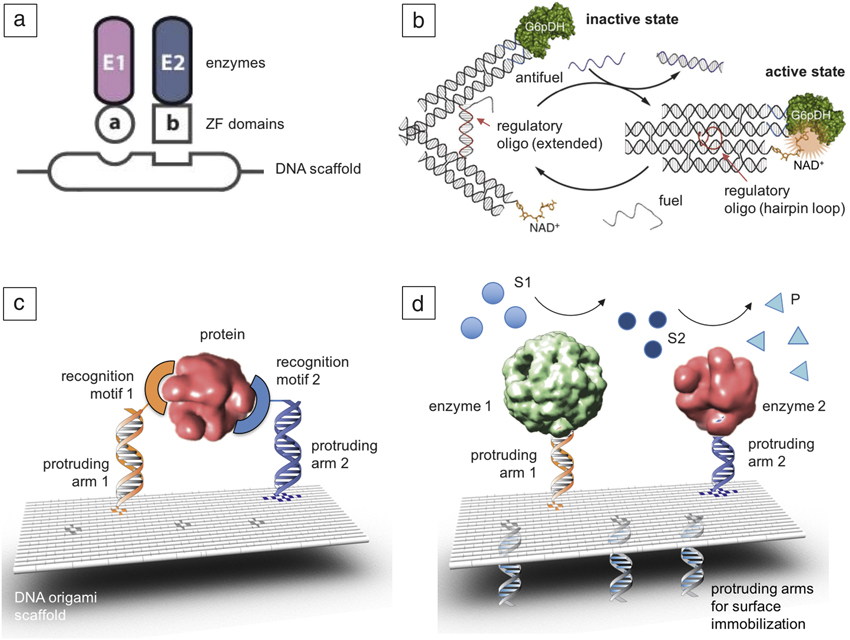
The simplest way to explore proximity effects occurring in natural multienzyme complexes is to place distinct enzymes along a monodimensional scaffold and measure their activity as a function of their intermolecular distance. In this way, enzymatic reactions have been efficiently coupled, Reference Muller and Niemeyer7 and the production of metabolic species has been increased ( Figure 1a). Reference Conrado, Wu, Boock, Xu, Chen, Lebar, Turnsek, Tomsic, Avbelj, Gaber, Koprivnjak, Mori, Glavnik, Vovk, Bencina, Hodnik, Anderluh, Dueber, Jerala and DeLisa8 Similarly, glucose oxidase enzymes have been modified with gold nanoparticles and scaffolded onto linear DNA molecules for the production of metallic nanoscale wires. Reference Wilner, Shimron, Weizmann, Wang and Willner9 In fact, the biocatalytic enlargement of gold nanoparticles via glucose oxidation in the presence of (AuCl4)– enabled the production of micrometer-long metallic wires, and could be further developed to obtain metallic nanocomponents of arbitrary shape and geometry.
Mono- and 2D DNA-scaffolded enzymes. (a) DNA scaffold-assisted assembly of metabolic pathways in E. coli. Schematic of a system developed for the biosynthesis of resveratrol, a type of natural phenol produced by several plants in response to injury or pathogens attack. E1 and E2 are the two enzymes that produce trans-resveratrol from 4-coumaric acid, genetically fused to zinc-fingers (ZF) domains, a and b, which bind specific sequences on the plasmid DNA scaffold. ZF domains are small protein motifs whose structures are stabilized by the coordination of one or more zinc ions. Adapted with permission from Reference Reference Conrado, Wu, Boock, Xu, Chen, Lebar, Turnsek, Tomsic, Avbelj, Gaber, Koprivnjak, Mori, Glavnik, Vovk, Bencina, Hodnik, Anderluh, Dueber, Jerala and DeLisa8. © 2012 Oxford University Press. (b) Schematic illustration of a DNA tweezer-regulated enzyme nanoreactor: a regulatory oligomer (shown in red) is designed to adopt a hairpin structure that holds the two arms of the tweezers close together, thus bringing the dehydrogenase enzyme (G6pDH) and its cofactor (NAD+) in close proximity (active state). The addition of a fuel strand—complementary to the regulatory loop—results in the formation of a DNA double helix between the tweezer arms that separate the enzyme/cofactor pair (inactive state). Cycling of the device is achieved by consecutive addition of fuel and antifuel strands. Adapted with permission from Reference Reference Liu, Fu, Hejesen, Yang, Woodbury, Gothelf, Liu and Yan10. © 2013 Macmillan Publishers Ltd. (c) Quasi-planar DNA nanostructures can be modified with single-stranded protruding arms (orange and blue helices) for DNA-driven immobilization of distinct protein recognition motifs (orange and blue symbols) at predefined intermolecular distances. This arrangement is then tested for the binding of multiple ligands to the same target enzyme (red). Adapted with permission from Reference Reference Rinker, Ke, Liu, Chhabra and Yan19. © 2008 Macmillan Publishers Ltd. (d) Alternatively, multiple enzymes (indicated in green and red) can be placed in spatial proximity for efficient substrate channeling. This generates a scaffolded enzyme cascade, where the substrate of the first enzyme (S1, light blue circles) is transformed into the substrate of the second enzyme (S2, dark blue circles), eventually leading to the reaction product (P, light blue triangles). Both systems in (c) and (d) can be modified for immobilization onto a solid surface (through the DNA strands shown in light blue). Adapted with permission from Reference Reference Fu, Liu, Liu, Woodbury and Yan20. © 2012 American Chemical Society.
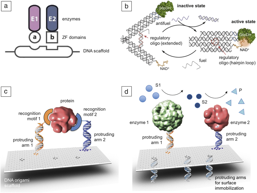
An interesting application of enzyme scaffolding consists of activating and inactivating enzymes in a user-controlled fashion. An example of such a system is represented by the cofactor-dependent dehydrogenase and its cofactor, whose mutual distance can be varied by attaching them to the extremities of a DNA tweezer (Figure 1b). Reference Liu, Fu, Hejesen, Yang, Woodbury, Gothelf, Liu and Yan10 Such tweezers are designed to switch from an open to a closed conformation upon detection of a specific DNA sequence, thus enabling full enzyme reconstitution and thereby conversion from the inactive to the active state. Note that a cofactor is a substance (other than the substrate) whose presence is essential for the activity of an enzyme. Similar strategies have also been applied to reversibly bind and release single protein molecules, Reference Zhou, Yang and Liu11 to regulate the efficiency of the cascade reaction catalyzed by the enzymes glucose oxidase (GOx) and horseradish peroxidase (HRP), Reference Xin, Zhou, Yang and Liu12 and in general to dynamically control protein activity via oligonucleotide-based triggers. Reference Engelen, Janssen and Merkx13
Enzymes confined onto 2D DNA surfaces
The key hallmark of self-assembled DNA nanostructures is the opportunity to achieve spatial control over molecular components with a resolution of a few nanometers. Sugiyama and co-workers provide a relevant example of how this feature can be strategically used to dissect biological events at the molecular level. Reference Endo, Katsuda, Hidaka and Sugiyama14–Reference Kobayashi, Misumi, Odahara, Ishibashi, Hirono, Hidaka, Endo, Sugiyama, Iwasaki, Kuroiwa, Shikanai and Nishimura17 In their study, they immobilized DNA recognition elements in the central cavity of a frame-shaped nanostructure and investigated the importance of DNA-strand relaxation and bending in DNA repair mechanisms Reference Endo, Katsuda, Hidaka and Sugiyama14 as well as the role of DNA topology on the outcome of the recombination process. Reference Suzuki, Endo, Katsuda, Ou, Hidaka and Sugiyama15,Reference Kobayashi, Misumi, Odahara, Ishibashi, Hirono, Hidaka, Endo, Sugiyama, Iwasaki, Kuroiwa, Shikanai and Nishimura17 In another important class of nanostructures, the DNA motifs are not processed or metabolized, but simply recognized by the incoming proteins, thus serving as molecular tags for biosensing applications Reference Chhabra, Sharma, Ke, Liu, Rinker, Lindsay and Yan18 or for the investigation of multivalent interactions (Figure 1c). Reference Rinker, Ke, Liu, Chhabra and Yan19
The opposite configuration, in which surface-bound enzymes are exposed to the bulk aqueous solution containing the substrate, has also been implemented to reconstruct multistep cascade reactions in vitro (Figure 1d). Reference Fu, Liu, Liu, Woodbury and Yan20–Reference Ke, Liu, Jiang, Qi, Yang, Wootten, Zhang, Zhu, Liu, Yang and Yan23 Yan and co-workers give one of the first examples of this kind. Reference Fu, Liu, Liu, Woodbury and Yan20 In their work, they organized discrete GOx/HRP enzyme pairs on planar DNA-based platforms with controlled inter-enzyme spacing and position. By measuring the activities of different enzyme-DNA complexes, they could identify distinct distance-dependent kinetic processes.
Scaffolded enzymatic cascades offer exciting perspectives for the development of more efficient enzymatic fuel cells—bioelectronics devices that use oxidoreductase enzymes to catalyze the conversion of chemical energy (e.g., from organic byproducts of agricultural and chemical industry) into electric energy. Reference Rasmussen, Abdellaoui and Minteer24 Such systems may considerably benefit from the nanometer-scale positional control and self-assembly properties of DNA nanostructures to more easily and efficiently organize sequential enzymes on the electrode surface. Reference Van Nguyen, Giroud and Minteer25
Enzymes confined within small DNA volumes
Since the very beginning of DNA nanotechnology, DNA nanocontainers of multiple shapes and sizes have been designed and characterized. Various static and dynamic wireframe structures have been constructed, Reference Chen and Seeman26 and in some cases, these were internally loaded with cargo molecules. Reference Banerjee, Bhatia, Saminathan, Chakraborty, Kar and Krishnan27,Reference Crawford, Erben, Periz, Hall, Brown, Turberfield and Kapanidis28
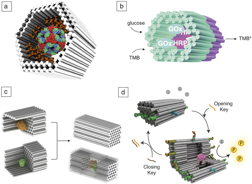
The invention of the DNA origami method Reference Rothemund5 has allowed rationally designed nanocompartments with solid walls to be built with the potential to fully enclose large biomolecules. Box-like nanocontainers with different sizes and dynamic mechanisms to control opening and closing processes have been realized Reference Andersen, Dong, Nielsen, Jahn, Subramani, Mamdouh, Golas, Sander, Stark, Oliveira, Pedersen, Birkedal, Besenbacher, Gothelf and Kjems29–Reference Perrault and Shih32 with one of the major goals being their use as advanced drug delivery systems. In some cases, the outer surface of DNA nanocontainers has been functionalized with lipid molecules to increase their biostability. Reference Perrault and Shih32 In other instances, the inner side of tubular DNA origami structures has been modified with protein recognition motifs for selective and noncovalent immobilization of a single protein molecule ( Figure 2a). Reference Sprengel, Lill, Stegemann, Bravo-Rodriguez, Schoneweiss, Merdanovic, Gudnason, Aznauryan, Gamrad, Barcikowski, Sanchez-Garcia, Birkedal, Gatsogiannis, Ehrmann and Sacca33 Instead, by using covalent DNA-protein conjugation, two distinct GOx- and HRP-loaded DNA chambers have been linked together to generate an enzyme cascade reaction inside the tubular construct (Figure 2b). Reference Linko, Eerikainen and Kostiainen34 Altogether, these studies demonstrate the potential of DNA nanostructures as biomimetic materials that can be shaped to mimic the structural features and encapsulation mechanisms typical of natural compartmentalization systems, such as viral capsids, Reference Douglas and Young35 lipid vesicles, Reference Nomura, Tsumoto, Hamada, Akiyoshi, Nakatani and Yoshikawa36 and membrane-bound organelles. Reference Zhu and Szostak37 Here again, robust chemical strategies are fundamental to achieve the desired DNA-conjugate in satisfactory yields (at least 60–70%) (see Gothelf et al. in this issue). Reference Gothelf6
DNA volume-confined enzymes. (a) A hollow DNA nanocontainer has been modified in its inner cavity with peptide recognition motifs (orange helices) pre-oriented toward the binding sites exposed on the surface of the DegP protease (multicolored sphere in the middle of the container), leading to selective encapsulation of a single protein molecule as a consequence of multivalent binding affinity and geometric matching of the two species. Please note that the DegP protease is an oligomeric protein, whose monomeric unit consists of a catalytic domain (red), a ligand-recognition domain (green), and a quaternary-structure inducing domain (blue). Adapted with permission from Reference Reference Sprengel, Lill, Stegemann, Bravo-Rodriguez, Schoneweiss, Merdanovic, Gudnason, Aznauryan, Gamrad, Barcikowski, Sanchez-Garcia, Birkedal, Gatsogiannis, Ehrmann and Sacca33. © 2017 Macmillan Publishers Ltd. (b) Two separately fabricated origami units equipped with biotinylated glucose oxidase (GOx, cyan) or horseradish peroxidase (HRP, purple), respectively, through biotin–avidin interaction. The units are linked together via base-pairing, resulting in a nanoreactor that is able to perform an enzyme cascade reaction. 3,3′,5,5′-tetramethylbenzidine (TMB) is oxidized into TMB diimine (TMB*) in the presence of glucose and oxygen. Adapted with permission from Reference Reference Linko, Eerikainen and Kostiainen34. © 2015 Royal Society of Chemistry. (c) Schematic representations of the assembly of a DNA nanocage encapsulating a pair of GOx (orange) and HRP (green) enzymes. Individual enzymes were first attached to half cages, followed by the addition of linker strands to combine the two halves into a full cage. Small pores (black holes of hexagonal shapes) were designed on the bottom of cages to facilitate the diffusion of substrate molecules into and out of the cage. Adapted with permission from Reference Reference Zhao, Fu, Dhakal, Johnson-Buck, Liu, Zhang, Woodbury, Liu, Walter and Yan38. © 2016 Macmillan Publishers Ltd. (d) Graphic representation of the DNA vault, Reference Grossi, Jepsen, Kjems and Andersen46 a 3D dynamic DNA nanocontainer, which encapsulates and fully encloses a protease molecule (shown in pink) in its inner cavity, thus shielding it from its substrate (gray circles) that is free in solution and preventing formation of the product (yellow circles). The opening and closing mechanism is triggered by the addition of specific DNA strands, which operate on the lock strands of the vault (green and blue). The opening and closing keys (in orange and brown, respectively), switch on or off, respectively, the enzymatic reaction by simply regulating the accessibility of the loaded enzyme to an external substrate.

In 2016, Yan and co-workers succeeded in fully enclosing active enzymes within a multilayered DNA nanocompartment, Reference Zhao, Fu, Dhakal, Johnson-Buck, Liu, Zhang, Woodbury, Liu, Walter and Yan38 demonstrating increased rate of substrate chemical conversion (turnover number) for both individual enzymes and co-localized enzyme cascades (Figure 2c). Interestingly, they observed an inverse correlation between the size of the encaged protein and its activity enhancement, which has been explained by the stabilizing effect of the negatively charged DNA surface on the active enzyme conformation through the action of a strongly bound hydration layer, in a chaperone-like fashion. Reference Gray, Wholey, Wagner, Cremers, Mueller-Schickert, Hock, Krieger, Smith, Bender, Bardwell and Jakob39 An alternative to this hypothesis was proposed by Hess and co-workers, who suggested that the pH near the surface of the DNA scaffold is lower than that in the bulk solution, resulting in a more optimal pH environment for the tethered enzyme. Reference Zhang, Tsitkov and Hess40 In other studies, however, the enhanced activity of the DNA-scaffolded enzyme has been attributed to the increased affinity of the substrate for the DNA template. Reference Gao, Roberts, Zhu, Lin, Chang and Wheeldon41,Reference Lin and Wheeldon42 The whole scenario is further complicated by the observation that these effects are often protein- and DNA sequence-dependent, Reference Glettenberg and Niemeyer43–Reference Timm and Niemeyer45 thus calling for further investigations into the role played by the DNA scaffold in the folding and activity of the anchored enzymes.
In natural systems, the activity of enzymes that are fully enclosed within nanocompartments is often controlled by physically separating such enzymes from their substrates. Inspired by this generally applicable approach, Andersen and co-workers have recently developed a dynamic DNA origami nanocontainer that encapsulates single enzyme molecules and controls their catalytic activity by compartmentalization. Reference Grossi, Jepsen, Kjems and Andersen46 The structure—named “DNA vault”—is loaded with a protease molecule and then closed, thanks to reversible hairpin locks (Figure 2d). By shielding the enclosed enzyme within its internal cavity, the structure hinders the encapsulated protease from interacting with free-diffusing substrate molecules, thus inhibiting de facto the enzymatic reaction. However, when a specific DNA sequence is added, the DNA vault opens up, exposing the enzyme to the external environment, therefore allowing enzyme–substrate interaction and consequent catalytic reactions to occur. Such an approach may offer a general method for taking an arbitrary enzyme for which there is no natural mechanism for turning its activity on and off and providing a robust DNA-switched control.
Conclusions
DNA-scaffolded enzymes represent ideal tools for the development of in-solution biosensors to detect molecules of interest. Reference Freeman, Sharon, Tel-Vered and Willner47,Reference Freeman, Sharon, Teller and Willner48 However, in order to realize cheaper, faster, and more easily readable biosensors, important issues must be still fully addressed, such as biostability and biocompatibility properties. Promising approaches have already been proposed, including chemical modifications of nucleobases or sugar-phosphate backbone, Reference Cassinelli, Oberleitner, Sobotta, Nickels, Grossi, Kempter, Frischmuth, Liedl and Manetto49,Reference Rajendran, Endo, Katsuda, Hidaka and Sugiyama50 different origami design approaches, Reference Benson, Mohammed, Gardell, Masich, Czeizler, Orponen and Hogberg51–Reference Veneziano, Ratanalert, Zhang, Zhang, Yan, Chiu and Bathe53 and DNA coating with protein- or lipid-based protective layers. Reference Perrault and Shih32,Reference Mikkila, Eskelinen, Niemela, Linko, Frilander, Torma and Kostiainen54 Enzyme immobilization is commonly used to facilitate recovery and reuse of catalysts, and to improve their stability, thus lowering overall costs in industrial enzymatic catalysis. Reference Rodrigues, Ortiz, Berenguer-Murcia, Torres and Fernandez-Lafuente55,Reference Geng and Paukstelis56
Although the higher stability of immobilized enzymes may result in increased apparent catalytic activity under extreme conditions, immobilized enzymes usually show lower activity compared to their native counterparts. This is mostly due to the hindered substrate accessibility of the enzyme, an issue that can be tackled by DNA nanotechnology approaches, which allow rational design of the specific enzyme–scaffold interaction.
Despite important progress in spatially restricting multiple enzymes, a major future challenge for the field lies in implementing control over the relative spatial orientation of enzymes. In fact, most natural protein assemblies enhance catalytic activities by orders of magnitude by achieving direct substrate channeling between catalytic sites, Reference Raushel, Thoden and Holden57 whereas DNA-scaffolded enzymes are usually organized by using flexible linkers, which have thus far limited our ability to achieve comparable results synthetically.
Acknowledgments
This work was supported by the Deutsche Forschungsgemeinschaft (CRC initiative 1093) (B.S.), and the European Research Council (ERC, Grant Number 683305) (E.S.A.).

Guido Grossi is a postdoctoral researcher in the Andersen Lab for Biomolecular Design at Aarhus University, Denmark. He studied biotechnology at Parma University, Italy, and he performed his PhD work in DNA nanotechnology at Aarhus University. His research interests include DNA and RNA nanotechnology, genetic engineering, and synthetic biology. Grossi can be reached by phone at +45 87156747 or by email at ggrossi@inano.au.dk.

Andreas Jaekel is a doctoral candidate in the Bionanotechnology group at the University of Duisburg-Essen, Germany. He studied biochemistry at the University of Greifswald, Germany, where he received his MSc degree in 2016. His current research interests focus on protein-DNA conjugation and the encapsulation of proteins in DNA origami-based cages. Jaekel can be reached by phone at +49 201 183 6911 or by email at andreas.jaekel@uni-due.de.

Ebbe Sloth Andersen has been head of the Biomolecular Design group at the Interdisciplinary Nanoscience Center, Aarhus University, Denmark, since 2012. He obtained his PhD degree at Aarhus University in 2006. He was a postdoctoral fellow at the Centre for DNA nanotechnology (CDNA), Aarhus University, and has been a visiting associate at California Institute of Technology. His research focuses on nucleic acid design principles and applications in biosensing, drug delivery, and synthetic biology. Andersen can be reached by phone at +45 41178619 or by email at esa@inano.au.dk.

Barbara Saccà has been head of the Bionanotechnology group at the University of Duisburg-Essen, Germany, since 2012. She obtained her PhD degree at the Max Planck Institute of Biochemistry, Germany, in 2002. After completing postdoctoral research in DNA biophysics, she moved to TU Dortmund, Germany, where she worked on DNA nanotechnology. Her research interests focus on the realization and manipulation of DNA-based biomimetic systems. Saccà can be reached by phone at +49 201 183 3395 or by email at barbara.sacca@uni-due.de.