Implications
The development of dental abnormalities is common in domestic horses due to congenital defects and modern management regimes. Prophylactic dentistry is aimed at minimising the potential damage and pain that such abnormalities may cause. Veterinary surgeons and lay dental technicians may endeavour to correct dental abnormalities such as enamel overgrowths. However, some extent of enamel overgrowths may be expected in all horses, whether extensively or intensively managed. The evidence from this study suggests that further research is required to potentially redefine what is considered normal dentition, particularly for Thoroughbred-type horses.
Introduction
The development of dental abnormalities is common in horses due to congenital defects and the management regime enforced on the horse through domestication (Lane, Reference Lane1994). The equine hypsodont dentition continuously erupts throughout the life of the adult horse at a rate of approximately 2.5 mm a year (Muylle et al., Reference Muylle, Simoens and Lauuwers1999), equating to the rate of attrition on the occlusal surface of the teeth as a result of high silica content in consumed grasses and vegetation. The horse, free grazing at grass, will graze for up to 16 h/day (Arnold, Reference Arnold1984), depending on seasonal daylight variations in a natural environment. When this behaviour pattern is compared to the eating behaviour of modern domesticated and stabled equines, it is easily understood why domestication may adversely affect the essential attrition of horses’ teeth.
There are few published results in the literature regarding the occurrence and prevalence of dental abnormalities in wild or semi-wild horses, the access to such populations being a limiting factor. However, Penzhorn (Reference Penzhorn1984) documented the post-mortem examination of 37 wild zebra skulls. Only 19% of the skulls exhibited any occurrence of dental abnormality, including misplaced incisors, cheek teeth wear abnormalities, diastemata and periodontitis. It was suggested that these abnormalities were incidental and did not cause the death of the zebra, and therefore were not preventing them from eating. This is compared to the 93% occurrence of dental disease in domesticated donkeys in a UK study (Du Toit et al., Reference Du Toit, Gallagher, Burden and Dixon2008a) and 62% in a Mexican study (Du Toit et al., Reference Du Toit, Burden and Dixon2008b). Domestication, and specifically the use of bits and bridles, has also been suggested to increase the occurrence of oral ulcers in horses, potentially caused by sharp enamel points (SEPs) on cheek teeth (Tell et al., Reference Tell, Egenvall, Lungstrom and Wattle2008).
Since it is likely that severe dental abnormalities may cause pain and discomfort while eating and during work, much prophylactic dentistry is aimed at minimising this outcome. However, there is also a body of work investigating potential improvement of various performance measures with such procedures. A ‘performance float’ is often performed whereby teeth edges and occlusal surfaces are routinely smoothed (Ralston et al., Reference Ralston, Foster, Divers and Hintz2001; Carmalt et al., Reference Carmalt, Carmalt and Barber2006). Some veterinary professionals feel that this process should be routine (Linkous, Reference Linkous2005). This is compared to ‘correction’ whereby dental abnormalities such as extreme SEPs are removed as a prophylactic measure (Ralston et al., Reference Ralston, Foster, Divers and Hintz2001).
The recent increase in equine dental research by veterinary surgeons has led to greater knowledge and understanding of the potential benefits of prophylactic treatment and care of horse dentition (Tremaine, Reference Tremaine2005; Galloway and Easley, Reference Galloway and Easley2008). Increased awareness and knowledge have initiated an increased demand for service from the professional field of equine dentistry. In part, this has resulted in a shift in the administration of dental treatment from qualified veterinary surgeons who previously carried out routine dental treatment as part of a holistic approach to the general maintenance of the horse to equine dental technicians (EDTs). Interestingly, a typical undergraduate veterinarian student spends relatively little time studying equine dentistry, particularly in the United Kingdom (Lowder, Reference Lowder1997). The recognition by horse owners of the need for regular dental prophylactic treatment in maintaining the welfare, comfort and condition of their horses has been well documented. A recent study records that 83% of horse owners, trainers and equestrian managers ensure that their horses receive regular dental treatment (Dixon et al., Reference Dixon, Andrew, Brannon, Burgess, Gibson, Little, Orange, Ross, Rudolph and Shaw2004).
This study examined one aspect of domestication of Equus caballus: the prevalence of dental abnormalities attributable to the feeding regime, which denies the horse its natural grazing and imposes a regime of high-energy, cereal-based feeds that are often consumed quickly with conserved forage combined with restriction of freedom to roam. The specific aims of the study were to investigate and compare the prevalence of common abnormalities in equine dentition between Thoroughbred-type horses when stabled and kept at grass. It is suggested that both forage- and cereal-based feeds have comparable levels of abrasive silica per weight (Cuddeford, Reference Cuddeford2005). However, due to the higher energy concentration of cereal-based feeds, it is likely that less will be fed, as is the case in this study, and therefore horses will spend less time masticating. Combined with less time spent grazing, it is hypothesised that stable-kept horses will have greater prevalence of dental abnormalities than grass-kept horses.
Method
Horses
In total, 60 horses were used, divided into two groups of 30 horses. All horses were between 5 and 15 years old, which is considered to be the period by which teeth are already mature, yet continue to erupt (Muylle et al., Reference Muylle, Simoens and Lauuwers1999). Both groups comprised both mares and geldings and were Thoroughbred or similar weight Thoroughbred cross.
Group 1, termed ‘free living’, was kept at a stock horse stud at Holbrook, New South Wales, Australia. These horses remained at pasture all year round with little human contact. Their diet comprised entirely of grassland consisting of native perennial grasses and a mixture of legumes and weeds. They were brought in from pasture only for veterinary treatment and weaning. Group 2, termed ‘stable kept’, was stabled in Gloucestershire, UK. These horses were stabled for the majority of the day on a bed of wood shavings with a rubber-matted floor and only removed from the stable for exercise once or twice a day. They were fed a diet consisting of a cereal-based compound mix twice a day (morning and evening) and one hay net at the same times, for at least 9 months preceding the examination. No horses in either group had received any prophylactic dentistry treatment for at least 9 months before the study.
Examination
The two groups underwent routine dental examination by a final-year undergraduate student studying Equine Dental Science, following the recommended procedures of the British Equine Veterinary Association. Importantly, this was the same person in both groups, and they were supervised by a qualified EDT. A generic dental chart was completed, which monitored oral health and dental condition and recorded the occurrence of 10 recognised dental abnormalities. These abnormalities are briefly defined as follows and have been previously described in the literature. The position of teeth is described using the modified Triadan numbering system, as described by Floyd (Reference Floyd1991). The examinations of both groups were conducted in empty, well-lit stables. A Millennium full-mouth speculum (Worldwide Dental Equipment, Glenns Ferry, ID, USA) was used to open the mouth and a head torch was used to illuminate the oral cavity.
1. Generalised incisor overgrowths (GIOs). The authors have included SEPs at position 03 in this category (abbreviations used in Figure 1; Dixon and Dacre, Reference Dixon and Dacre2005).
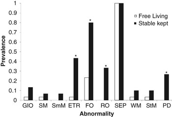
Figure 1The prevalence of each dental abnormality; asterisk indicates a significant difference (χ 2) between groups (P < 0.01).

2. Slant mouth (SM), an abnormality of the incisors whereby the occlusal surface of the incisors wears in a slanted direction, often caused by a horse masticating in only one direction (Dixon and Dacre, Reference Dixon and Dacre2005).
3. Smile mouth (SmM), an abnormality of the incisors whereby the occlusal surface of the incisors wears unevenly to a downwardly curved surface (Du Toit et al., Reference Du Toit, Burden and Dixon2009). The authors have, by extension, also included a dorsal curvature of the incisors in this category.
4. Exaggerated transverse ridges (ETRs) of the occlusal surface of the cheek teeth, as previously described by Dixon (Reference Dixon2002), and beyond the natural ridging of the occlusal surface.
5. Focal overgrowths (FOs) of the cheek teeth (Penzhorn, Reference Penzhorn1984), which frequently occur as overgrowths at positions 106/206 and 311/411 (Baker, Reference Baker1979; Dixon, Reference Dixon2002) and are often referred to as ‘hooks’ (Tell et al., Reference Tell, Egenvall, Lungstrom and Wattle2008).
6. Ramped overgrowths (ROs) of the cheek teeth whereby the opposing cheek teeth at position 06 wear in a sloped manner (Du Toit et al., Reference Du Toit, Gallagher, Burden and Dixon2008a)
7. SEPs on the buccal and lingual edges of the cheek teeth (Du Toit et al., Reference Du Toit, Burden and Dixon2009)
8. Wave mouth (WM) whereby the occlusal surface of the cheek teeth wears unevenly and becomes undulating (Dixon et al., Reference Dixon, Tremaine, Pickles, Kuhns, Hawe, McCann, McGorum, Railton and Brammer2000; Du Toit et al., Reference Du Toit, Burden and Dixon2009). WM is described by Dixon (Reference Dixon2002) as being a severe example of ETRs.
9. Step mouth (StM) whereby a cheek tooth overgrows, often because the equivalent tooth on the opposing arcade is missing or damaged (Dixon et al., Reference Dixon, Tremaine, Pickles, Kuhns, Hawe, McCann, McGorum, Railton and Brammer2000; Du Toit et al., Reference Du Toit, Burden and Dixon2009).
10. Periodontal disease (PD) includes occurrence of gingivitis, inflammation of the gums and/or periodontitis, inflammation of the underlying tissue (Baker, Reference Baker1979).

Statistical analysis
The prevalence of each dental abnormality was calculated as a proportion of the group size (n = 30). Where the occurrence of the dental abnormality was frequent enough for statistical analysis (in the case of ETRs, FOs and ROs of the cheek teeth, SEPs and PDs) the difference between groups was analysed using a χ 2 test using Microsoft Office Excel 2003 (Microsoft Corporation). The difference in the total number of abnormalities per horse in each group was analysed using a Mann–Whitney test using SPSS version 16 (SPSS Inc.).
Results
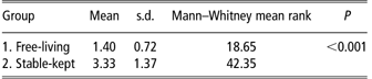
The prevalence of each dental abnormality is shown in Figure 1. The result of the analysis of difference between the total number of abnormalities for each horse per group is shown in Table 1 in which the mean is 30. A count of 1 was given for recognition of any and each of the abnormalities listed above, regardless of severity. For example, a horse showing SEPs and ETRs will be given a count of 2.
There are clear numerical differences between stable-kept and free-living horses in the prevalence of all disorders except SEPs of the cheek teeth. All 10 abnormalities were observed in the stable-kept group and the prevalence was greater than in free-living horses, except in the case of SEPs on the cheek teeth. SmM, ROs of the cheek teeth and PD were not observed in any free-living horses. The difference in occurence was confirmed to be significant in the case of ETRs, FOs of the cheek teeth, ROs of the cheek teeth and PD (P < 0.01 in all cases). Unfortunately, the occurrence of abnormalities was insufficient in the remainder of cases to perform the statistical analysis. In total, significantly more dental abnormalities were observed in stable-kept horses when compared with free-living horses (Table 1; P < 0.001).
Analysis of the difference in number of observations per horse, in each group

Discussion
Until now, there has been little research comparing the dentition of free-living with stable-kept horses. The above observations are of importance as they add weight to the anecdotal argument that a modern equine lifestyle and diet, based on energy-dense cereals as opposed to forage, may adversely affect the dentition of the horse, by increasing the prevalence of dental abnormalities. This is particularly interesting when considered with the observation that there is no difference between the prevalence of SEPs (P = 1) between free-living and stable-kept horses. SEPs are considered to be detrimental and would commonly be removed during dental correction (Ralston et al., Reference Ralston, Foster, Divers and Hintz2001). The result of our study potentially suggests that this dental ‘abnormality’ is to be expected. Other recent studies also challenge the opinion that SEPs on the buccal and lingual edges of the cheek teeth are abnormal features of the horse’s teeth. In two recent studies, such enamel was seen in 98% of 203 donkeys (Du Toit et al., Reference Du Toit, Burden and Dixon2008b) and 96% of 300 horses (Simhofer et al., Reference Simhofer, Griss and Zetner2008). Surprisingly, in the latter study, the horses showed no signs of discomfort, even when there was associated buccal ulceration (Simhofer et al., Reference Simhofer, Griss and Zetner2008). However, another study involving donkeys found SEPs on only 26.9% of the 357 donkeys examined (Du Toit et al., Reference Du Toit, Burden and Dixon2009). The authors suggested, however, that the animals were managed in an extensive system, with a forage-based diet, which may have attributed to this relatively low prevalence. As a result, ‘dental disease’ is a term used by the University of Edinburgh group to describe the equine dental condition. The term includes abnormalities such as those above, but excludes SEPs, but does include diastemata and missing teeth (Du Toit et al., Reference Du Toit, Gallagher, Burden and Dixon2008a and Reference Du Toit, Burden and Dixon2009). Clearly, this is still a threat to animal health, with donkeys exhibiting dental disease significantly more likely to have a lower body condition score (Du Toit et al., Reference Du Toit, Burden and Dixon2009). As horses and donkeys age, it is increasingly more important to monitor dental health as an increase in the prevalence of abnormalities is observed with age (Baker, Reference Baker1979; Du Toit et al., Reference Du Toit, Burden and Dixon2008b and Reference Du Toit, Burden and Dixon2009).
The results of our study may also suggest that further study is required to decide whether routine removal of sharp edges is actually a necessity and that this process is not removing an anatomical feature that is required by the horse. Studies are needed to monitor sharp edges seen in such horses, and whether they develop into a more serious problem, or whether they are maintained in a way that is beneficial, or at least not detrimental, to the horse. There is evidence that routine dental equilibration of the occlusal tooth surface does not necessarily improve performance (Carmalt et al., Reference Carmalt, Carmalt and Barber2006). The same group suggests that ‘floating’ can correct an abnormality with rostrocaudal mobility of the mandible (Carmalt et al., Reference Carmalt, Townsend and Allen2003), but that this does not increase feed digestibility and weight gain (Carmalt et al., Reference Carmalt, Townsend, Janzen and Cymbaluk2004; Carmalt and Allen, Reference Carmalt and Allen2006). There is no increase in the digestibility of crude protein, fibre or dry matter, when small ‘hooks’ and enamel points on pre-molars are removed (Ralston et al., Reference Ralston, Foster, Divers and Hintz2001), and no relationship between occlusal surface morphology and feed digestibility or particle size was found (Carmalt and Allen, Reference Carmalt and Allen2008) . Therefore, it appears that there is no distinct benefit, in terms of feed digestion, in routine smoothing of the occlusal surface. Schumacher (Reference Schumacher2001) suggests that dental abnormalities are often overlooked as the clinical signs are minimal or non-existent. Potentially, this is evidence that these abnormalities often do not cause the horse any harm. This evidence, along with the results of this study, may bring into question the necessity for removing some abnormalities found in certain types of horse and revising what the veterinary dentistry world considers as normal dentition. There is the potential for damage to teeth during prophylactic dentistry (Kempson et al., Reference Kempson, Davidson and Kacre2003; Wilson and Walsh, Reference Wilson and Walsh2005), and it may also be distressing for the horse. One study found that 51% of horses were being sedated for routine dentistry in one area of the United Kingdom (Dixon et al., Reference Dixon, Andrew, Brannon, Burgess, Gibson, Little, Orange, Ross, Rudolph and Shaw2004). This being said there is clearly merit in preventing and removing such abnormalities that begin to impinge on the soft tissues of the mouth, causing pain and potentially leading to infection; qualified EDTs and veterinary surgeons clearly have a key role to play in monitoring and assessing potential dental problems in domesticated equines. There seems to be little evidence in the literature on the percentage of abnormalities that leads to this situation. This kind of research would potentially have huge welfare and ethical implications and would need to be carefully executed to prevent knowingly causing neglect.
By their very nature, Thoroughbreds are descended from a small selection of 17th and 18th century foundation stallions. It is possible that their normal dentition has developed some extent of sharp edges through in-breeding. Thoroughbreds have been shown to have significantly different head measurements in comparison with other breeds (Evans and McGreevy, Reference Evans and McGreevy2006), and it is also suggested that the subtle conformational structures of teeth vary with specific breeds (Friker et al., Reference Friker, Dias and Zeiler2006). Often, it is likely that severe dental abnormalities that result from anatomical problems, and which would not correct themselves, would be fatal in free-living horses. Therefore, it is likely that horses in free-living populations do not exhibit so many abnormalities, as many of the problems are bred out by natural selection. It is not possible to suggest this as the reason for the lack of abnormalities in the current free-living population, as they had some history of dental treatment. However, historically this theory may be applied to the phenotype, leading to a lesser prevalence of abnormalities through time.
A scoring system that indicates the severity of a dental abnormality could be developed, incorporating a cut-off point at which routine procedures are necessary. It is routine for owners to know their horse’s basal respiration, temperature and heart rate, and it would potentially be useful to add ‘dental norm’ to this list.
Acknowledgements
The authors would like to thank Mr Martin Brookes, the Grills Family of Berragoon Australian Stock Horse Stud and Mr Hitchman of Beaufort Polo Club.