Implications
Since their discovery, recombinant fragments from heavy-chain antibodies (HCAbs) have been considered to be a new platform to produce target inhibitory Ab fragments, and many groups have devoted to characterize them. Nevertheless, our work is one of the few aimed to understand the correlation between these findings and the role of HCAbs during in vivo immune responses. Our results show that HCAbs contribute only in a modest manner to whole serum titer, and that enzyme inhibition is not as easily achieved as previous works seem to suggest. Therefore, the natural role of these Abs remains an issue that deserves further research.
Introduction
In the last decades, the knowledge about the structure and function of antibodies (Abs) has shown a marked progress. As part of it, several new Ab classes have been described and cataloged as ‘non-conventional’ because of their structural and biochemical properties. One of the most relevant discoveries in this field is the existence of functional immunoglobulin (Ig) isotypes devoid of light chains, named ‘Heavy-Chain Antibodies’ (HCAbs) as a consequence of their homodimeric structure. These Abs were first reported by Hamers-Casterman et al. (Reference Hamers-Casterman, Atarhouch, Muyldermans, Robinson, Hamers, Songa, Bendahman and Hamers1993), and they have been extensively described for all the members of the Camelidae family (dromedaries, camels, llamas, guanacos, vicunas and alpacas; Muyldermans et al., Reference Muyldermans2001; Nguyen et al., Reference Nguyen, Su, Muyldermans and van der Loo2002; Su et al., Reference Su, Nguyen and Nei2002). In these studies, it was reported that conventional Abs (IgG1 and IgM) constitute about 60% to 80% of total mass of serum Igs, and HCAbs (IgG2 and IgG3) the remaining 20% to 40% (De Simone et al., Reference De Simone, Saccodossi, Ferrari and Leoni2008).
Since the discovery of HCAbs, many researchers have focused their work on the biophysical properties of the recombinant variable fragments (rVHHs) of HCAbs, as well as on their possible biotechnological applications (Saerens et al., Reference Saerens, Ghassabeh and Muyldermans2008; Muyldermans et al., Reference Muyldermans2009; Doña et al., Reference Doña, Urrutia, Bayardo, Alzogaray, Goldbaum and Chirdo2010). Among the unique features that were described, it was found that these rVHHs display an unusually reversible unfolding when exposed to high temperature and denaturing agents (van der Linden et al., Reference van der Linden, Frenken, de Geus, Harmsen, Ruuls, Stok, de Ron, Wilson, Davis and Verrips1999; Pérez et al., Reference Pérez, Renisio, Prompers, van Platernik, Cambillau, Darbon and Frenken2001; Ewert et al., Reference Ewert, Cambillau, Conrath and Plückthun2002). In addition, it has been reported that the non-canonical structure of complementary determining regions leads to a preferential cleft recognition, allowing rVHHs to interact with epitopes that are cryptic for conventional Abs, similar to the active site of enzymes (Martin et al., Reference Martin, Volpari, Steinkuhler, Dimasi, Brunetti, Biasiol, Altamara, Cortese, De Francesco and Sollazzo1997; Lauwereys et al., Reference Lauwereys, Ghahroudi, Desmyter, Kinne, Hölzer, De Genst, Wyns and Muyldermans1998; De Genst et al., Reference De Genst, Silence, Decanniere, Conrath, Loris, Kinne, Muyldermans and Wyns2006). Therefore, these rVHHs have been proposed as an ideal source of specific small inhibitory molecules.
Although the reports on rVHH's properties are outstanding and have already given us fundamental knowledge for future biomedical technologies, a question arises on whether this inhibitory capacity can also be obtained by raising polyclonal HCAbs (pHCAbs) during in vivo immune responses. This issue has been partially addressed by Lauwereys et al. (Reference Lauwereys, Ghahroudi, Desmyter, Kinne, Hölzer, De Genst, Wyns and Muyldermans1998), who succeeded in demonstrating enzyme modulation by rVHH fragments for two enzymes (lysozyme and α-amylase); however, it is not clear whether these results may be extrapolated to all the other enzymes, or are a consequence of a special feature of the selected antigens. The relevance of this matter is reinforced by our own preliminary reports concerning β-lactamase activation by pHCAbs from a single serum sample of a llama (Lama glama; Ferrari et al., Reference Ferrari, Rodríguez, Power, Weill, De Simone, Gutkind and Leoni2007).
For the reasons mentioned above, in this work we further explored the interaction between pHCAbs and two microbial enzymes, characterizing this interaction during the time-course of a short and a long immunization schedule. The enzymes that were selected have their active sites buried in clefts (Matagne et al., Reference Matagne and Frère1995; Yang et al., Reference Yang, Teplyakov and Quail1997; Page et al., Reference Page and Laws1998). Both the enzymes belong to families of well-characterized proteins, with known sequence and resolved crystallographic structure. In addition, their catalytic activity can be measured by simple methods. The ability to specifically recognize the enzymes and modulate their enzymatic activity in vitro was studied for total serum, the most abundant conventional Ig subisotype (IgG1) and the most abundant HCAb subisotype (IgG3). The properties of these three fractions were followed and compared within the immunization schedule in order to compose a time-dependent profile based on the behavior of specific Abs raised against the enzymes.
Material and methods
Aspartic protease (APM) purification
An APM from Mucor miehei was purified from a commercial solid preparation (DSM Food Specialties, Delft, the Netherlands), by size-exclusion chromatography. A solution containing 400 mg/ml of the solid was prepared in phosphate buffered saline (PBS), and 500 μl were loaded into a Sephadex G-150 column (GE Healthcare, Milwaukee, USA) (100 cm × 1 cm) using PBS as the mobile phase. Fractions of 1 ml were collected, and protein purity was assessed by 12% SDS-PAGE. All fractions containing >95% of APM were pooled, protein concentration was measured using the BCA Protein Assay kit (Pierce Biotechnology Inc., Rockford, IL, USA), and aliquots were stored at −20°C until use.
β-lactamase expression and purification
A recombinant Escherichia coli clone with a plasmid encoding for a CTX-M-2 like β-lactamase (CTX-M-2) from Morganella morganii, was kindly provided by Dr Gutkind (Facultad de Farmacia y Bioquímica, Universidad de Buenos Aires). Cloning, expression and purification were performed as previously described (Power et al., Reference Power, Galleni, Ayala and Gutkind2005; Ferrari et al., Reference Ferrari, Rodríguez, Power, Weill, De Simone, Gutkind and Leoni2007).
Animals and immunization schedules
Two adult llamas were inoculated with APM (L1APM and L2APM), and two adult llamas were inoculated with CTX-M-2 (L1CTX and L2CTX). All animal experimentations have been carried out in accordance with the EC Directive 86/609/European Economic Community for animal experiments, and the Guiding Principles of the Exact and Natural Sciences School, University of Buenos Aires, Argentina.
Preimmune and immune sera were obtained by jugular vein puncture. All the animals were immunized subcutaneously using a mixture of equal parts of the immunogen solution (700 μg of total mass) and Complete Freund Adjuvant. Animals immunized with APM were inoculated once a week, and serum samples were collected at days 0 (preimmune), 8, 29 and 42. Animals immunized with CTX-M-2 were inoculated every 2 weeks and serum samples were collected at days 0 (preimmune), 60, 75, 90, 150, 170 and 215.
Serum titration by ELISA
The titration of total serum Igs was performed as previously described (Ferrari et al., Reference Ferrari, Rodríguez, Power, Weill, De Simone, Gutkind and Leoni2007). Briefly, 96-well microplates (Nunc Maxisorp, Roskilde, Denmark) were coated with 0.1 μg of purified APM or CTX-M-2 per well, and blocked with 3% non-fat milk diluted in PBS. Coated microplates were incubated for 1 h at 37°C with serum samples in twofold dilutions, and then with a peroxidase labeled secondary Ab (Horse Radish Peroxidase goat anti-llama IgG; Bethyl Laboratories Inc., Montgomery, TX, USA) diluted 1/4000 in PBS. Immunoreactivity was developed with O-phenylenediamine 2 mg/ml and H2O2 0.1% v/v during 10 min. Optical density (OD) was measured in a Multiscan EX microplate reader (Thermo Electron Corporation, Massachusetts, USA) and ELISA titers were expressed as the inverse of the dilution of the central value of the sigmoidal curve (EC50).
Purification of llama Igs
IgG1 and IgG3 were purified from the selected serum samples as previously described (De Simone et al., Reference De Simone, Saccodossi, Ferrari, Leoni and Leoni2005; Ferrari et al., Reference Ferrari, Rodríguez, Power, Weill, De Simone, Gutkind and Leoni2007), using a HiTrap-protein G column (GE Healthcare, Piscataway, NJ, USA) and low pH elution buffers (pH 3.5 for IgG3 and pH 2.7 for IgG1). All Ab fractions were neutralized with Tris 1 M, dialyzed against PBS and protein purity was assessed by 10% SDS-PAGE. Samples were stored at −20°C until use. An additional fraction containing total Igs was obtained by serum precipitation with 38% (NH4)2SO4, and used to assess the modulatory capacity of APM by total Abs; this procedure was performed in order to eliminate non-Ig proteins, as it was demonstrated that serum proteins comprised an interference for the activity modulation assays of APM (data not shown). Control experiments proved that proteins do not interfere with the activity measurement of CTX-M-2.
Titration of purified Igs
Purified IgG1 and IgG3 from all animals were titrated as described for the whole sera, starting at 10 μg/ml of Ig concentration, using twofold dilutions.
APM activity modulation assays
The enzymatic activity of APM was determined by the hydrolysis of azocasein (Sigma Chemical Co., St Louis, MO, USA) at 37°C, as it has been previously described (Bendicho et al., Reference Bendicho, Martí, Hernández and Martín2002). Briefly, APM was incubated in the presence of azocasein (5 mg/ml) in acetate buffer 0.1 M pH 5.0, for 90 min at 37°C. Non-hydrolyzed azocasein and other proteins were precipitated with 50 μl of 20% trichloroacetic acid and then centrifuged at 10 000×g during 10 min at 4°C. To measure the concentration of soluble peptides produced by azocasein hydrolysis, 200 μl of each sample were transferred to a 96-well microplate, and OD at 405 nm was measured using a microplate reader.
The activity modulation capacity of total Igs from serum samples was determined by preincubating APM with a fixed mass of total Igs, composed of different proportions of immune/preimmune Igs obtained by SO4(NH4)2 precipitation. The activity modulation capacity of purified llama subisotypes was determined by preincubating APM with a fixed mass of Igs obtained by affinity chromatography, composed of different proportions of immune/preimmune Igs.
Ig degradation due to APM activity during the experiments was checked by SDS-PAGE at different incubation times, and considered negligible (results not shown).
CTX-M-2 activity modulation assays
The enzymatic activity of purified CTX-M-2 was determined by the hydrolysis of cephalothin (Ivax Argentina S.A., Buenos Aires, Argentina) at 37°C during 150 s, and measuring UV absorption at 273 nm  in a thermostatized spectrophotometer (Beckman Coulter DU 7400, Fullerton, CA, USA).
The activity modulation capacity of serum samples was determined by preincubating CTX-M-2 with a fixed volume of serum, composed of different proportions of immune/preimmune sera. The activity modulation capacity of purified llama subisotypes was determined by preincubating CTX-M-2 with a fixed mass of Ig, composed of different proportions of immune/preimmune Igs.
Nonlinear regression analysis of modulation assays
Nonlinear regression analyses were performed using GraphPad Prism 4.0 (GraphPad Software, San Diego, CA, USA). A ‘one phase exponential decay/association’ curve was fitted to residual enzymatic activity data, and two parameters were obtained: the maximum modulation (MM) achieved, and the Ab concentration that produces 50% of MM (Modulatory Capacity 50, MC50). An arbitrary score was calculated for each serum sample, according to expression (1):

This score assigns an arbitrary relative weight to each parameter, compensating their differences in magnitude and units. It was empirically constructed, and it is expected to facilitate the analysis of the modulation assays. The f MW terminus is a correction factor applied to compensate the differences in molecular weight between conventional (f MW = 1.0) and non-conventional Abs (f MW = 1.5).
Statistical analyses
Titration assays were further analyzed by ANOVA, and a Dunnet post test was performed, comparing each condition v. the condition without immune Igs. Differences were considered significant when P < 0.05.
Results
Antigen purification
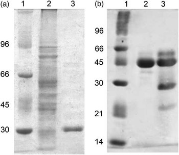
Both the antigens were purified as described, and their purity was verified by SDS-PAGE, as shown in Figure 1. Purity was demonstrated to be at least 95%, as demonstrated by a densitometry analysis.
Verification of antigen purity by SDS-PAGE. Panel (a) shows purity of CTX-M-2: molecular weight standards (lane 1), bacterial lysate (lane 2) and purified protein (lane 3). Panel (b) shows purity of APM: molecular weight standards (lane 1), purified protein (lane 2) and commercial solid preparation (lane 3).

Serum titration by ELISA
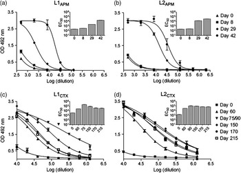
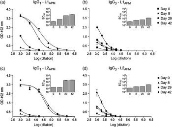
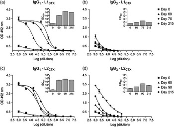
All llamas elicited a significant antigen-specific immune response. Serum titers of animals L1APM and L2APM began to rise from day 8 until the end of the immunization schedule; titers of L1CTX and L2CTX reached a maximum at days 75 (L1CTX) or 90 (L2CTX), and then decreased to a lower and more stable value until the end of their immunization schedule (Figure 2).
Serum titration by ELISA. Curves correspond to titration assays, and bar graphs are the corresponding EC50 values for each curve. Error bars represent the standard deviation. All differences were significant by a Dunnet post test, except the ones corresponding to day 8 from L1APM and L2APM.
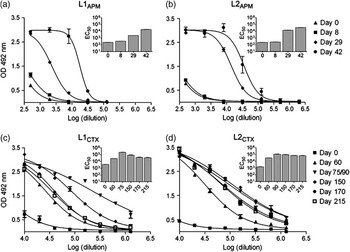
According to these results, three different serum samples from animals L1CTX and L2CTX were selected for the catalytic modulation assays, and all serum samples from animals L1APM and L2APM were used. All further Ig purification and activity modulation assays were performed with these samples.
Activity modulation assays with total Igs
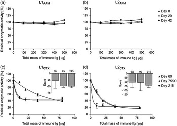
Total Igs precipitated from L1APM and L2APM samples produced no significant enzyme modulation, and therefore neither the curve fitting nor the modulatory score could be calculated. On the contrary, serum samples from L1CTX and L2CTX had increasing inhibitory power during the schedule (Figure 3). The modulatory score of both animals immunized with CTX-M-2 had a moderate tendency to increase within the immunization schedule.
Aspartic protease and CTX-M-2 like β-lactamase modulation assays by total serum immunoglobulins (Igs). Curves correspond to activity modulation assays, and were constructed plotting the residual enzymatic activity v. the absolute mass of immune Ig used in the reaction mixture. Total serum Igs were considered to be at a concentration of 10 mg/ml. Bar graphs are the corresponding score values for each curve; error bars represent the standard error, as calculated by the error propagation method.

Purification and titration of Igs
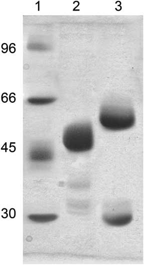
Purification of IgG1 and IgG3 was performed as described, and their purity was assessed by SDS-PAGE (Figure 4). All fractions were proved to have a purity of at least 95%, as demonstrated by a densitometry analysis. Titrations, as shown in Figures 5 and 6, indicate that purified IgG1 and IgG3 from all animals have increasing titers during the immunization schedule. IgG1 titers had much higher values than those of IgG3 in all animals, and its progress pattern resembles the behavior of total Igs. This difference in the relative contribution of each subisotype is reinforced by the fact that normal serum IgG3 concentration, although being the most abundant HCAb isotype, has been previously reported to be 5 to 10 times lower than that of IgG1 (De Simone et al., Reference De Simone, Saccodossi, Ferrari and Leoni2008).
Purity of purified IgG1 and IgG3. Lanes correspond to molecular weight standards (lane 1), IgG3 (lane 2) and IgG1 (lane 3), under reducing conditions. The figure shows that IgG3 is composed of a single sort of chain (47 kDa) and that IgG1 is composed of heavy (50 kDa) and light (30 kDa) chains.

Titration of purified immunoglobulins (Igs) from animals L1APM and L2APM. Curves correspond to ELISA titration assays, and bar graphs are the corresponding EC50 values for each curve. Error bars represent the standard deviation. All differences were significant by a Dunnet post test, except the one corresponding to IgG1 from day 8 (L2APM).

Titration of purified immunoglobulins (Igs) from animals L1CTX and L2CTX. Curves correspond to ELISA titration assays, and bar graphs are the corresponding EC50 values for each curve. Error bars represent the standard deviation. All differences were significant by a Dunnet post test.

Activity modulation assays with purified Igs
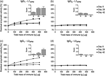
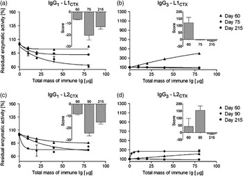
IgG1 from both L1APM and L2APM produced a positive modulation of APM activity (Figure 7); on the contrary, IgG3 had no effect on the enzyme. In contrast, purified IgG1 from animals L1CTX and L2CTX produced an intense negative modulation of CTX-M-2 activity, as shown in Figure 8. This modulation had a progress pattern with less intense values than that of serum, and a maximum at days 75 (L1CTX) or 90 (L2CTX). Purified IgG3 from both animals immunized with CTX-M-2, not only failed to produce a negative modulation of the enzyme, but also induced a strong positive modulation of its catalytic activity. This positive modulation had a maximum at days 60 (L1CTX) or 90 (L2CTX), and then decreased to a slight negative modulation.
Aspartic protease activity modulation assays by purified immunoglobulins (Igs). Curves correspond to activity modulation assays, and were constructed plotting the residual enzymatic activity v. the absolute mass of immune Ig used in the reaction mixture. Bar graphs are the corresponding score values for each curve; error bars represent the standard error, as calculated by the error propagation method.

CTX-M-2 like β-lactamase activity modulation assays by purified immunoglobulins (Igs). Curves correspond to activity modulation assays, and were constructed plotting the residual enzymatic activity v. the absolute mass of immune Ig used in the reaction mixture. Bar graphs are the corresponding score values for each curve; error bars represent the standard error, as calculated by the error propagation method.

Discussion
Previous reports have stated that rVHHs have a special ability to interact with certain epitopes that are cryptic to conventional Abs, similar to the active site of enzymes. These reports also show that because of this interaction, rVHHs have a natural ‘preference’ to negatively modulate the catalytic activity of enzymes (Lauwereys et al., Reference Lauwereys, Ghahroudi, Desmyter, Kinne, Hölzer, De Genst, Wyns and Muyldermans1998; De Genst et al., Reference De Genst, Silence, Decanniere, Conrath, Loris, Kinne, Muyldermans and Wyns2006). Nevertheless, it is not clear whether these results depend or not on the structure and chemical properties of the selected antigen. Furthermore, preliminary studies performed by our group on a single serum sample had suggested that polyclonal IgG3 may be unable to inhibit an enzymatic immunogen, producing instead an unexpected strong activation.
In order to analyze whether significant quantities of HCAbs that negatively modulate enzymes can be obtained and purified from polyclonal sera, two enzymes were used to immunize llamas, and the modulatory response was characterized during the time-course of the immunization schedule. Total Ig titers indicate that all animals elicited a significant humoral immune response. Although the immunization schedule with APM yielded the expected ELISA titers, total Igs failed to modulate the catalytic activity of APM. In contrast, serum from animals immunized with CTX-M-2 produced a strong negative modulation of the enzyme, which progressed with time. The results obtained with the two groups of animals are not fully comparable, considering that they were immunized with different enzymes under different schedules; nevertheless, they indicate that both strategies yielded high titers of specific Abs.
IgG1 and IgG3 were purified from all animals. ELISA titers showed that the total Ig titer is mainly due to IgG1, as the progress profile of this subisotype has similar pattern to total Igs. The modulatory capacity of purified Igs was then studied. These assays revealed that IgG1 from animals immunized with APM produced a moderate positive modulation of the catalytic activity, and that IgG3 failed to produce any enzyme modulation. In contrast, IgG1 from animals immunized with CTX-M-2 produced a strong negative modulation and IgG3 produced an intense positive modulation, which reverted to a subtle negative effect at the end of the immunization schedule.
The results presented in this work confirm our previous findings, now with two different enzymatic systems and during the whole immunization schedule. Total pHCAbs failed to inhibit the enzymes used as immunogens, either because no inhibitory HCAbs are produced or because these are produced in combination with activatory HCAbs. Indeed, total pHCAbs not only did not inhibit the enzymes but also activated one of them – in accordance with Saerens et al. (Reference Saerens, Kinne, Bosmans, Wernery, Muyldermans and Conrath2004), who reported prostate-specific antigen activation by rVHH – suggesting that production of polyclonal inhibitory HCAbs during in vivo immune responses is not a straightforward strategy as the production of rVHHs. Considering our results and recent reports on llamas and camels (van der Linden et al., Reference van der Linden, de Geus, Stok, Bos, van Wassenaar, Verrips and Frenken2000; Cook et al., Reference Cook, Samarasekara, Wagstaff, Kinne, Wernery and Harrison2010), the nature of the selected antigen and the immunization plan could play a main role in biasing the humoral response toward conventional or HCAbs.
Acknowledgments
The authors thank Dr Gabriel Gutkind, Dr Pablo Power and Biochemist María M. Rodríguez, for their help with CTX-M-2. This work was granted by Agencia de Promoción Científica y Tecnológica (ANPCyT), PICT 2004 No. 26162 and Consejo Nacional de Investigaciones Científicas y Técnicas (Conicet).
Additional data published after this submission
After the submission of our article, an article was published in the PLoS Pathogens (Stijlemans et al., Reference Stijlemans, Caljon, Natesan, Saerens, Conrath, Pérez-Morga, Skepper, Nikolaou, Brys, Pays, Magez, Field, De Baetselier and Muyldermans2011) that might contribute to solving the apparent discrepancy between our results on polyclonal heavy-chain antibodies (pHCAbs) and the current references on recombinant variable fragments (rVHHs). In that article the authors describe a trypanolytic activity of rVHHs, that cannot be revealed in reconstituted heavy-chain antibodies (HCAbs; by means of a molecular biology strategy), but can be demonstrated in polyclonal VHHs obtained by controlled proteolysis. These results suggest that some of the features described for variable fragments of HCAbs might not be detectable in whole molecules, and are consistent with our results.