Introduction
Microbial synthesis of nanoparticles has emerged as a promising field of research known as a branch of Nanobiotechnology that interconnects biotechnology and nanotechnology.[Reference Narayanan and Sakthivel1] The biological synthesis approaches for nanoparticles are safe, cost-effective, and furthermore it is eco-friendly as compared to the typical chemical processes that demand stringent conditions and often involve toxic chemicals. Many microorganisms produce a variety of inorganic materials either intra- or extracellularly. For example, Konishi et al.[Reference Konishi, Tsukiyama, Tachimi, Saitoh, Nomura and Nagamine2] reported that Shewanella algae reduced gold ions on bacterial surfaces at pH 2.8 extracellularly. Prasad et al.[Reference Prasad, Jha and Kulkarni3] demonstrated lactobacillus-assisted synthesis of Titanium spherical nanoparticles having a size of 40–60 nm. Mao et al.[Reference Mao, Solis, Reiss, Kottmann, Sweeney, Hayhurst, Georgiou, Iverson and Belcher4] used the virus-based scaffold for the synthesis of semiconducting (single-crystal ZnS and CdS) and magnetic (CoPt and FePt) nanowires.
Considerable amount of research on microbial carbonate precipitation (MCP) has been done using urealytic bacteria.[Reference Millo, Ader, Dupraz, Guyot, Thaler, Foy and Ménez5, Reference Chahal, Rajor and Siddique6] Most commonly studied system for MCP is the urea hydrolysis via the enzyme urease in a calcium-rich environment. This enzyme catalyzes the hydrolysis of urea to CO32− and NH4+ ions. These results in an increase of the pH and carbonate concentration in the bacterial environment and is able to influence precipitation of the calcium carbonate.
 $$\eqalign{& {\rm CO}\lpar {\rm NH}_2\rpar _2 + 2{{\rm H}_2}{\rm O} \to {\rm 2NH}_ 4^ {+} + {\rm CO}_3^{2 - }\comma \; \cr & {\rm Ca}^{2 +} + {{\rm CO}_ 3}^{2 - } \to {{\rm CaCO}_ 3}.}$$
Similar pathways for transition metal ions precipitation are an area of considerable interest for synthesis of the metal oxide structures. The present research aims to obtain Ni precipitates of various structures produced from Ni solution using bacteria as templates. Related research has been done by Magyarosy et al.[Reference Magyarosy, Laidlaw, Kilaas, Echer, Clark and Keasling7] who reported nickel oxalate rectangular crystals by Aspergillus niger on cell walls and inside the cell.
NiO is an inorganic compound of technological importance in fuel cell, lithium-ion batteries, supercapacitors and exhibits magnetic and catalytic properties.[Reference Park and Cairns8–Reference Ren, Yang, Sun, Hui, Dong, Wang and Su12] These properties are determined by the size, shape, and crystallinity of the nanoparticles. NiO hollow materials with different shapes such as spheres and polyhedron are known in the literature. Wang et al.[Reference Wang, Song, Hu and Fu13] reported the synthesis of hollow NiO spheres using surface of styrene-acrylic acid copolymer latex particles to form core/shell structures. Qian et al.[Reference Qian, Lin, Zhang, Gunawan and Xu14] also used urea as a precipitant for the synthesis of hollow nanostructured metal oxides with carbonaceous saccharide nanospheres as hard templates. Wang et al.[Reference Wang, Yu, Hu and Yuan15] reported the synthesis of single-crystalline hollow NiO crystals with octahedral morphology using a template-assisted carbothermal method.
Fabrication of hollow nanostructures has attracted interest in recent years because of their well-defined interior voids, low-density, large surface area, stability, and surface permeability.[Reference Wang, Tang, Ozawa, Chen, Mukherji, Zhu, Zou, Cheng and Lu16] Such hollow structures may find applications as chemical reactors, drug-delivery carriers, energy-storage devices, catalysis, and others.[Reference Fei, Cui, Yan, Qi, Yang, Wang, He and Li17, Reference Lou, Yuan, Zhang and Archer18] Various template and template-free methods are reported for the formation of hollow-shaped micro- and nanosized crystals. The reliable technique for construction of hollow materials with well-defined shapes, however, is still a challenge to material scientists. Self-propelled devices are of interest in biomedical micro/nanorobots.[Reference Ghosh and Fischer19, Reference Pak, Gao, Wang and Lauga20] Helically shaped micro/nanoscale propulsion has also attracted considerable attention due to its promise for biomedical applications such as targeted drug delivery.[Reference Ghosh and Fischer19] The magnetically driven helical devices can be used as wireless manipulators for medical and biological applications under three-dimensional control in fluid environments.
In this report, we present an eco-friendly pathway for the formation of hollow cylinder and helically shaped NiO micro/nanostructure using Bacillus pasteurii as a synthesis director under urea hydrolysis conditions. To the best of our knowledge there have been no reports on the synthesis of hollow cylinder and helical NiO materials. A formation mechanism of these NiO structures is also proposed.
Experimental procedures
Materials and characterization
NiCl2·6H2O (Choneye Pure Chemicals), yeast (Becton Dickinson and Company), ammonium sulfate (J.T. Baker), Tris–HCl (J.T. Baker), and Milli-Q water were used in all experiments
Bacteria used were B. pasteurii
Samples of the isolated strain were cultivated under sterile aerobic batch conditions in a medium consisting of 20 g/L yeast, 10 g ammonia sulfate, and 20.48 g Tris–HCl at pH of 8.5. The bacterial solution had an optical density of 1.2 (107 cells/mL). Biomass concentration was determined by measuring optical density of bacterial suspension with a spectrophotometer at a wavelength of 600 nm.
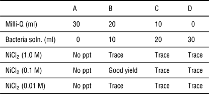
Table I presents the experimental conditions and the results obtained from the synthesis experiments are explained in the section of ‘Results and discussion’. The required amount of NiCl2.6H2O, milli-Q, and urea were mixed to form homogeneous solution in a screw-capped glass tube and bacterial solution was added. The tube was then kept in an orbital shaker (200 rpm) at 35 °C for 24 h. The precipitate obtained was separated by centrifugation at 3700 rpm for 15 min. The precipitates were washed several times with water and ethanol, respectively, and dried in an air oven for 6 h at 50 °C. The precipitates were finally calcined at a temperature of 550 °C for 6 h. After calcination, the light green precipitate became a dark color.
Detailed experimental conditions to obtain Ni precipitate.

Reaction conditions: urea, 1.0 M; temperature 35 °C; time, 24 h; bacterial solution, OD 1.2; total volume, 30 mL.
The amount of nickel recovered from the solution after precipitation with bacteria were determined by titration techniques, which included a colorimetric titration with 0.1 N hydrochloric acid standard solution combined with an indicator methyl orange.
The obtained powder was collected for the following characterizations. Powder X-ray diffraction (XRD) patterns for Ni(OH)2 and NiO were recorded on a Shimadzu X-ray diffractometer (model LabX XRD-6000) equipped with Ni-filtered CuKα (λ = 0.1541 nm, 4 kVA, 30 mA) radiation and a graphite crystal monochromator. Scanning electron microscope (SEM) measurements were carried out on a Leica Stereoscan-440 SEM)equipped with a Phoenix EDAX attachment. The microstructure of the samples was analyzed by transmission electron microscope (TEM) using a JEOL 3000F high-resolution transmission electron microscope (HRTEM) operated at an accelerating voltage of 200 kV. Fourier transmission infrared spectroscopy (FTIR) spectra of the samples were recorded on a Shimadzu 8300 FTIR spectrometer.
Results and discussion
Metal plays an important biological role in the life processes; nickel is one of the essential nutrients for the microorganism. However, high concentration of nickel is toxic to organism. A series of experiments were performed to find out the optimized conditions such as concentration of metal ions and the amount of biological material required for the precipitation by bacteria during urea hydrolysis. The results are presented in Table I. Using only milli-Q water as sole solvent no precipitation took place, also pure bacterial solution did not yield good product. However, the appropriate combination of water (20 mL) and bacterial solutions (10 mL, OD 1.2) were required to precipitate the nickel product under the urea hydrolysis conditions under our experimental conditions. The mechanism of urea hydrolysis is reported elsewhere.[Reference Krajewska21, Reference Millo, Ader, Dupraz, Guyot, Thaler, Foy and Ménez5] These experiments clearly demonstrate that both bacteria and water were required to precipitate nickel from solution. By trace precipitate (Table I) we mean that the material obtained was sufficient for characterization and did not produce good isolated yield. However, a good isolated yield of 35% was obtained using 0.1 M NiCl2, 20 mL milli-Q water, and 10 mL bacterial solution (1.2 × 10⁷ cells/mL). The initial pH of 6.7 increased to 7.4, suggesting urease activity and the hydrolysis of urea. The present work reports characterization of this crop of Ni precipitate.
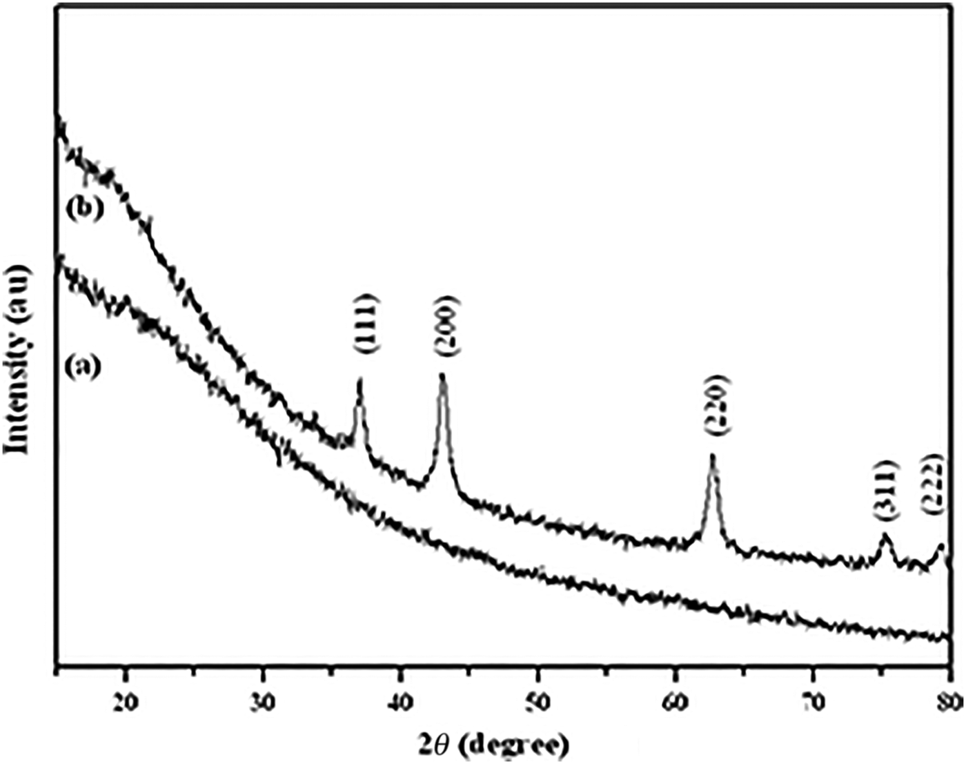
There are several reports on the synthesis of Ni(OH)2 using urea as a precipitant material. Synthesis methods such as hydrothermal, sonochemical, microwave-assisted hydrothermal reported the formation of α-nickel hydroxide phase.[Reference Acharya, Subbaiah, Anand and Das22–Reference Zhu, Wei, Liu and He24] There are two polymorphs of nickel hydroxide α-Ni(OH)2 and β-Ni(OH)2. The α-Ni(OH)2 phase displays a more disordered and larger interlayer spacing as the interlamellar space contains anions, e.g., NO3−, CO32−, SO42−, and water molecules. The characteristics of the Ni precipitate obtained with bacteria were determined. The Ni precipitate, as shown in Fig. 1(a), did not show any XRD peaks, indicating the amorphous nature of the substance. It may be noted that the sample was comprised dead bacterial organic matter (discussed in IR studies), which could be one of the reasons of not showing the XRD peaks. Thus, the exact nature of Ni precipitate (either nickel hydroxide or nickel carbonate) could not be ascertained. However, thermal decomposition of Ni precipitate yields NiO: seen in Fig. 1(b) that showed the XRD peaks of the calcinated product. The calcination process increased crystallinity of the Ni precipitate (conversion to NiO) with the removal of bacterial template. On calcinations the bacterial template is removed which can be seen from IR studies (Fig. 2) show no signals of nucleic acid, cell-wall constituents, and protein which indicated bacterial template is removed on calcinations. On the removal of organic components, Ni precipitate transforms to NiO and showed the XRD peaks.
XRD of nickel products: (a) Ni precipitate, (b) calcination of Ni precipitate at 550 °C for 6 h (NiO).
FTIR patterns of Ni precipitate and NiO powders.
All peaks could be indexed to the crystalline structure of NiO. The diffraction peaks can be perfectly indexed to the face-centered cubic crystalline structure of NiO (JCPDS no. 78-0643). The diffraction peaks are not sharp suggesting that the sample is composed of smaller crystallite grains. The average crystallite size of NiO was calculated from the intense XRD peak using Scherrer formula. The crystallite size of NiO was found to be 11.45 nm. The elemental composition was confirmed with energy-dispersive X-ray (EDX) analysis. The results of the EDX analysis confirmed that the product consisted solely of nickel, carbon, and oxygen, a chemical composition that is not in agreement with that of pure NiO. The presence of carbon in the final product is due to the decomposition of organic bacterial remains. Only the peaks of cubic NiO could be observed, indicating amorphous nature of the carbon in the NiO composite. The EDX figure of Ni precipitate and NiO is shown in Figs. S1 and S2, respectively.
Figure 2 displays the FTIR pattern of Ni precipitate and its calcined product, NiO. It may be noted that as-obtained material has dead bacterial constituents, which are comprised nucleic acid structures, proteinaceous structures, and aliphatic structural units. Filip et al.[Reference Filip, Herrmann and Kubat25] reported the detailed FTIR studies on Bacillus subtilis grown under different culture conditions. The IR spectrum of Ni precipitate showed a very broad vibrational band from 3600 to 2400 cm−1 and from 1800 to 1200 cm−1 in addition to bands at 1075, 837, and 723 cm−1. Based on the results as described by Filip et al.[Reference Filip, Herrmann and Kubat25] we assign FTIR spectra peaks. The broad peak about 3300 cm−1 is due to nucleic acid structures, cell-wall constituents at 3000–2800 cm−1, and proteinaceous structures at 1800–1200 cm−1. The peak at 1075 cm−1 is due to aliphatic structural units. On calcination many of these peaks disappear and reduced in intensity; the calcined sample showed the characteristics of NiO comprising a broad band about 3424 cm−1 which are due to OH-stretching vibrations, and the broad absorptions about 1631 cm−1 can be assigned to existence of the water. A strong band below 600 cm−1 can be attributed to the Ni–O stretching band.
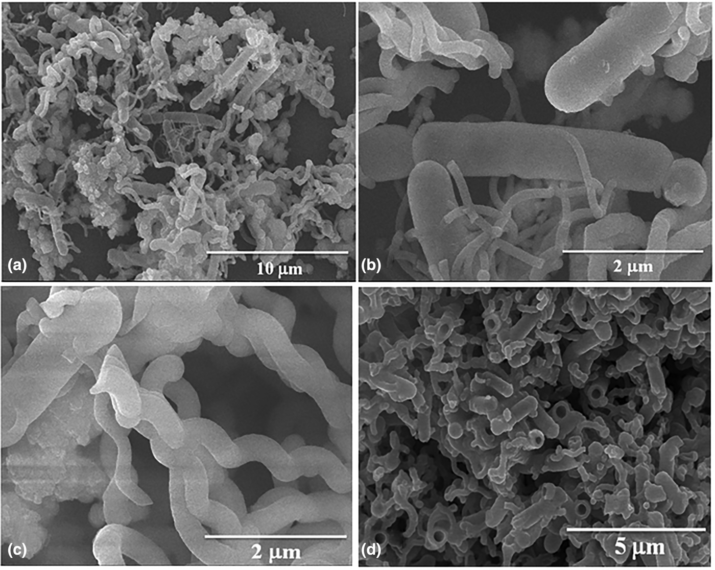
The morphology and dimension of the Ni precipitate and annealed products were examined by an SEM. Figure 3 shows the SEM images of hollow cylinder and helical structures of the as-obtained nickel product. Figure 3(a) shows the panoramic view containing both the cylinder and helical structures. Figure 3(b) shows a single-cylinder structure. Figure 3(c) shows the helical structure and Fig. 3(d) shows the cracked cylinder with hollow cavity. These shapes were preserved even after the calcination at 550 °C for 6 h. Figure S3 shows the SEM images of NiO. The average wall width of hollow cylinder was found to be ~0.65 µm. The SEM image of NiO obtained from the reaction is composed of a large quantity of well-dispersed nickel oxide having definite internal diameters and thickness of the wall varied from each other. The surface is not smooth suggesting that they might be made from aggregation of smaller particles (Fig. S3). The central hollow cavity shows the presence of both interior and exterior surfaces. The length of each cylinder varied and was non-uniform; as long as 7 µm could be found. Similarly the helical structures were also found to be non-uniform in lengths. Bacteria have flagella that propel them toward the favorable conditions. Flagellum has helical form,[Reference Samatey, Matsunami, Imada, Nagashima, Shaikh, Thomas, Chen, DeRosier, Kitao and Namba26] helical NiO structures may have originated from bacterial flagella-shaped structures or other proteinaceous structures; however, detailed studies are required to ascertain its origin. The explanation of helical structure formation is beyond the scope of this paper. Ghosh and Fischer[Reference Ghosh and Fischer19] reported SiO2 helices; these propellers were typically 200–300 nm in width and about 1–2 µm long. These helices were fabricated with the aid of sophisticated instruments involving vapor deposition and computer-controlled substrate manipulator. Zhang et al.[Reference Zhang, Abbott, Dong, Kratochvil, Bell and Nelson27] reported artificial bacterial flagella based on the self-scrolling technique, which represent the demonstration of microscopic artificial swimmers. Our method is an easy way to produce hollow-shaped NiO helical macrostructures.
SEM images of the as obtained Ni precipitate: (a) panoramic view showing formation of both cylinder and helical structures, (b) magnified image of a cylinder, (c) magnified image of helical structure, (d) fractured images of cylinder rods showing its hollow cavity.
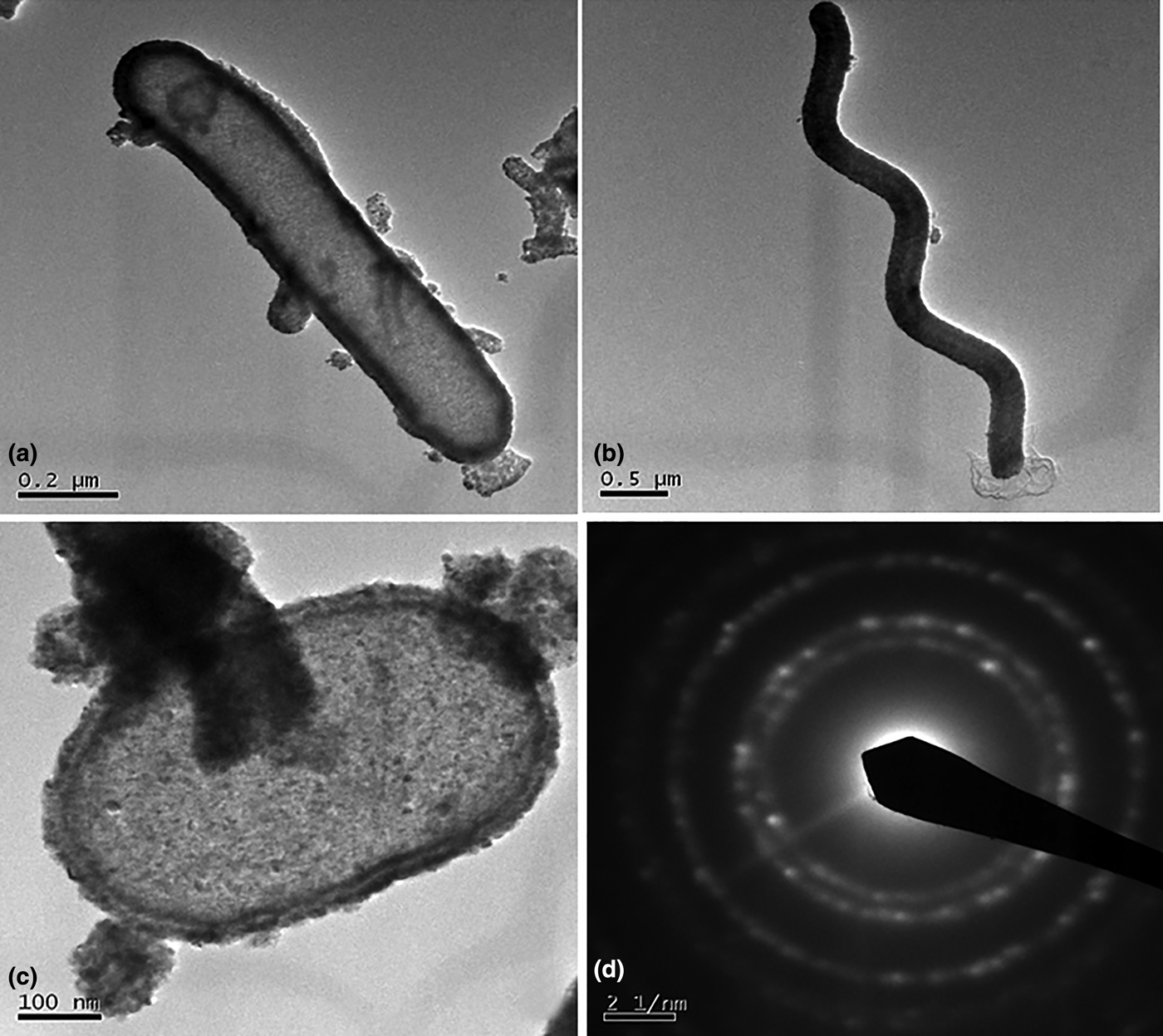
The products were further examined by TEM and HRTEM. Figure 4 shows the TEM images of both hollow cylinder and helical structure. High magnification TEM reveals that nanopolycrystalline grains are deposited on the cell-wall-like structure of bacteria [Fig. 4(c)]. Concentric rings like spotted electron diffraction patterns in Fig. 4(d) also indicate the polycrystalline nature of cubic-phased NiO. The annealed material clearly shows that individual nanoparticles are arranged along the tubular structure of the cylinder. The hollow cylinders clearly show hemi-spherical ends [Fig. 4(c)]. The inner and outer walls of the cylinder show double-shell structure (TEM images shows sharp contrast between the dark walls and pale centers in between them). The walls were intact after heating and appear porous. For porous nanoparticles it is expected that the external surfaces to be irregular. Figure S4 shows that NiO samples exhibited irregular external surfaces for both the hollow cylinder and helices.
TEM images of hollow NiO: (a) cylinder, (b) helical structures, (c) magnified image of a cylinder showing the cell-like structure, (d) selected area electron diffraction (SAED) pattern associated with polycrystalline NiO.
Plausible growth mechanism of formation of cylinder and helical structures
Urease in calcium carbonate biomineralization is well documented in the literature.[Reference Krajewska21, Reference Dhami, Reddy and Mukherjee28] One mole of urea is hydrolyzed intracellularly to 2 M of ammonia and 1 M of carbonic acid. These products equilibrate in water to form ammonium & hydroxide ions and bicarbonate ions. By increasing pH and providing carbonate ions, in the presence of Ca2+ ions gives rise to the precipitation of carbonates.
 $${\rm H}_2 {\rm N} - {\rm CO} - {\rm NH}_2 + 2 {\rm H}_2 {\rm O} + {\rm Ca}^{2+} + \mathop{\longrightarrow}\limits^{{\rm Urease}} {\rm 2NH}_4^{+} + {\rm CaCO}_3 \downarrow$$
The role of bacteria in the process is thought to be 3-fold, namely: (i) to increase the alkalinity of the environment that favors the precipitation of CaCO3; (ii) to increase the concentration of dissolved inorganic carbon; and (iii) to serve as crystal nucleation sites.
Based on the above literature reports and our results a possible mechanism for the formation of hollow NiO cylinder and helices can be explained. The cell wall of bacteria consists of sugar, phosphate, and amino acid residues which give an anionic charge density and make it an appropriate system for the interaction of nickel metals in solution. The initial binding reaction might take place by stoichiometric interaction of soluble metal and reactive sites within the wall. During hydrolysis of urea there is an increase in the pH that favors carboxylate ion (–COO−) to bind with the nickel ion. The results in Table I showed that without bacteria and keeping all other reaction conditions same did not yielded any precipitate. However, the proper combination of water and bacteria with urea yielded good product yield. The initial pH of 6.7 increased to 7.4, suggesting urease activity and the hydrolysis of urea. During urea hydrolysis it decomposes and reacts with nickel ions to form Ni precipitate on the surface of bacterial template. It is also reported that the bacterial cell surface can induce mineral deposition by providing a nucleation site.[Reference Stocks, Galinat and Bang29] We suppose that this is the initial formation of Ni precipitate on the cell-wall structure. The proposed formation mechanism is illustrated in Fig. 5. As a result, the nucleation stages follow and organize the precipitates uniformly on the bacterial structure for the formation of hollow cylinder and helical structures. SEM/TEM analyses show that the surfaces of these structures are not smooth suggesting the initial formation of a nanocrystallite followed by the aggregation of smaller particles. The thermal decomposition of the Ni precipitate resulted in the formation of NiO but the biological morphology of hollow structures was preserved.
Schematic illustration of the formation of NiO cylinder and helical micro/nanostructures.
Conclusions
In summary, a facile one-step approach for the synthesis of hollow cylinder and helical shape NiO has been achieved by B. pasteurii under ambient conditions. The advantages of this method lie in its simplicity, cheapness, and mild reaction conditions. The engineering of growth mechanism could favor either for rod shape or for helical hollow structures by suitable reactions. The method is suitable for a large-scale synthesis of hollow metal oxide structures and useful for the fundamental study on the shape characteristics of microorganisms. We are currently investigating the synthesis and applications of other metal oxides using other bacterial (other bacillus shaped and spiral bacteria) shaped templates.
Acknowledgment
The authors are very grateful to the National Science Council of Taiwan for sponsoring this research.
Supplementary materials
For supplementary material for this article, please visit http://dx.doi.org/10.1557/mrc.2014.24