INTRODUCTION
The Silent Information Regulator (Sir) proteins, also known as sirtuins, are a family of NAD+-dependent protein deacetylases that present an evolutionarily conserved catalytic domain (Frye, Reference Frye2000). The deacetylase activity is unique for the sirtuin proteins that cleave the co-substrate NAD+ in a reaction that removes an acetyl group from the lysine side chain of a protein substrate, generating the deacetylated protein, O-acetyl-ADP-ribose, and nicotinamide as the final products (Tanner et al. Reference Tanner, Landry, Sternglanz and Denu2000). In addition, sirtuins are also involved in a large variety of biological processes that include DNA recombination and repair, apoptosis, metabolic regulation and aging. Moreover, they show variations in their subcellular localization, which is probably due to their interactions with different substrates (reviewed by Michan and Sinclair, Reference Michan and Sinclair2007; Schwer and Verdin, Reference Schwer and Verdin2008; Imai and Guarente, Reference Imai and Guarente2010; Monteiro and Cano, Reference Monteiro and Cano2011).
The genus Leishmania comprises several species of parasites among which are the causative agents of leishmaniasis, a worldwide disseminated and neglected disease that can be expressed in different clinical forms. The disease shows distinct degrees of morbidity and mortality depending on the parasite species and the host immune system (Desjeux, Reference Desjeux2004; Nylén and Gautam, Reference Nylén and Gautam2010). LmSir2-Related Protein 1 (RP1), the first sirtuin homologue described in Kinetoplastida protozoa (Yahiaoui et al. Reference Yahiaoui, Taibi and Ouaissi1996), is localized in cytoplasmic granules, is present among the parasite's excreted/secreted antigens (Zemzoumi et al. Reference Zemzoumi, Sereno, François, Guilvard, Lemesre and Ouaissi1998), and predisposes eukaryotic cells to Leishmania infection when expressed (Sereno et al. Reference Sereno, Vanhille, Vergnes, Monte-Allegre and Ouaissi2005). The L. infantum Sir2RP1 homologue, LiSir2RP1, the first Leishmania sirtuin characterized as an NAD+-dependent deacetylase (Tavares et al. Reference Tavares, Ouaissi, Santarém, Sereno, Vergnes, Sampaio and Cordeiro-da-Silva2008), is localized in the cytoplasm. Both LmSir2RP1 and LiSir2RP1 are implicated in parasite cell survival, virulence and in the protection from apoptosis (Vergnes et al. Reference Vergnes, Sereno, Madjidian-Sereno, Lemesre and Ouaissi2002, Reference Vergnes, Vanhille, Ouaissi and Sereno2005).
Despite its medical relevance, the genome of L. amazonensis, the parasite focused on in this study, has not yet been fully sequenced, making investigations into its genetic organization more challenging. Considering the essential cellular and developmental roles described for other Leishmania's Sir2RP1 proteins, we were interested in identifying and characterizing the L. amazonensis Sir2RP1 homologue. In this work, we present the identification of the LaSir2RP1 gene, the cloning and the characterization of the recombinant protein, rLaSir2RP1, as a NAD+-dependent deacetylase. Moreover, LaSir2RP1 was immunodetected in total protein extracts, in cytoplasmic granules, and in the secreted material of both promastigotes and lesion-derived amastigotes. We also provide evidence demonstrating that the LaSir2RP1 is post-translationally modified by glycosylation.
MATERIALS AND METHODS
Parasites
Leishmania amazonensis MHOM/BR/73/M2269 promastigotes were cultivated in exponential growth in Medium 199 (Cultilab, Campinas, SP, Brazil) supplemented with 40 mm HEPES, 0·1 mm adenine, 10% heat inactivated fetal bovine serum (FBS), and 1 X antibiotic/antimycotic solution (Life Technologies) at 26°C for 72 h in 25 cm3 culture flasks. Amastigotes were isolated from BALB/c mice with active skin lesions, as previously described (Barbiéri et al. Reference Barbiéri, Giorgio, Merjan and Figueiredo1993) and used immediately after isolation.
In silico analysis
The GeneDB identifiers for the primary sequence of proteins used in this study are: for Leishmania mexicana (Lmx) Sir2RP1, LmxM26.0210; for Sir2RP2, LmxM23.1210 and for Sir2RP3, LmxM33.2140; for Leishmania braziliensis (Lbr) Sir2RP2, LbrM23.V2.1310 and for Sir2RP3, LbrM20.V2.1640; for Leishmania infantum (Li) Sir2RP1, LinJ26.V3.0200; for Sir2RP2, LinJ23.V3.1900 and for Sir2RP3, LinJ34.V3.1900 and for Leishmania major (Lm) Sir2RP1, LmjF26.0210; for Sir2RP2, LmjF23.1210 and for Sir2RP3, LmjF34.2140. Multiple sequence alignments were made using ClustalW (http://www.ebi.ac.uk/Tools/clustalw/) and were manually edited using BioEdit (v7.0.9.0). The presence of O-glycosylation and/or phosphorylation sites in LaSir2RP1 was analysed using the YinOYang 1.2 predictor (http://www.cbs.dtu.dk/services/YinOYang), which predicts O-β-GlcNAc attachment sites in eukaryotic protein sequences and also utilizes the NetPhos 2.0 phosphorylation predictor (http://www.cbs.dtu.dk/services/NetPhos) to identify possible phosphorylated sites and hence predict sites that could be reversibly and dynamically modified by O-β-GlcNAc and/or phosphate groups. The presence and the position of the putative signal anchor were predicted using SignalP3.0 (http://www.cbs.dtu.dk/services/SignalP/). The leucine-rich sequence of the putative Nuclear Export Signal (NES), LX3LXLX3L, was manually identified as previously described (Sereno et al. Reference Sereno, Vanhille, Vergnes, Monte-Allegre and Ouaissi2005).
Molecular biology
Total RNA was extracted from approximately 1×10⁹ log phase promastigotes using Trizol Reagent (Life Technologies) and the concentration was estimated using the absorptivity measurement at 260 nm (A260). mRNA was extracted from approximately 1 mg of total RNA using Message Maker Reagent Assembly (Life Technologies) according to the instructions of the manufacturer. Five μg of each RNA and mRNA were fractionated in a denaturing formaldehyde-agarose gel. Double-stranded cDNA was synthesized from ∼3 μg of mRNA using random hexamers in combination with oligo (dT), according to Superscript Choice System for cDNA synthesis (Life Technologies). In order to separate different populations of double-stranded cDNA, samples were size-fractionated, followed by adapter addition and phosphorylation. Five ng of ds cDNA of size ⩾1·0 kb were used to amplify an internal fragment of LaSir2RP1 (LaSir2RP1 probe). For this reaction, 10 μ m of the forward primer, SirIntF (5′ GGCATCCCAGACTTTCGCTCATC 3′), and 10 μ m of the reverse primer, SirIntR (5′ CAGACATCGGGGTGGGCGTAGTA 3′), were used in the presence of 1X Platinum Taq High Fidelity buffer containing 0·8 mm dNTP mix (Amersham Biosciences), 2 mm MgCl2, and 1 U of Platinum Taq High Fidelity. PCR conditions were: 94°C/3 min, followed by 35 cycles of 94°C/30 sec, 55°C/45 sec and 74°C/90 sec. Primers were designed based on leisir2a (GI:1203986), which encodes the L. major Sir2RP1 homologue, to amplify a genomic fragment of 839 nucleotides corresponding to the central portion of the gene and comprising the deacetylase catalytic domain.
Promastigotes were lysed by incubation in TELT buffer (10 mm Tris-HCl, pH 7·5, 5 mm EDTA, pH 8·0, 100 mm lithium chloride and 0·1%Triton X-100) for 5 min at RT (room temperature). Genomic DNA (gDNA) was extracted by phenol:chloroform followed by phenol:chloroform:isoamyl alcohol (25:24:1) and ethanol precipitation. RNA was eliminated by incubating the solution with 10 μg of RNase A for 15 min at 37°C and the resulting purified DNA was precipitated by ethanol and re-suspended in TE (Tris-HCl 10 mm, pH 7·5, EDTA 1 mm, pH 8·0) according to Cotrim et al. (Reference Cotrim, Paranhos, Mortara, Wanderley, Rassi, Camargo and da Silveira1990). Aliquots of 10 μg of DNA were digested with restriction enzymes (MluI, Hind III, PstI and SalI; Amersham Biosciences) chosen according to the restriction map of LaSir2RP1 from GeneRunner software. For Southern blotting, digested DNA was fractionated in 1% agarose gels in 0·5 X TAE buffer and transferred to Hybond N+ membranes. For the LaSir2RP1 probe, 200 ng was labelled using an ECL random primer labelling kit (GE Healthcare). Membranes were pre-hybridized with hybridization solution containing dextran sulphate, according to the manufacturer's instructions. The denatured labelled LaSir2RP1 probe (20 ng/ml) was added to 10 ml of hybridization solution, and the membranes were incubated overnight at 60°C. The hybridization of L. amazonensis genomic DNA with LaSir2RP1 was detected using the ECL detection systems, version II (GE Healthcare). After hybridization, membranes were exposed to Hyper film (GE Healthcare).
The entire LaSir2RP1 coding region (1122 pb) was obtained by PCR amplification using 200 ng of gDNA, and 10 μ m of the forward primer, SirF (5′ CATATGACAGGGTCTCCGAGAG 3′), and 10 μ m of the reverse primer, SirR (5′ TCACCATGTTGGCAGCAATTC 3′), both designed based on leisir2a (GI:1203986) and encoding for the L. major Sir2RP1 homologue (see above). For the heterologous expression of the protein into Escherichia coli, strain BL21(DE3), the LaSir2RP1 gene was cloned into a pET28a+ expression vector (Novagen). The expression of the recombinant protein was induced by the addition of 1 mm IPTG when A600 was 0·6–0·8, followed by further incubation for 3 h at 30°C. Cells were harvested and the bacterial pellet was collected, lysed in buffer A (50 mm Tris-HCl, pH 8·0, containing 150 mm NaCl) and the soluble fraction was loaded onto an affinity Ni-NTA Sepharose column (Qiagen) and eluted with buffer A containing 100 mm imidazole. Fractions containing the recombinant protein were pooled and loaded onto a Superdex 200 size exclusion column. Purified recombinant LaSir2RP1 (rLaSir2RP1) was eluted from the column with buffer A. For circular dichroism (CD) spectroscopy assays (see below) rLaSir2RP1 was eluted with 100 mm sodium phosphate buffer, pH 8·0, containing 100 mm NaCl.
Biophysical experiments
For circular dichroism (CD) spectroscopy analysis, the protein concentration used was 9 μg/ml in 100 mm sodium phosphate, pH 8·0, and 100 mm NaCl. For emission fluorescence spectroscopy analysis, the protein concentration was 5 μg/ml in 50 mm Tris-HCl, pH 8·0, and 150 mm NaCl. A JASCO model J-810 CD spectropolarimeter (Jasco) equipped with a thermoelectric sample temperature controller (Peltier system) was used to record CD spectra (Correa and Ramos, Reference Correa and Ramos2009) and an Aminco Bowman® Series 2 (Slm-Aminco) fluorimeter was used to record emitted fluorescence spectra, using quartz cuvettes with a 1 cm×1 cm optical path length. Each CD measurement was taken from 260 to 200 nm at 25°C and averaged over 32 times. Tryptophan emitted fluorescence of protein samples was measured using excitation at 295 nm, and emission was collected from 315 to 370 nm. The fluorescence of the HDAC substrate 1 (a fluorogenic acetylated peptide derived from a regulatory acetylation site of p53, BPS Bioscience, USA) was measured using excitation at 360 nm, and emission was collected from 455 to 465 nm. All tryptophan fluorescence experiments were taken at 25°C using a band-pass of 4 nm and 8 nm for excitation and emission measurements, respectively. Fluorescence data were analysed by their maximum wavelength (λ max) and centre of spectral mass ( ), as described by the equation:

where λ i is each wavelength and F i is the fluorescence intensity at λ i (Silva et al. Reference Silva, Miles and Weber1986).
Multi-angle laser light-scattering coupled with size-exclusion chromatography (SEC-MALLS) was used to analyse the distribution of masses of purified rLaSr2RP1. Data analyses were performed by chromatographic separation of 10 μ m of the protein using a 1 cm×60 cm Superdex 200 (GE Healthcare) column, at RT, at a flow rate of 0·5 ml/min in 50 mm Tris-HCl, pH 8·0, and 150 mm NaCl. The experiment was performed continuously on the column eluate as it passed through a DAWN EOS System (Wyatt Technology Corp, USA) and data were analysed using the Astra software package (version 5.3.4.14).
Deacetylase activity assays
Deacetylase activity was tested using 3 μ m of rLaSir2RP1 and 200 μ m of substrate, a fluorogenic acetylated peptide derived from a regulatory acetylation site of p53 (HDAC substrate 1, BPS Bioscience, USA), in activity buffer (50 mm Tris-HCl, pH 8·0, 137 mm NaCl, 2·7 mm KCl and 1 mm MgCl2). Assays were done in the presence of increasing concentrations of NAD+ (nicotinamide adenine dinucleotide, oxidized form; Sigma), ranging from 0 to 2 mm, for 1 h at 37°C. Subsequently, a stop buffer (10 mg/ml trypsin and 20 mm nicotinamide diluted in activity buffer) was added and the mixture was incubated for 15 min at RT. The samples were then analysed by fluorescence spectroscopy as described above. Alternatively, 20 μg of total protein extract from promastigote forms was used as substrate in a reaction containing 1 mm NAD+ in the presence and in the absence of 3 μ m of rLaSir2RP1. The reaction products were separated using 10% SDS-PAGE and analysed by Western blot analysis with anti-acetyl lysine serum as a probe.
Immunoassays
Polyclonal anti-rLaSir2RP1 serum was produced by immunization of 2 rabbits with the denatured protein (Celula B UFRGS, Brazil). Human anti-tubulin monoclonal antibody (Sigma Aldrich) was a gift from Professor Dr Robert Ivan Schumacher (IQ/USP, Brazil). Rabbit polyclonal anti-acetyl lysine serum was purchased from Cell Signaling Technology (USA), and rabbit polyclonal anti-LaRPA-1 serum was obtained as previously described (Siqueira Neto et al. Reference Siqueira Neto, Lira, Giardini, Khater, Perez, Peroni, dos Reis, Freitas-Junior, Ramos and Cano2007).
Proteins for Western blot assays were separated in reducing conditions using 10% SDS-PAGE and electro-transferred to nitrocellulose membranes. Membranes were blocked for 1 h at RT, with TBS-T (Tris-buffered saline containing 0·03% Tween-20) supplemented with 1% BSA, and incubated with the respective primary serum for 2 h at RT. After 3 washes with TBS-T, membranes were incubated for 1 h at RT with the secondary antibody (anti-mouse or anti-rabbit IgG coupled to alkaline phosphatase, from Bio-Rad) and detected using an Alkaline Phosphatase Conjugate Substrate Kit (Bio-Rad).
For immunofluorescence assays 1×10⁴ promastigotes were added to each well of a 12-well glass slide previously coated with poly-D-lysine. Parasites were fixed with 2% paraformaldehyde in PBS for 1 h at RT (Tavares et al. Reference Tavares, Ouaissi, Santarém, Sereno, Vergnes, Sampaio and Cordeiro-da-Silva2008). After 3 washes with PBS, parasite membranes were permeabilized for 30 min at room temperature with blocking solution (0·05% Triton X-100 (v/v) and 1% BSA (w/v) in PBS). Parasites were then incubated for 1 h at RT with a specific serum diluted in blocking solution, followed by 3 washes with PBS. Parasite cells were then incubated for 1 h at RT with anti-rabbit or anti-mouse IgG labelled with Alexa Fluor 594 or 488, respectively (Invitrogen). After 3 washes with PBS, mounting solution (glycerol:PBS 1:1(Zemzoumi et al. Reference Zemzoumi, Sereno, François, Guilvard, Lemesre and Ouaissi1998) containing 1 μg/ml Hoescht 33342 – Invitrogen) was added and slides were sealed with a glass cover-slip. Images were analysed with a Nikon Eclipse 50i fluorescence microscope and captured with a digital camera (Nikon DXM1200-F). When necessary, images were superimposed using ACT-1 imaging software (Nikon).
All animal experiments were conducted in accordance with Institutional guidelines in compliance with the recommendations of the Guide for Care and Use of Laboratory Animals. The experimental protocol was approved by the University of Campinas (UNICAMP) Animal Ethics Committee.
Parasite subcellular extracts
Three different procedures were used to obtain subcellular fractions from parasites. (a) Digitonin fractionation. In order to isolate the mitochondrial fraction (digitonin insoluble fraction) from other cellular components (soluble fraction), L. amazonensis promastigotes were subjected to cell lyses using digitonin (Hide et al. Reference Hide, Ritleng, Brizard, Monte-Allegre and Sereno2008). Briefly, 5×10⁸ parasites were washed with PBS and re-suspended in THB (25 mm Tris-HCl, pH 7·8, 1 mm EDTA, 0·6 m sucrose, 1 mm DTT) containing a protease inhibitor cocktail (Calbiochem). Then, 0·25 mg of digitonin was added per mg of total protein extract and samples were incubated for 2 min at 37°C and centrifuged at 10 000 g for 10 min at 4°C. Soluble (supernatant) and insoluble (mitochondria-enriched pellet) fractions were isolated and used for Western blot assays. (b) Igepal fractionation. In order to separate cytoplasmic soluble components (Igepal soluble fraction) from other cellular components (Igepal insoluble fraction), L. amazonensis promastigotes were subjected to cell fractionation using the protocol described by Shapira and Pinelli (Reference Shapira and Pinelli1989). Briefly, Nonidet-P40 was substituted for Igepal, as follows: 5×10⁸ parasites were washed with PBS and re-suspended in 5 volumes of cold Dignam's buffer (10 mm HEPES, pH 7·9, 1·5 mm MgCl2, 10 mm KCl, 0·5 mm DTT) and incubated at 4°C for 10 min. Furthermore, Igepal was added to a final concentration of 0·5% and cells were incubated for additional 10 min at 4°C followed by centrifugation at 2000 g for 10 min. The Igepal soluble (supernatant, cytosol) and insoluble (pellet, nuclei and cytoskeleton enriched) fractions were isolated and used for Western blot assays. (c) Isolation of the microsomal fraction. Leishmania amazonensis promastigotes were submitted to subcellular fractioning in water/ice bath as previously described (Zaverucha do Valle et al. Reference Zaverucha do Valle, Gaspar, Souza-Lemos, Souza, Zamora Márquez, Beatas-da-Cruz, d'Escofier, Côrte-Real, Calabrese and Gonçalves da Costa2007). Parasites were washed with PBS+2% glucose, re-suspended with 20 mm Tris-HCl, pH 7·4, 12·5 mm sucrose, incubated for 20 min, and mechanically disrupted by a Douncer homogenizer in the presence of 10% non-ionic detergent Lubrol PX to produce the total protein extract (ET). After cell lyses sucrose was added to 0·25 m and the material was centrifuged at 750 g for 90 s to remove nucleus and intact cells. Further debris was eliminated by a 2-step centrifugation at 10 000 g for 10 min and 27 000 g for 30 min. Supernatant (6 ml) was layered on top of 2 ml of buffer containing 20% sucrose, 10 mm MgCl2, 25 mm KCl , and 10 mm Tris-HCl, pH 7·4, and centrifuged at 100 000 g for 30 min to produce the microsomal fraction (micF), which contains endoplasmic reticulum-derived vesicles, including ribosomes (Zaverucha do Valle et al. Reference Zaverucha do Valle, Gaspar, Souza-Lemos, Souza, Zamora Márquez, Beatas-da-Cruz, d'Escofier, Côrte-Real, Calabrese and Gonçalves da Costa2007).
Secretion assays
Secretion assays as described by Silverman et al. (Reference Silverman, Chan, Robinson, Dwyer, Nandan, Foster and Reiner2008) and Cuervo et al. (Reference Cuervo, de Jesus, Saboia-Vahia, Mendonça-Lima, Domont and Cupolillo2009) were used to analyse the secreted/excreted material from promastigotes and from amastigotes isolated from mice with active skin lesions. Briefly, 1×10⁹ parasites were washed with secretion medium (Medium 199 supplemented with 40 mm HEPES and 0·1 mm adenine), transferred to 1 ml of fresh secretion medium supplemented with 2x protease inhibitors cocktail (Calbiochem) and incubated for 2 h at 26°C. Supernatants containing secreted/released proteins from the parasites were collected after 2 cycles of centrifugation at 4°C, first at 650 g for 10 min, and then at 10 000 g, for 10 min. Following centrifugation, proteins in the supernatant were precipitated with TCA (10% v/v) for 30 min at RT and washed with 100% acetone at 14 000 g for 15 min. The pellet was re-suspended in 1X SDS-PAGE loading buffer, separated in 10% SDS-PAGE, and electro-transferred to nitrocellulose membranes for Western blot assays (see below).
Concanavalin A-affinity purification of parasite proteins
Promastigotes or amastigotes of L. amazonensis were lysed with 0·5 X RIPA buffer (150 mm Tris-HCl, pH 7·5, 150 mm NaCl, 1% Triton X-100, 0·1% SDS) and 500 μg of a total protein preparation (determined using the Bio-Rad Protein Assay, using BSA as standard) was incubated with 100 μl of Concanavalin A-Sepharose (Sigma) previously equilibrated in buffer A (25 mm Tris-HCl, pH 8·0, 150 mm NaCl, 0·5% Igepal) for 30 min at RT. Following incubation, samples were centrifuged and the supernatant (flow through) was removed. The pellet containing the resin was washed 4 times with 1 ml of buffer A, and proteins were eluted with buffer A containing 0·1 m glucose and 0·1 mα-methyl-D-mannopyranoside. Proteins in the eluted fractions were precipitated using TCA (10% v/v) for 30 min at RT and washed with acetone at 14 000 g for 15 min. The pellet was separated using 10% SDS-PAGE and subsequently prepared for Western blot analysis.
RESULTS
LaSir2RP1 is a single copy gene within the genome of Leishmania amazonensis
A PCR-based cloning strategy, which used primers designed based on the sequence of leisir2a (GI:1203986) from L. major, was performed to identify for the first time a sirtuin-related gene in the L. amazonensis genome. The gene sequence was deposited in the GenBank database under Accession No. gi|24635164|gb|AAN63359.1. Sequence analysis of the LaSir2RP1 open-reading frame identified a polypeptide of 343 amino acid residues long with a predicted molecular mass of 40·4 kDa and a putative Sir2 domain (Fig. 1). SignalP3.0 analysis indicated that LaSir2RP1 contained a predicted N-terminal signal anchor localized in the first 39 residues. By manual comparison with LmSir2RP1 (Sereno et al. Reference Sereno, Vanhille, Vergnes, Monte-Allegre and Ouaissi2005), LaSir2RP1 also showed a putative leucine-rich LX3LXLX3L nuclear export-like signal (NES-like) within its C-terminus (Fig. 1).
Amino acid and nucleotide sequences of LaSir2RP1. Nucleotide: the fragment used as a probe in Southern blot experiments is in bold and the 2 restriction sites of the SalI endonuclease are double-underlined. Amino acid residues corresponding to the putative signal anchor are underlined; the catalytic deacetylase site is highlighted in grey and the putative nuclear export signal (NES) is in bold and double-underlined.

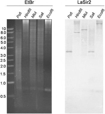
In order to gain insight into the genomic organization of LaSir2RP1, a Southern blot assay was performed on L. amazonensis promastigote genomic DNA (gDNA) digested with restriction enzymes selected based on the absence (PstI, HindIII, MluI, EcoRI) or presence (SalI) of restriction sites within the gene according to the virtual digestion of the LaSir2RP1 gene using GeneRunner software. The Southern blot was probed with a LaSir2RP1 internal fragment (comprising nucleotides 139-977) and a single reactive band was revealed under all conditions tested (Fig. 2). This result is in agreement with GeneRunner analysis, which predicted that LaSir2RP1 would not be digested by PstI, HindIII, MluI and EcoRI but would be digested in 2 different positions by SalI. However, note that in the autoradiogram shown in Fig. 2 only 1 SalI-digested band is seen. This is explained by the fact that one of the SalI sites is localized downstream of the probe annealing position (see Fig. 1 for details).
LaSir2RP1 is a single copy gene in the genome of Leishmania amazonensis. Genomic DNA from promastigotes of L. amazonensis was extracted and digested with specific restriction enzymes as indicated at the top of the gel. Digested DNA was fractionated on a 0·8% agarose gel and stained with ethidium bromide (panel EtBr) or Southern blotted and hybridized with a LaSir2 probe (panel LaSir2). The presence of a single reactive band resulting from all of the restriction enzymes tested indicates that LaSir2RP1 is a single copy gene. Left, molecular mass standard in kb.

Characterization of LaSir2RP1
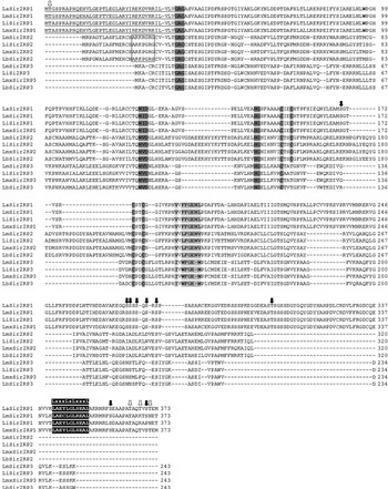
Sirtuin proteins from Leishmania are classified into 3 groups named Sir2RP1 to Sir2RP3, according to their molecular mass and the presence or absence of specific signal sequences (Ivens et al. Reference Ivens, Peacock, Worthey, Murphy, Aggarwal, Berriman, Sisk, Rajandream, Adlem, Aert, Anupama, Apostolou, Attipoe, Bason, Bauser, Beck, Beverley, Bianchettin, Borzym, Bothe, Bruschi, Collins, Cadag, Ciarloni, Clayton, Coulson, Cronin, Cruz, Davies, Gaudenzi, Dobson, Duesterhoeft, Fazelina, Fosker, Frasch, Fraser, Fuchs, Gabel, Goble, Goffeau, Harris, Hertz-Fowler, Hilbert, Horn, Huang, Klages, Knights, Kube, Larke, Litvin, Lord, Louie, Marra, Masuy, Matthews, Michaeli, Mottram, Müller-Auer, Munden, Nelson, Norbertczak, Oliver, O'Neil, Pentony, Pohl, Price, Purnelle, Quail, Rabbinowitsch, Reinhardt, Rieger, Rinta, Robben, Robertson, Ruiz, Rutter, Saunders, Schäfer, Schein, Schwartz, Seeger, Seyler, Sharp, Shin, Sivam, Squares, Squares, Tosato, Vogt, Volckaert, Wambutt, Warren, Wedler, Woodward, Zhou, Zimmermann, Smith, Blackwell, Stuart, Barrell and Myler2005). To determine within which group the L. amazonensis sirtuin homologue should be classified, an amino acid multiple sequence alignment was performed between it and the putative members of Leishmania spp. sirtuin family. The analysis revealed that it belongs to the Sir2-Related Protein 1 group and, therefore it was named LaSir2RP1 (Fig. 3).
Amino acid multiple sequence alignments of Leishmania spp. sirtuins. Alignments were executed using ClustalW followed by manual editing. The predicted signal anchor present in all Sir2RP1 proteins is underlined. A box indicates the putative mitochondrial target signal for the Sir2RP2 group. A putative NES (highlighted in black) is represented by a conserved C-terminal leucine-rich region (indicated above the alignment). GAG and NID motifs, the 4 cysteine residues from the putative C4 zinc-finger domain, and conserved HG residues, are in grey. Residues predicted to interact with acetylated peptides are highlighted in light grey (Cosgrove et al. 2006). LaSir2RP1 residues with a high score (>0·5) prediction for O-glycosylation are indicated by white arrows, and those with prediction either for O-glycosylation or phosphorylation are indicated by black arrows.

LaSir2RP1 shares all the conserved features found in most of the eukaryote sirtuin protein family. LaSir2RP1 contains a conserved fFGEnl motif that has been shown to be responsible for binding to an acetylated substrate and motifs and amino acid residues responsible for interaction with NAD+ and deacetylase activity. In addition, LaSir2RP1 presents the conserved HG motif that is important for ADP ribosyltransferase activity. It also contains the GAG and NID motifs and the C4 zinc-finger domain that are individually essential for the transcriptional silencing activity present in all Sir2-related proteins (Fig. 3). Lastly, all Leishmania Sir2RP1 proteins share the manually identified NES (Nuclear Export Signal), which appears to be a common regulatory element for other sirtuins described so far (Wilson et al. Reference Wilson, Le, Zimmerman, Marmorstein and Pillus2006).
Using classical predictors found in the Expasy Proteomics Server (www.expasy.org/tools) it was also possible to verify that LaSir2RP1 and its orthologues could be post-translationally modified at conserved sites by glycosylation and/or phosphorylation. The analyses showed that LaSir2RP1 has positive predictions for O-glycosylation, but not for N-glycosylation or for GPI attachment sites. According to YinOYang analysis, the best scored sites (cut off >0·5) of O-glycosylation (white arrows) and the sites predicted to be glycosylated and/or phosphorylated (black arrows) in LaSir2RP1 are shown in Fig. 3.
Recombinant LaSir2RP1 is folded and monomeric
In order to biochemically and biophysically characterize LaSir2RP1, the recombinant protein (rLaSir2RP1) was expressed and purified with a 6xHis tag at its N-terminus using an E. coli expression system. The protein was purified to homogeneity after 2 chromatographic steps (data not shown) and used to immunize rabbits. The obtained immune serum (anti-rLaSir2RP1) was able to recognize a protein band with an estimated molecular mass of about 43 kDa in a Western blot assay (data not shown), corresponding to the purified recombinant protein. We performed multi-angle laser light-scattering in conjunction with size exclusion chromatography (SEC-MALLS) experiments to determine whether rLaSir2RP1 was monomeric or multimeric. Figure 4 shows a plot of the calculated molecular masses versus elution time for the selected scattering peaks, which corresponds to the UV monitored elution peak. An average molecular mass of 44±1 kDa was obtained (Fig. 4) which is similar to the predicted 42·6 kDa (from amino acid sequence) for monomeric rLaSir2RP1.
SEC-MALLS analysis revealed that rLaSir2RP1 is monomeric. The protein was analysed using a MALLS detector during the elution from size exclusion chromatography, and the major peak presented a polydispersity of 1·0±0·1 and an average molecular mass of 44±1 kDa.

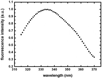
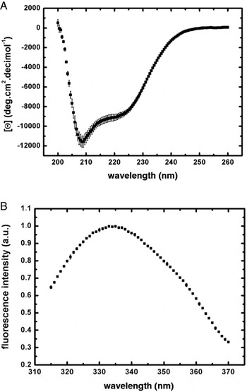
In order to gain insight into the secondary structure of rLaSir2RP1, and confirm that the purified recombinant protein was not denatured, we performed circular dichroism analysis on rLaSir2RP1. Figure 5A shows that the protein is properly folded with a spectrum presenting 2 minima at approximately 209 and 221 nm, and values of residual molar elipticity [Θ] of −11 500 and −9000 deg.cm2.dmol−1, respectively, presenting a predicted α-helical content of about 20%. Emission fluorescence spectroscopy was used to access the environment of the single tryptophan residue, thus as a probe for local folding present at rLaSir2RP1. As shown in Fig. 5B, the protein displayed an emitted fluorescence spectrum with λ max at 335 nm, indicating that the tryptophan is buried in the apolar interior of the protein. These data strongly indicate that rLaSir2RP1 was produced folded, and is a monomeric species under the conditions tested.
Spectroscopic characterization of rLaSir2RP1. (A) The circular dichroism spectrum is characteristic of a folded protein. Analysis using the signal at 222 nm (Correa and Ramos, Reference Correa and Ramos2009) indicates that the protein has an alpha-helical content of about 20%. (B) rLaSir2RP1 was excited at 295 nm and the emitted fluorescence spectrum had λ max at 335 nm and a centre of mass at 337 nm, suggesting that the single tryptophan is buried within the apolar interior of the protein.

rLaSir2RP1 is an NAD+-dose dependent deacetylase
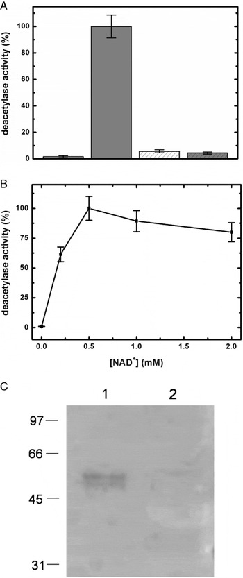
To evaluate whether rLaSir2RP1 displays deacetylase activity, an acetylated fluorogenic peptide derived from the protein p53 was used in the absence or in the presence of 1 mm NAD+ in a deacetylase reaction (Fig. 6A, white and grey bars, respectively). As expected for a member of the Leishmania Sir2RP1 family, rLaSir2RP1 displayed NAD+-dependent activity, which was completely abrogated when the protein activity was assayed under denaturing conditions (Fig. 6A, hatched bars). The deacetylase activity of rLaSir2RP1 reached a plateau at ∼0·5 mm NAD+ concentration and, at high NAD+ concentrations, the deacetylase activity was inhibited (Fig. 6B). Additionally, we detected that rLaSir2RP1 was also able to fully deacetylate a major acetylated protein in promastigote extracts, corresponding to a ∼50 kDa protein band, as shown by a Western blot probed with anti-acetyl lysine serum (Fig. 6C). This ∼50 kDa protein band is likely the most abundant acetylated protein in the parasite extracts, which might explain why other acetylated proteins that should also be recognized by the anti-acetyl lysine serum are not seen in the gel. In addition, rLaSir2RP1 appears to display substrate specificity because, under the experimental conditions used, it was not able to accept acetylated BSA as a substrate (not shown), but it did deacetylate the fluorogenic peptide derived from p53 and the ∼50 kDa protein from promastigote extracts as shown in Fig. 6A and C.
rLaSir2RP1 is an NAD+-dependent deacetylase. (A) rLaSir2RP1 was tested for deacetylase activity using a p53-derived peptide as an acetylated substrate in a reaction medium without (white) or containing 1 mm NAD+ (grey) in the absence (smooth) or in the presence (hatched) of 4 m urea. (B) rLaSir2RP1 deacetylase activity measured as a function of increasing NAD+ concentrations. Error bars correspond to the standard deviation, and the line was drawn to guide the eye. (C) Western blot analysis of promastigote extracts incubated with1 mm NAD+ in the absence (lane 1) or in the presence (lane 2) of 10 μ m of rLaSir2RP1 probed with an anti-acetyl lysine polyclonal serum. Note the presence of a major ∼50 kDa lysine acetylated protein that was recognized as a substrate by LaSir2RP1 deacetylase (lane 2). Left, molecular mass standards in kDa.

LaSir2RP1 is expressed in both promastigote and amastigote developmental forms
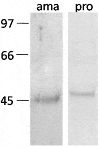
To assess whether LaSir2RP1 is variably expressed in different life stages of L. amazonensis, we performed a Western blot analysis on extracts obtained from promastigotes and lesion-derived amastigotes. LaSir2RP1 was detected in both extracts when the blot was probed with a specific serum produced against rLaSir2RP1. As shown in Fig. 7, amastigote LaSir2RP1 has an apparent molecular mass of about 45 kDa, which appears to be slightly smaller than that of the promastigote, which has an apparent molecular mass of about 48 kDa. Also, the native proteins migrated with a molecular mass slightly higher than the recombinant protein (data not shown) in a denaturing gel, suggesting that LaSir2RP1 could be post-translationally modified by the covalent attachment of 1 or more functional groups which will increase its apparent molecular mass (see below).
LaSir2RP1 is expressed in both amastigote and promastigote forms of Leishmania amazonensis. Total parasite extracts from promastigotes (pro) or lesion-derived amastigotes (ama) were analysed by Western blot probed with an anti-LaSir2 serum. Left, molecular mass standards in kDa. LaSir2RP1 from amastigotes had an apparent molecular mass of approximately 45 kDa whereas LaSir2RP1 from promastigotes had an apparent molecular mass of approximately 48 kDa.

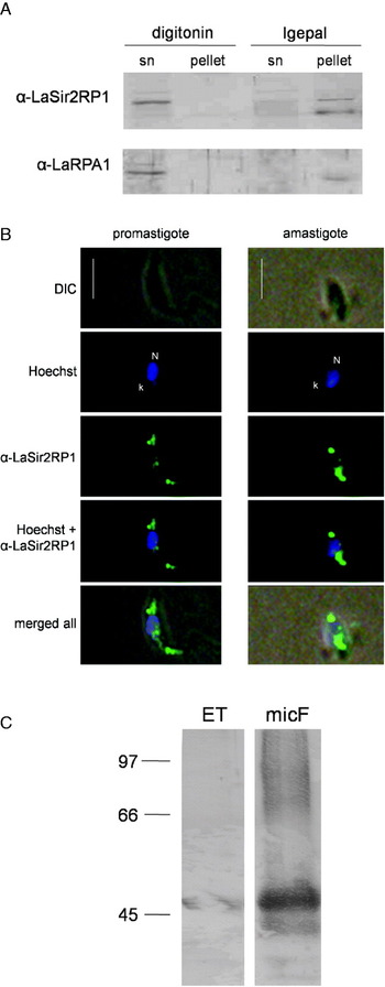
To gain insight into the subcellular localization of LaSir2RP1, three distinct cell fractionation experiments using L. amazonensis promastigote cells were made. A fraction containing mitochondria (digitonin-insoluble) isolated from the other cellular components (digitonin-soluble fraction) (Hide et al. Reference Hide, Ritleng, Brizard, Monte-Allegre and Sereno2008) was obtained by the first approach, while soluble components of cytoplasm (Igepal-soluble fraction) from the other cellular components (Igepal-insoluble fraction) (Shapira and Pinnelli, Reference Shapira and Pinelli1989) were obtained by the second experimental procedure. Subsequently, the obtained subcellular fractions were analysed by a Western blot probed with anti-rLaSir2RP1 serum and the results demonstrated that, under the experimental conditions used, LaSir2RP1 was not detected in the mitochondria-enriched fraction or in the soluble cytosolic fraction (Fig. 8A). In contrast, both LaSir2RP1 and LaRPA-1, a known L. amazonensis nuclear protein (Siqueira Neto et al. Reference Siqueira Neto, Lira, Giardini, Khater, Perez, Peroni, dos Reis, Freitas-Junior, Ramos and Cano2007) used here as a control, were found in the soluble fraction of the digitonin extract, which contains most of the cellular content but not mitochondria (Hide et al. Reference Hide, Ritleng, Brizard, Monte-Allegre and Sereno2008), and in the nuclei-cytoskeleton-enriched Igepal fraction (Fig. 8A). Since LaSir2RP1 was found to be virtually excluded from the soluble components of the cytoplasm (Fig. 8A) and displayed a vesicular subcellular distribution pattern (Fig. 8B), the microsomes isolated from promastigotes were analysed by Western blot probed with anti-rLaSir2RP1, and found that LaSir2RP1 was highly enriched into the microsomal fraction (Fig. 8C). Altogether, these results suggest that LaSir2RP1 is neither soluble in the cytoplasm, nor in the mitochondria of the parasite but is part of the vesicular content of the cell.
Subcellular distribution of LaSir2RP1. (A) Digitonin and Igepal-derived subcellular fractions of Leishmania amazonensis promastigotes were submitted for Western blot analysis. The detergent soluble (sn) and non-soluble (pellet) fractions were assayed with anti-rLaSir2RP1 and anti-RPA sera as probes. (B) Indirect immunofluorescence assay of paraformaldehyde-fixed promastigotes and amastigotes. Parasites were stained with rabbit anti-rLaSir2RP1 followed by anti-rabbit IgG labelled with Alexa Fluor 488 (green). Hoechst 33342 (blue) was used to stain DNA in the nucleus (N) and in the kinetoplast (k). 40X magnification. Scale bars=5 μm. (C) LaSir2RP1 is enriched in microsomes of L. amazonensis promastigotes. Aliquots of 100 μg of total extract (ET) and 100 μg of microsomal fraction (micF) from promastigotes were submitted for Western blot analysis, using an anti-rLaSir2RP1 serum as probe.

LaSir2RP1 is distributed in cytoplasmic vesicles and is a secreted/excreted protein in both L. amazonensis developmental forms
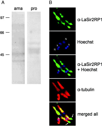
An indirect immunofluorescence assay using anti-rLaSir2RP1 as a probe in L. amazonensis promastigotes and amastigotes was used to complement the fractionation studies on the subcellular localization of LaSir2RP1. As shown in Fig. 8B, LaSir2RP1 displays a punctuate distribution throughout the cytoplasm in both parasitic forms. LaSir2RP1 was also detected by Western blot assays in conditioned media from both amastigote and promastigote forms (Fig. 9A) but other protein bands that migrate with different apparent molecular mass, both higher and lower than the predicted, were also recognized by anti-laSir2RP1 serum. Since these secreted fractions were not purified, we suggest the following explanations to this result. (i) The protein can undergo different levels of post-translational modification such as the covalent attachment of multiple functional groups which might increase the apparent molecular mass of the resultant modified protein species. (ii) Protein degradation may have occurred resulting in polypeptides with apparent molecular mass lower than predicted. These events are not mutually exclusive and may be occurring concurrently.
LaSir2RP1 is a secreted protein in both Leishmania amazonensis amastigotes and promastigotes. (A) Promastigotes and lesion-derived amastigotes were washed and incubated for 3 h at 26°C with serum-free medium. Culture supernatants, containing the secreted material from parasites were analysed by a Western blot probed with anti-rLaSir2RP1 serum. (B) Indirect immunofluorescence assays of paraformaldehyde-fixed promastigotes and amastigotes. Parasites were stained with rabbit anti-rLaSir2RP1 and mouse anti-human tubulin followed by incubation with an anti-rabbit IgG labelled with Alexa Fluor 488 (green) and anti-mouse IgG labelled with Alexa Fluor 594 (red). Hoechst 33342 (blue) was used to stain DNA in the nucleus (N) and in the kinetoplast (k). An overexposure of the fluorescent signal revealed extracellular LaSir2RP1 (green) near the flagellar pocket (arrows).

Additionally, LaSir2RP1 could also be observed in extracellular vesicle-like structures near the flagellar pocket of promastigote forms (Fig. 9B). Collectively, these results indicate that LaSir2RP1 is secreted/excreted from both L. amazonensis developmental forms.
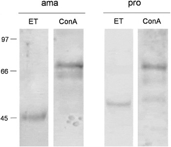
LaSir2RP1 is a glycoprotein
Several predictors suggest the presence of putative glycosylation sites in LaSir2RP1 translated protein. Therefore, we asked whether the parasite protein was post-translationally modified, specifically by glycosylation. To test this hypothesis, we analysed Concanavalin A (Con A)-affinity purified promastigote and amastigote extracts in a Western blot probed with anti-rLaSir2RP1. The results showed that in the glucose/α-D-mannopyranoside eluted fractions, obtained from both Con-A purified extracts, anti-rLaSir2RP1 serum recognizes a major ∼66 kDa protein band (Fig. 10, lanes ConA). Since other experiments shown in this work (see above) identified protein species with molecular mass ranging from 45 to 48 kDa, which are lower than that of the protein recognized in the Con-A purified extracts, and Con A is a lectin able to bind glycosyl and/or mannosyl residues, we suggest that the 66 kDa protein band represents the main glycosylated form of LaSir2RP1, which was enriched in (Con A)-affinity purified promastigotes and amastigotes extracts. Curiously, an additional protein band of ∼64 kDa was recognized in the detergent-insoluble fraction (cytoskeleton pellet) from L. infantum promastigotes in a Western blot assay probed with anti-LiSir2RP1 serum (Tavares et al. Reference Tavares, Ouaissi, Santarém, Sereno, Vergnes, Sampaio and Cordeiro-da-Silva2008).
LaSir2RP1 from both amastigotes and promastigotes forms are modified by glycosylation. Parasite proteins were solubilized with 0.5 X RIPA buffer (ET) and affinity-purified using Concanavalin A-Sepharose beads. After exhaustive washing, proteins were eluted with 0·1 m glucose mixed with 0·1 mα-methyl-D-mannopyranoside (ConA) and were analysed by a Western blot probed with anti-rLaSir2RP1 serum. Left, molecular mass standards in kDa. A major reactive ∼66 kDa protein band was enriched in both lectin-purified extracts, suggesting that LaSir2RP1 is glycosylated in vivo.

DISCUSSION
Despite the sequencing of the entire genomes of L. major, L. infantum, and L. braziliensis, little is known about L. amazonensis genomic organization. Considering that sirtuin proteins have been identified in other species of the Leishmania genus and are essential to the parasite biology, we hypothesize that a sirtuin orthologue is also present within the genome of L. amazonensis. Here, we present for the first time, the identification of a single copy sirtuin-related gene in the L. amazonensis genome, dubbed LaSir2RP1.
In order to biochemically and biophysically characterize LaSir2RP1, we expressed and purified recombinant LaSir2RP1 (rLaSir2RP1). The protein was produced pure, folded as a monomeric species and functional. The circular dichroism spectrum, a probe of global secondary structure (Correa and Ramos, Reference Correa and Ramos2009), was characteristic of an α-helical protein, and the emitted tryptophan fluorescence spectrum, a probe of local tertiary structure, indicated that the residue was well buried in the interior of the protein (Ramos, Reference Ramos2004). The folded monomeric conformation was confirmed by SEC-MALLS experiments and, as expected, LaSir2RP1 showed the deacetylase activity previously reported for the L. infantum Sir2RP1 orthologue (Tavares et al. Reference Tavares, Ouaissi, Santarém, Sereno, Vergnes, Sampaio and Cordeiro-da-Silva2008). In addition, LaSir2RP1 deacetylase activity was inhibited at high NAD+ concentrations, probably due to the release of nicotinamide, a subproduct of the deacetylation reaction, and a known inhibitor of other Sir2-related proteins (Bitterman et al. Reference Bitterman, Anderson, Cohen, Latorre-Esteves and Sinclair2002; Avalos et al. Reference Avalos, Bever and Wolberger2005).
Since some members of the mammalian sirtuin family appear to be post-translationally modified (North and Verdin, Reference North and Verdin2007; Yang et al. Reference Yang, Fu, Chen, Olashaw, Zhang, Nicosia, Bhalla and Bai2007), we considered the possibility that LaSir2RP1 may also be modified, and decided to analyse the Leishmania protein sequence with an algorithm that predicts possible types of post-translational modifications, such as glycosylation. As a matter of fact, by Western blot analysis, the apparent molecular mass of LaSir2RP1 from promastigotes and amastigotes was higher than that estimated based on its amino acid composition alone, suggesting that LaSir2RP1 could be post-translationally modified by the covalent attachment of one or more functional groups, which will increase its apparent molecular mass. Also, the Con A-affinity purified extracts from both promastigotes and amastigotes probed with anti-LaSir2RP1 serum by Western blot, showed the presence of a major protein band of approximately 66 kDa in addition to proteins with lower molecular masses. We suggest that the ∼66 kDa protein may represent a glycoform of LaSir2RP1, which contains mannosyl and/or glucosyl residues accounting for the ∼20 kDa of mass augmentation. Also, the proteins with lower molecular masses may represent intermediate forms of LaSir2RP1 glycosylation. Since Concanavalin A binds specifically to mannosyl and/or glucosyl residues of glycoproteins (Goldstein et al. Reference Goldstein, Hollerman and Smith1965), the absence of the ∼45–48 kDa LaSir2RP1 band may not necessarily mean that these forms are not glycosylated, since they could be glycoforms that are not specific for lectin binding.
Moreover, in this work we show that LaSir2RP1 is expressed in cytoplasmic vesicles of both promastigote and amastigote developmental forms and is secreted/excreted, which could correlate with protein glycosylation. Our results are in good agreement with those from Zemzoumi et al. (Reference Zemzoumi, Sereno, François, Guilvard, Lemesre and Ouaissi1998) showing that LiSir2RP1 can be detected in the supernatant fraction of [35S] methionine-labelled L. major promastigote cultures. We provide evidence that LaSir2RP1 is secreted from both developmental stages of L. amazonensis. Thus, it would be interesting to determine whether LaSir2RP1 is secreted in a soluble form, either inside of or anchored to the membrane of vesicles/exosomes (Silverman et al. Reference Silverman, Clos, de Oliveira, Shirvani, Fang, Wang, Foster and Reiner2010), since it does not present the classical XDXL motif at the C-terminus, as characteristic for ER/Golgi resident proteins from protozoa to mammals (Munro and Pelham, Reference Munro and Pelham1987; Ilgoutz et al. Reference Ilgoutz, Mullin, Southwell and McConville1999), but was found dispersed in the parasite within vesicle-like structures. Accordingly, the secretory apparatus in parasitic kinetoplastids is organized in a similar way to those in mammalian cells and sequencing of the genes encoding some proteins of the kinetoplastid secretory pathway revealed that the presence of an N-terminal anchor signal is an important feature shared by these proteins (Sakaguchi et al. Reference Sakaguchi, Tomiyoshi, Kuroiwa, Mihara and Omura1992; McConville et al. Reference McConville, Mullin, Ilgoutz and Teasdale2002). Recently, Silverman et al. (Reference Silverman, Clos, de Oliveira, Shirvani, Fang, Wang, Foster and Reiner2010), demonstrated that Leishmania spp. use an exosome-based machinery as a general mechanism for protein secretion. Therefore, here we show significant evidence in favour of a secreted LaSir2RP1, such as (1) a putative signal anchor at the N-terminus, (2) a subcellular localization in cytoplasmic vesicules and in enriched microsomal fractions, (3) presence in the secreted/excreted material of both developmental forms of the parasite and (4) modification by glycosylation.
In conclusion, this is the first report that identifies and characterizes a sirtuin-related protein from L. amazonensis, LaSir2RP1. Biophysical studies showed that the recombinant protein was folded as a monomeric species. It was also shown that LaSir2RP1 shares classical features with other proteins within the Sir2RP1 family, such as NAD+-deacetylase activity, and was predominantly found in cytoplasmic vesicles. Additionally, evidence was presented indicating that a Sir2RP1 from Leishmania could be glycosylated in vivo, which may represent a regulatory tool and/or a secretory signal conserved within the Leishmania genus. Recent efforts are being directed in order to identify differences between the catalytic site of Leishmania's sirtuin and its human orthologues (Kadam et al. Reference Kadam, Kiran and Roy2006; Kaur et al. Reference Kaur, Shivange and Roy2010), in order to design novel pharmaceutical agents against Leishmania infections. The possibility that LaSirRP1 can undergo post-translational modifications represents an additional feature on sirtuin proteins that drugs can target.
ACKNOWLEDGMENTS
The authors would like to thank to Professor Dr R. I. Schumacher (IQ/USP) for the anti-tubulin antibody; Dr H. Aoyama (IB/Unicamp) for the Concanavalin-A Sepharose supply and Dr D. Mokry for critical reading of the manuscript. This research was supported by grants from Fundação de Amparo a Pesquisa do Estado de São Paulo (FAPESP), and Conselho Nacional de Pesquisa e Desenvolvimento (CNPq). M.R.F. thanks the FAPESP for a Fellowship.