1. Introduction
Protein phosphorylation regulates most cellular activities. The post-translational modification is catalysed by the protein kinases. The protein kinases transfer the γ-phosphate of ATP to the hydroxyl group of a serine, threonine or tyrosine residue on the target protein. Phosphorylation results in a number of diverse responses in different proteins (reviewed by Johnson & Lewis, Reference Johnson and Lewis2001). It may result in the activation of enzyme activity (e.g. many protein kinases themselves require phosphorylation for activity) or the inhibition of enzyme activity (e.g. glycogen synthase), create a protein recognition site for another protein (e.g. phosphorylation of the insulin receptor cytoplasmic region creates a recognition site for the insulin receptor substrate), or result in either an order-to-disorder or a disorder-to-order transition (e.g. K+ channel inactivation domain or the tumour suppressor pRb protein response to phosphorylation). Each of these events allows a phosphorylated protein to signal to proteins downstream initiating a cascade of reactions. There are a variety of conformational responses to phosphorylation. Conformational changes can be large [e.g. phosphorylation of glycogen phosphorylase results in a shift of the phosphorylase serine residue (Ser14) by 40 Å and changes in the subunit/subunit interactions] or minimal (e.g. isocitrate dehydrogenase where the phosphoryl group acts as a steric and electrostatic blocker). Phosphorylation as a biological control process has the advantage that it is reversible. The phosphate group can be removed by one of the phosphatases, allowing different specificities to be used in the forward and reverse reactions. The biological processes may be switched on or off to meet different demands.
The protein kinases are classified as serine/threonine or tyrosine or dual substrate kinases based on the receiving amino acid of their substrates. Analysis of the human genome showed that there are 518 protein kinases (~1·7% of proteins encoded), of which only 90 are tyrosine kinases, and these include 58 receptor tyrosine kinases (Manning et al. Reference Manning, Whyte, Martinez, Hunter and Sudarsanam2002). Comparison of the kinase chromosomal map with known disease loci indicated that 164 kinases map to loci seen frequently in tumours and ~80 kinases map to loci implicated in other major diseases. A screen of the kinase coding exons in 210 human cancers revealed a diverse number and pattern of mutations (Greenman et al. Reference Greenman, Stephens, Smith, Dalgliesh, Hunter, Bignell, Davies, Teague, Butler, Stevens, Edkins, O'Meara, Vastrik, Schmidt, Avis, Barthorpe, Bhamra, Buck, Choudhury, Clements, Cole, Dicks, Forbes, Gray, Halliday, Harrison, Hills, Hinton, Jenkinson, Jones, Menzies, Mironenko, Perry, Raine, Richardson, Shepherd, Small, Tofts, Varian, Webb, West, Widaa, Yates, Cahill, Louis, Goldstraw, Nicholson, Brasseur, Looijenga, Weber, Chiew, Defazio, Greaves, Green, Campbell, Birney, Easton, Chenevix-Trench, Tan, Khoo, Teh, Yuen, Leung, Wooster, Futreal and Stratton2007). Most somatic mutations are likely to be passengers that do not contribute to oncogenesis, but ~120 kinase genes had evidence for ‘driver’ mutations that could function as cancer agents. Second in the list was B-RAF, a protein kinase that had been identified from a previous screen as an oncogene (Davies et al. Reference Davies, Bignell, Cox, Stephens, Edkins, Clegg, Teague, Woffendin, Garnett, Bottomley, Davis, Dicks, Ewing, Floyd, Gray, Hall, Hawes, Hughes, Kosmidou, Menzies, Mould, Parker, Stevens, Watt, Hooper, Wilson, Jayatilake, Gusterson, Cooper, Shipley, Hargrave, Pritchard-Jones, Maitland, Chenevix-Trench, Riggins, Bigner, Palmieri, Cossu, Flanagan, Nicholson, Ho, Leung, Yuen, Weber, Seigler, Darrow, Paterson, Marais, Marshall, Wooster, Stratton and Futreal2002a). Somatic mutations in B-RAF are associated with 60% of malignant melanomas and with other cancers. Mutations in the kinases involved in the Jun N-terminal kinase and fibroblast growth factor signalling pathways also corresponded to proteins recognised in previous cancer screens. However, there were some surprises. Titin kinase, a giant molecule (gene >300 kb), had the most mutations, and yet, from its role as a molecular ruler and stress response mediator in muscle, mutations in titin are unlikely to cause tumour growth. Only two of the 63 mutations in titin were in the kinase domain, and the structure shows that these have no apparent links to the catalytic site. The possible role of titin as a cancer gene will require direct biological evaluation.
Until the late 1990s, anti-cancer drugs were directed towards metabolic enzymes (e.g. methotrexate) or to DNA (cisplatin, gemcitabine, cyclophosphamide) or, with increasing success, to DNA topoisomerases (doxorubicin, etoposide, irinotecan, topotecan), to hormonal signalling pathways via the nuclear hormone receptors for breast (e.g. tamoxifen) and prostate cancer (e.g. flutamide) and to microtubule stabilisation (e.g. paclitaxel). In the present century, protein kinases, especially the tyrosine protein kinases, have become prime targets for cancer intervention. Most extracellular signals that drive cell growth and survival are amplified and transduced inside cells by three processes: through receptor-activated tyrosine kinases, through receptor coupling to GTP binding proteins, or through cytokine receptors (Pawson & Nash, Reference Pawson and Nash2000; Schlessinger, Reference Schlessinger2000). All of these, either directly or indirectly, involve protein kinases. Cancer can arise through a variety of cellular mechanisms that may include overexpression of growth factors such as vascular endothelial growth factor (VEGF) or transforming growth factor α (TGF-α) or growth factor receptors [e.g. platelet-derived growth factor receptor (PDGFR) or epidermal growth factor receptor (EGFR)], deregulation of kinases (e.g. ABL or EGFR), deregulation of kinase activity by activation of oncogenes (e.g. the RAS/RAF pathway), or loss of tumour suppression functions (e.g. mutations in p53 or pRb leading to activation of the cell cycle).
Knowledge of signalling pathways and the ways in which a cell responds to external signals have led to protein kinases becoming important targets for anti-cancer drug design. Protein kinases have proved a druggable target class, especially in cases where the role of a particular protein kinase has a direct causal relevance to cancer through its inappropriate activation. In the treatment of cancer, a single agent is seldom effective, and the combination therapies that target both the kinase and other targets such as DNA have often proved more effective than a single compound. Almost always (with the exception of chronic myeloid leukaemia, or CML; discussed below), no single mutated gene lies at the heart of cancer, but it is the cumulative mutations in a number of genes that lead to the diseased state. However, these genes often work together so that the number of pathways affected may be smaller, giving hope that targeting a pathway can prove effective for treatment of disease.
Protein kinases are also targets for treatment of inflammatory disease, diabetes and neurodegenerative diseases (Bain et al. Reference Bain, Plater, Elliott, Shpiro, Hastie, McLauchlan, Klevernic, Arthur, Alessi and Cohen2007). There has been progress in these areas (Sebolt-Leopold & English, Reference Sebolt-Leopold and English2006), but as yet there are no drugs in clinical use, possibly because of the complexity of the signalling pathways that involves much cross-talk between pathways. Of particular interest is the diarylurea compound BIRB-796, which is a potent and specific inhibitor of p38 kinase and which has been in clinical trials for rheumatoid arthritis. The structure of this compound with p38α (Pargellis et al. Reference Pargellis, Tong, Churchill, Cirillo, Gilmore, Graham, Grob, Hickey, Moss, Pav and Regan2002) showed that it bound with large conformational changes to an inactive conformation of the kinase at a site that only partially overlapped the ATP binding site, an early example of an allosteric kinase inhibitor. For neurodegenerative diseases, inhibition of the kinases that phosphorylate the tau protein could be beneficial. For these indications, drugs must cross the blood–brain barrier, but for other indications such as cancer (except brain cancers), it is advantageous that compounds do not cross the blood–brain barrier to avoid neurological side effects. Kinases are also possible targets for drugs against parasitic diseases. For example, knowledge of the malaria genome has allowed characteristic sequences that are specific to the parasite and different from the human host to be discerned. Those kinases that are essential for the parasite life cycle could be exploited for drug design from structural knowledge (Leroy & Doerig, Reference Leroy and Doerig2008). Protein kinase inhibitors play an important role in cell biology. For example, the specific inhibitors of FGFR kinase (SU5402), mitogen-activated protein kinase kinase 1 (MEK1) (PD184352) and GSK3β (CHIR99021) are found to maintain the pluripotent state of embryonic stem cells (Ying et al. Reference Ying, Wray, Nichols, Batlle-Morera, Doble, Woodgett, Cohen and Smith2008).
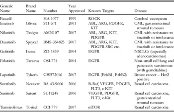
There are 10 protein kinase inhibitors that are approved and in clinical use (Table 1), with many more compounds (>50) in clinical trials. In this review, I consider the contribution that structural biology has made to the design of protein kinase drugs listed in Table 1 and give a brief account of some recent developments. This review builds on previous reviews (Cohen, Reference Cohen2002; Fabbro et al. Reference Fabbro, Ruetz, Buchdunger, Cowan-Jacob, Fendrich, Liebetanz, Mestan, O'Reilly, Traxler, Chaudhuri, Fretz, Zimmermann, Meyer, Caravatti, Furet and Manley2002; Noble et al. Reference Noble, Endicott and Johnson2004; Fabian et al. Reference Fabian, Biggs, Treiber, Atteridge, Azimioara, Benedetti, Carter, Ciceri, Edeen, Floyd, Ford, Galvin, Gerlach, Grotzfeld, Herrgard, Insko, Insko, Lai, Lelias, Mehta, Milanov, Velasco, Wodicka, Patel, Zarrinkar and Lockhart2005; Liu & Gray, Reference Liu and Gray2006; Sebolt-Leopold & English, Reference Sebolt-Leopold and English2006; Tintelnot-Blomley & Lewis, Reference Tintelnot-Blomley and Lewis2006; Liao, Reference Liao2007). In addition to small molecule inhibitors, there are also successful approaches that have resulted in products for clinical use using humanised monoclonal antibodies generated against the extracellular domain of transmembrane receptor protein kinases [e.g. trastuzumab (Herceptin) that targets Her2 (ErbB2) receptor for treatment of breast cancer or cetuximab (Erbitux) that targets EGFR for treatment of metastatic colorectal cancer] or against the growth factors themselves such as bevacizumab (Avastin) that targets VEGF for treatment of metastatic colorectal cancer. Structures are known for each of these complexes [trastuzumab (Cho et al. Reference Cho, Mason, Ramyar, Stanley, Gabelli, Denney and Leahy2003) PDB 1N8Z; cetuximab (Li et al. Reference Li, Schmitz, Jeffrey, Wiltzius, Kussie and Ferguson2005) PDB 1YY9; bevacizumab (Fuh et al. Reference Fuh, Wu, Liang, Ultsch, Lee, Moffat and Wiesmann2006) PDB 2FJG]. The structures have contributed to an understanding of the mechanisms of action, but a detailed discussion is outside the scope of this review.
Clinically approved protein kinase targeted inhibitors

2. Protein kinase structures
To date, there are more than 118 protein kinase structures deposited in the protein data bank (Marsden & Knapp, Reference Marsden and Knapp2008). The structures provide coverage of most of the seven different classes of protein kinases identified from sequence (Manning et al. Reference Manning, Whyte, Martinez, Hunter and Sudarsanam2002) [AGC (containing PKA, PKG, PKC families), CAMK (calcium calmodulin-dependent kinase families), CK1 (casein kinase 1 family), CMGC (containing CDK, MAPK, GSKs, CLK families), STE (homologs of yeast sterile kinases), TK (tyrosine kinases) and TKL (tyrosine kinase-like)] (Fedorov et al. Reference Fedorov, Marsden, Pogacic, Rellos, Muller, Bullock, Schwaller, Sundstrom and Knapp2007b). The structures also map reasonably well to those protein kinases that have been identified as drug targets so that there are representative structures of most of the classes that are relevant to disease (Vieth et al. Reference Vieth, Sutherland, Robertson and Campbell2005).
Protein kinases share a common catalytic domain as predicted from their sequence similarities, but there are a variety of different regulatory mechanisms. The kinase can exist in an active or an inactive state. The kinase may be present as a single domain or it may be covalently linked to other regulatory domains. Examples of the latter include SRC, which contains regulatory SH3 and SH2 domains in addition to the kinase catalytic domain, and cyclic AMP-dependent protein kinase (PKA), whose N-terminal and C-terminal extensions help hold the kinase in the active conformation. Several kinases, such as the cyclin-dependent protein kinases (CDKs), require an additional subunit, the cyclin, for activation and their activity is regulated by the expression of such subunits. Receptor tyrosine kinases (e.g. EGFR) have extracellular regions that recognise and bind growth factors and signal through the membrane to the intracellular kinase domain. Growth factor binding promotes receptor dimerisation that leads to activation of the kinase domain. Kinases are often regulated by post-translational modifications. To exhibit activity, many kinases require phosphorylation by an upstream kinase on a residue(s) in the activation segment (discussed below). Other phosphorylation reactions (e.g. on a tyrosine residue in the glycine-rich loop in CDK2) inhibit kinase activity. Further post-translational modifications (such as myristylation in ABL) may regulate activity, or other domains or binding partners can achieve regulation by targeting the kinase to its correct location in the cell (e.g. mTOR).
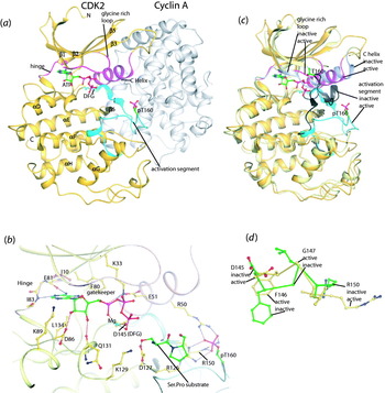
The fold of the catalytic kinase domain, comprising ~300 amino acid residues, consists of a smaller N-terminal lobe of about ~80 residues that contains a five-stranded β-sheet with one α-helix, the C-helix, which is important in regulation, and a ~200 residue C-terminal lobe that is mostly α-helix with a few short stands of β-sheet (Fig. 1a). A hinge region links the two lobes. The ATP-binding pocket of the catalytic site is formed by a groove at the interface between these two lobes. Non-polar side chains from both lobes (e.g. residues Ile10 and Leu134 in CDK2) enclose the adenine (Fig. 1b). The contacts also include three important regions: the flexible glycine-rich loop between strands β1 and β2 containing the motif GXGXΦG (using the single-letter code, where Φ is usually a tyrosine or phenylalanine); the C-helix (indirectly), where a glutamate (Glu51) holds in place a lysine (Lys33) from strand β3, which contacts the α and β phosphates of the triphosphate moiety of ATP; and the hinge region between the lobes. ATP binding is characterised by two hydrogen bonds from the adenine moiety to main chain groups of the hinge region (Fig. 1b). The ribose contacts a polar region from the C-terminal lobe and makes hydrogen bonds to the carboxylate of an acidic residue (Asp86 in CDK2 at the start of β6) and to a main chain carbonyl oxygen (from Gln131 in CDK2 from the start of β8). The triphosphate moiety is stabilised by contacts to a metal ion (magnesium) that is bound by an aspartate, Asp145 in CDK2, which is part of the DFG motif (using the single-letter amino acid code) at the start of the activation segment (discussed below), to a lysine residue, Lys33 in CDK2, and to main chain atoms from the glycine-rich loop between strands β1 and β2 (Fig. 1b).
Structural features of protein kinases illustrated by CDK2. (a) CDK2 in the active conformation (gold) associated with cyclin A (grey). Secondary structural elements, the glycine-rich loop (magenta), C-helix (magenta), hinge region (magenta) and activation segment (cyan) are labelled. ATP is shown bound between the N-terminal and C-terminal lobes of the structure. The phospho-threonine pT160, which is essential for activation, is shown. (b) Details of the ATP and substrate binding sites. The colour code is as in (a). See text for further details. (c) The active and inactive conformations of CDK2. CDK2 in the active (gold) and inactive (pale yellow) conformations have been superimposed to show the shifts in the glycine-rich loop, the C-helix and the activation segment (shown in black in the inactive conformation). (d) The conformation of the DFG motif in the active (yellow) and inactive (green) conformations. The view is similar to (b) where D145 is partially obscured by the γ-phosphate of ATP. The shift in the DFG to the active conformation allows Arg150 to contact the phospho-threonine. From PDB IDs 1QMZ and 1HCK.

The protein substrate-binding site involves the region known as the activation segment. The activation segment is composed of about 20–30 residues between the conserved DFG motif and the conserved APE motif (just before helix αF) in the C-terminal lobe. The correct conformation of the activation segment is crucial for providing a platform against which the protein substrate is docked and which confers substrate specificity. In most but not all kinases, the catalytically competent conformation is promoted by phosphorylation of a threonine or tyrosine residue in the activation segment (Johnson et al. Reference Johnson, Noble and Owen1996). This phosphorylation acts as an organising centre to coordinate structural elements that yield the active kinase conformation. In CDK2, for example, interactions between the phospho-threonine (Thr160) and three basic residues (Arg50, Arg126 and Arg150) stabilise the active conformation (Fig. 1b). In inactive kinase conformations, the activation loop may block the catalytic site, interfering with ATP substrate binding and preventing protein substrate binding, as in CDK2, or it may be disordered (Fig. 1c). In active kinases, part of the activation segment lies parallel to helices E and F, and the catalytic site is accessible. Some protein kinases are constitutively active without the need for phosphorylation [e.g. phosphorylase kinase (PDB 1PHK), casein kinase 1 (PDB 1CKJ) and ROCK (PDB 2ESM) (Jacobs et al. Reference Jacobs, Hayakawa, Swenson, Bellon, Fleming, Taslimi and Doran2006)]. In the absence of interactions with a phosphorylated residue, other interactions stabilise the loop conformation. These interactions significantly increase the surface area buried by docking the activation segment against the kinase.
In serine/threonine kinases, catalysis is promoted by a conserved aspartate residue (Asp127 in CDK2) acting in concert with a conserved lysine (Lys129 in CDK2) (Fig. 1b). (In tyrosine kinases, an arginine four residues from the catalytic aspartate plays the role of the basic group). The key event is the correct localisation of ATP triphosphate through contacts to the magnesium and the lysine (Lys33) side chain and the localisation of the substrate through interactions with the activation segment and a few key side chains providing specificity. The phosphotransfer reaction of the γ-phosphate of ATP to the hydroxyl of the serine (or threonine) is facilitated through a direct in-line mechanism. The lone pair electrons from the hydroxyl are directed to the ATP γ-phosphorus in line to the phosphorus βγ bridging oxygen direction. As the reaction progresses, the aspartate (Asp127 in CDK2) accepts a proton from the hydroxyl group, which is subsequently transferred to the departing phosphate dianion. Lys129 in proximity to the ATP γ phosphate helps to stabilise the transition state of the pentaco-ordinated phosphorane, in which the apical bonds (attacking and departing) are long and weak.
All kinases exhibit a similar conformation in the active state, but in the inactive state, there are several differences (Huse & Kuriyan, Reference Huse and Kuriyan2002; Nolen et al. Reference Nolen, Taylor and Ghosh2004). These are characterised (Fig. 1c) by (i) a different orientation of the two lobes that reflects flexibility about the hinge region in the kinase fold; (ii) different orientations of the C-helix, which in many inactive states is swung out removing the contact from the glutamate to the lysine that contacts to the triphosphate of ATP and creating an extra pocket for inhibitor groups; (iii) displacement of the activation segment, which in many inactive kinases is disordered; and (iv) a different conformation of the DFG motif at the start of the activation segment. In the active conformation, the aspartate of the DFG points in to the ATP site and binds the magnesium, the counter ion for the triphosphate (Fig. 1b, d). The phenylalanine of the DFG is buried in a hydrophobic pocket and is not accessible. In the inactive conformation, the aspartate of the DFG motif points out so that it can no longer bind the magnesium ion and the phenylalanine is directed towards the ATP site where it can contact inhibitor groups (Fig. 1d). The DFG loop has been identified as a switch between active and inactive kinase conformations where the phenylalanine in the active conformation is part of a hydrophobic cluster including groups from the C-helix and the loop that contains the catalytic aspartate (Asp127 in CDK2) (Kornev et al. Reference Kornev, Haste, Taylor and Eyck2006; Liao, Reference Liao2007). The glycine of the DFG, which can adopt conformations not accessible to other amino acids, is a critical pivot point for the conformation of the following activation segment.
3. Protein kinase inhibitors in clinical use
3.1 Targeting the ATP site
All the kinase inhibitors that are in clinical use (Table 1) bind at the ATP site, with the exception of temsirolimus that targets mTOR (discussed later). The ATP site is common to all the protein kinases, and hence, it is remarkable that targeting this site can result in inhibitors that are specific for just a few kinases. Selectivity is engineered by targeting pockets adjacent to the ATP site with groups that have chemical diversity.
Structural analyses of compounds bound to the ATP site have revealed five (or at least five) pockets that can be exploited for binding different chemical groups (reviewed by Liao, Reference Liao2007) (Fig. 1b). (i) The adenine-binding pocket that is lined by aliphatic and aromatic hydrophobic groups with the potential for two or more hydrogen bonds to the hinge region. Many groups with planar geometry based on sp2 hybridisation bind here provided that they also contain at least one group that can have a hydrogen bond with the hinge region. (ii) The ribose-binding pocket that has several polar groups. (iii) The so-called p38 pocket (recognised in the p38α/BIRB-796 complex and in the ABL/imatinib complex) above the adenine, which is guarded by a small residue (usually threonine) known as the ‘gatekeeper’ in p38 and other kinases such as ABL and RAF but is blocked by a more bulky residue [e.g. phenylalanine (Phe80) in CDK2] in other kinases. (iv) The pocket below the adenine, the Lys89 pocket in CDK2 or the hydrophobic pocket in other kinases, which allows targeting by a number of different groups. (v) The type II inhibitor pocket formed between the C-helix and the activation segment, which accommodates some large inhibitors that target the inactive conformation (Liu & Gray, Reference Liu and Gray2006).
There have been exciting developments in the generation of allosteric inhibitors. BIRB-796 in complex with P38α was an example of a compound that only partly overlapped with the ATP site and bound to the inactive conformation, eliciting a slow conformational change (Pargellis et al. Reference Pargellis, Tong, Churchill, Cirillo, Gilmore, Graham, Grob, Hickey, Moss, Pav and Regan2002). The MEK1 inhibitors, PD184352 and the more potent compound PD0325901, are more striking. The relatively small PD184352 binds to a site tucked under the C-helix that is adjacent to the ATP site in the inactive conformation. It does not overlap ATP and acts as an allosteric inhibitor (Ohren et al. Reference Ohren, Chen, Pavlovsky, Whitehead, Zhang, Kuffa, Yan, McConnell, Spessard, Banotai, Mueller, Delaney, Omer, Sebolt-Leopold, Dudley, Leung, Flamme, Warmus, Kaufman, Barrett, Tecle and Hasemann2004). These compounds have progressed in clinical trials but have not yet received approval (Sebolt-Leopold, Reference Sebolt-Leopold2008). Because of their potency and selectivity, they have been recommended in cellular studies for elucidating the roles of signalling pathways (Bain et al. Reference Bain, Plater, Elliott, Shpiro, Hastie, McLauchlan, Klevernic, Arthur, Alessi and Cohen2007).
3.2 Targeting other sites
To date, there have been no compounds in clinical use that exploit the protein substrate specificity site. This site is solvent exposed with fewer identifying hydrophobic slot features and, hence, it is difficult to design high-affinity compounds. Some protein kinases use a remote docking site for substrate recognition (e.g. MAPK family kinases and CDK2/cyclin A). There has been progress in the design of constrained peptides or peptide mimics that recognise the cyclin A recruitment site and inhibit CDK2 kinase activity against substrates that require a docking site (Kontopidis et al. Reference Kontopidis, Andrews, McInnes, Cowan, Powers, Innes, Plater, Griffiths, Paterson, Zheleva, Lane, Green, Walkinshaw and Fischer2003; McInnes et al. Reference McInnes, Andrews, Zheleva, Lane and Fischer2003). In PDK1, as in most kinases of the AGC group, there is a so-called PIF pocket in the N-terminal region that is used to bind a phosphorylated hydrophobic motif on the substrate to both localise the substrate and activate PDK1. Allosteric inhibitors have been designed to target this pocket (Engel et al. Reference Engel, Hindie, Lopez-Garcia, Stroba, Schaeffer, Adrian, Imig, Idrissova, Nastainczyk, Zeuzem, Alzari, Hartmann, Piiper and Biondi2006). They successfully imitate the activation but prevent substrate recognition. Allosteric compounds that target the plextrin homology domain and inhibit AKT have also been achieved (Barnett et al. Reference Barnett, Defeo-Jones, Fu, Hancock, Haskell, Jones, Kahana, Kral, Leander, Lee, Malinowski, McAvoy, Nahas, Robinson and Huber2005). In kinases that require binding of another subunit, there is also scope for interfering with the protein–protein interactions that govern this association. There has been success in the design of compounds that interfere with the association between the α and the β subunits of CK2 (Laudet et al. Reference Laudet, Barette, Dulery, Renaudet, Dumy, Metz, Prudent, Deshiere, Dideberg, Filhol and Cochet2007). The scope for interfering with protein–protein interactions has also been suggested for regulation of EGFR by imitating the action of the MIG6 (mitogen-induced gene 6) protein that inhibits EGFR by binding to the kinase C-terminal domain and blocking the heterodimer association that is a key feature of EGFR activation (Zhang et al. Reference Zhang, Pickin, Bose, Jura, Cole and Kuriyan2007). Temsirolimus, a derivative of rapamycin, which has been approved for clinical use, appears to inhibit mTOR by interfering with mTOR targeting to its substrate, although its mode of action is not fully understood (discussed later). As Nagar et al. (Reference Nagar, Hantschel, Young, Scheffzek, Veach, Bornmann, Clarkson, Superti-Furga and Kuriyan2003) have commented in connection with the myristoyl binding site of ABL, other opportunities apart from the ATP site do exist for developing small molecule inhibitors for protein kinases. There are many opportunities for exploiting the diversity of regulatory mechanisms that control protein kinase action, but except temsirolimus, all inhibitors so far are of low affinity and none have made it for clinical use.
3.3 Selectivity
Selective inhibition of only a few kinases is essential to minimise side effects when a drug is administered to patients. Kinase inhibitors are routinely screened against a library of kinases, and a degree of discrimination of at least 100-fold over most other kinases is usually required for the compound to move forward as a drug candidate. Such screens have shown that many inhibitors were much less specific than at first reported (Davies et al. Reference Davies, Reddy, Caivano and Cohen2000; Bain et al. Reference Bain, Plater, Elliott, Shpiro, Hastie, McLauchlan, Klevernic, Arthur, Alessi and Cohen2007). Staurosporine, for example, is a potent inhibitor of at least 44 protein kinases and is too toxic for clinical use. A small modification gives a more specific compound, 7-hydroxy-staurosporine (UCN-01), which entered clinical trials as a Chk1 inhibitor (Tse et al. Reference Tse, Carvajal and Schwartz2007). Other staurosporine analogues (e.g. lestauritinib, midostaurin and enzastaurin) are in advanced clinical trials. A recent screen of 156 validated kinase inhibitors, many of which are in clinical trials, against a library of 60 Ser/Thr protein kinases using a thermal stability shift assay revealed many unexpected cross-reactivities, some of which could indicate other targets for compounds that had passed toxicity tests (Fedorov et al. Reference Fedorov, Marsden, Pogacic, Rellos, Muller, Bullock, Schwaller, Sundstrom and Knapp2007a). Using an alternative approach of ATP site-dependent competition binding assay, Fabian et al. (Reference Fabian, Biggs, Treiber, Atteridge, Azimioara, Benedetti, Carter, Ciceri, Edeen, Floyd, Ford, Galvin, Gerlach, Grotzfeld, Herrgard, Insko, Insko, Lai, Lelias, Mehta, Milanov, Velasco, Wodicka, Patel, Zarrinkar and Lockhart2005) showed a broad specificity for 20 kinase inhibitors when screened against a panel of 119 protein kinases. For example, the potent and specific Ser/Thr kinase p38 inhibitor BIRB-796 was found to bind to the imatinib-resistant tyrosine kinase ABL, and the aurora kinase inhibitor VX-680/MK-0457 also has activity against drug-resistant ABL, with applications in clinical trials (Giles et al. Reference Giles, Cortes, Jones, Bergstrom, Kantarjian and Freedman2007).
Bantscheff et al. (Reference Bantscheff, Eberhard, Abraham, Bastuck, Boesche, Hobson, Mathieson, Perrin, Raida, Rau, Reader, Sweetman, Bauer, Bouwmeester, Hopf, Kruse, Neubauer, Ramsden, Rick, Kuster and Drewes2007) have used a chemical proteomics approach in which immobilised non-selective kinase inhibitors (termed kinobeads – a mixed kinase inhibitor affinity resin) were used to bind ~200 protein kinases from cell lysates. The proteins were identified using mass spectrometry. Binding targets of drugs were determined by measuring the competition by selected drugs with the affinity matrix. Imatinib (Glivec) was comparatively specific. Of 142 kinases, imatinib bound to ABL, to the ABL family-related kinase ARG, to the tyrosine receptor kinase DDR1, and to a non-kinase, the quinone reductase NQO2. Dasatinib (Table 1) exhibited a much broader spectrum. In addition to the four proteins identified for imatinib, dasatinib bound to 35 other kinases including SRC family kinases, BTK and SYK, which signal downstream of immune receptors and three serine/threonine kinases. KIT was also identified as a target but only when cells in culture were used and only when KIT was phosphorylated. A striking outcome of the inhibitor profiling studies is that compounds designed to target a particular kinase, in this case breakpoint cluster region (BCR)-ABL, and administered for the same indication (CML), display different target profiles (Bantscheff et al. Reference Bantscheff, Eberhard, Abraham, Bastuck, Boesche, Hobson, Mathieson, Perrin, Raida, Rau, Reader, Sweetman, Bauer, Bouwmeester, Hopf, Kruse, Neubauer, Ramsden, Rick, Kuster and Drewes2007; Hantschel et al. Reference Hantschel, Rix and Superti-Furga2008). In the most comprehensive analysis of selectivity to date, a screen of 38 kinase inhibitors across a panel of 317 kinases (>50% of the human kinome) using a competition binding assay revealed a wide diversity of hitherto unsuspected interactions (Karaman et al. Reference Karaman, Herrgard, Treiber, Gallant, Atteridge, Campbell, Chan, Ciceri, Davis, Edeen, Faraoni, Floyd, Hunt, Lockhart, Milanov, Morrison, Pallares, Patel, Pritchard, Wodicka and Zarrinkar2008). Some approved drugs (e.g. sunitinib, dasatinib) had relatively low selectivity but are nevertheless effective for clinical use. Knowledge of target profiles should allow careful evaluation as to which drug or drug combination should be used in a particular situation to better exploit each drug's full potential.
3.4 Active versus inactive conformations
The differences between active and inactive kinases at the ATP site have provided scope for engineering selectivity. Conventional kinase assays by their nature identify inhibitors of the active kinase conformation and can establish if the inhibitor is competitive with ATP. However, binding studies, such as those used in two of the analyses described above, can identify binding to either the active or an inactive conformation. Of the compounds listed in Table 1, fasudil, dasatinib, gefitinib and erlotinib bind to the active conformations, whereas imatinib, lapatinib and sorafenib bind to the inactive conformation of their respective kinases. Targeting an inactive conformation has attractions because this is more likely to represent a conformation that is unique to that protein kinase. For example, residues that contact imatinib in ABL are conserved in SRC; the latter is not inhibited by imatinib because SRC cannot access the inactive conformation of ABL. A further advantage is that the inactive conformation may have a reduced affinity for ATP and therefore represent a more druggable target because the inhibitor would be more effective when competing with cellular concentrations of ATP. However, targeting regions that may not be directly involved in catalytic function allows the possibility of mutations that lead to drug resistance. In contrast, the active conformation requires conservation of both key residues and tertiary structure and is less likely to be tolerant of mutations. However, drug resistance mutations are a potential risk for both conformational states. Where disease occurs through loss of regulation leading to an inappropriately active protein kinase, targeting the active conformation can be advantageous, as in the case of gefitinib (discussed below). In practice, targeting either the inactive or the active conformation has proved successful for drug design.
4. Structural features of protein kinase inhibitors
4.1 Fasudil and Rho kinase (ROCK)
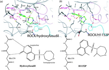
Fasudil was the first protein kinase inhibitor to be used clinically. Developed in Japan and using the structural information from PKA, the first protein kinase structure to be solved in 1991 (Knighton et al. Reference Knighton, Zheng, Ten Eyck, Ashford, Xuong, Taylor and Sowadski1991a, Reference Knighton, Zheng, Ten Eyck, Xuong, Taylor and Sowadskib), the compound is based on an isoquinolinesulfonamide scaffold. Fasudil was approved in Japan in 1995 for the treatment of cerebral vasospasm after surgery for haemorrhage (Ono-Saito et al. Reference Ono-Saito, Niki and Hidaka1999). It is an inhibitor of the Rho-associated coiled coil kinase ROCK, a kinase that regulates the activity of muscle myosin regulatory light chain proteins by direct phosphorylation and by inhibitory phosphorylation of myosin phosphatase. ROCK activity leads to increased levels of phosphorylated myosin light chain and subsequent muscle contraction. The structure of PKA in complex with fasudil demonstrated the characteristics of binding at the ATP site, including the role of a phenylalanine from the C-terminal region of the kinase (Phe368 in ROCK; Fig. 2) that seals part of the adenine site and is typical of ATP interactions in AGC family kinase members (Breitenlechner et al. Reference Breitenlechner, Gassel, Hidaka, Kinzel, Huber, Engh and Bossemeyer2003). However, fasudil is relatively non-specific and also inhibits PKC-related kinases 1 and 2 and a number of other kinases (Bain et al. Reference Bain, Plater, Elliott, Shpiro, Hastie, McLauchlan, Klevernic, Arthur, Alessi and Cohen2007). Structures of ROCK in complex with fasudil and fasudil-related compounds have revealed some features resulting in selectivity (Jacobs et al. Reference Jacobs, Hayakawa, Swenson, Bellon, Fleming, Taslimi and Doran2006) (Fig. 2). Overall, the PKA and ROCK ATP sites are very similar, presenting a challenge for selectivity. Fasudil is metabolised to hydroxyfasudil through the oxidation at the isoquinoline C-1 position, and the physiologically significant state is shown in Fig. 2a. Hydroxyfasudil (oxo tautomer) has an increase in potency of more than 10-fold for ROCK over PKA (K i values of hydroxyfasudil for ROCK, 0·15 μM, and for PKA, 2·2 μM). The addition of the two methyl groups in the compound H1152P results in a 6-nM inhibitor of ROCK and a 0·34-μM inhibitor of PKA (Fig. 2b). H1152P binds in a reversed binding mode compared with hydroxyfasudil (Fig. 2b, a) and in a similar orientation to fasudil itself. Part of the explanation for selectivity appears to reside in residue Ala215 in ROCK, which is a threonine (Thr183), in PKA. The smaller side chain in ROCK allows the homopiperazine methyl group to be accommodated in its preferred conformation (Jacobs et al. Reference Jacobs, Hayakawa, Swenson, Bellon, Fleming, Taslimi and Doran2006) (Fig. 2b). The work demonstrates the contribution that structure can make to understanding and guiding drug design. More recently, the role of ROCK in cell motility has been noted, and it was also shown that fasudil is an inhibitor of vascular endothelial cell motility and angiogenesis through its ability to inhibit vascular endothelial growth factor receptor (VEGFR) (Yin et al. Reference Yin, Morishige, Takahashi, Hashimoto, Ogata, Tsutsumi, Takata, Ohta, Kawagoe, Takahashi and Kurachi2007). The anti-angiogenic properties of fasudil suggest that it may also be relevant for the treatment of cancer.
Details of ROCK kinase in complex with hydroxyfasudil and with H1152P. In this and subsequent figures, the kinase ATP site is shown with residues within 4·5 Å of contact distance from the inhibitor. Residues from the glycine-rich loop, the C-helix and the hinge are in magenta, the activation segment in cyan and the remainder of the chain in grey. The ion pair from the glutamate on the C-helix to the lysine from β3 strand and the aspartate and phenylalanine of the DFG motif are shown for reference. The schematic secondary structure is shown for orientation. Inhibitor carbon atoms are in green. In the chemical structure drawings of the inhibitor, the peptide hinge region of the kinase and the hydrogen bonds are shown. The inhibitor has been drawn to be in a similar orientation to the 3D diagram. (a) ROCK in complex with hydroxyfasudil. The isoquinoline ring makes two hydrogen bonds to the hinge region. One is through the protonated nitrogen to the main chain oxygen of Glu154 while the other involves the oxygen, which is added on metabolisation of fasudil, which forms hydrogen bonds to the main chain nitrogen of Met156. The homopiperazine secondary amine interacts with the side chain of Asp216, the aspartate of the DFG motif. From PDB ID 2ETK. (b) ROCK in complex with H1152P. The position of the isoquinoline is reversed compared with hydroxyfasudil and is similar to that observed for fasudil. The isoquinoline nitrogen accepts a hydrogen bond from Met156 main chain nitrogen. The aromatic C1 can donate a hydrogen bond to the carbonyl oxygen of Glu154. There are no other hydrogen bonds, but the additional two methyl groups restrict the conformation of the homopiperazine and make additional contacts to ROCK; the methyl on the isoquinoline contacts the side chains of Ile82 and Phe368 (from the C-terminal region) and the methyl on the homopiperazine contacts the side chains of Ala215 and Asp216. From PDB ID 3D9V.

4.2 ABL inhibitors: imatinib (Glivec), nilotinib (Tasigna) and dasatinib (Sprycel)
CML is a haematological stem cell disorder characterised by excessive myeloid proliferation. It is a relatively rare cancer affecting about 1–2 persons per 100 000. (The common cancers of breast, lung, colorectal and prostate, occur with more than 30 000 cases per year in the UK.) CML is associated with the Philadelphia chromosome characterised by a reciprocal translocation between chromosomes 9 and 22 that results in the fusion of the ABL tyrosine kinase gene with BCR. In the fusion protein BCR-ABL, ABL lacks the N-terminal region that carries a myristoyl modification. In the native kinase, the myristoyl group binds to the kinase C-terminal region and induces conformational changes that allow the regulatory SH2 domains to dock and autoinhibit (Nagar et al. Reference Nagar, Hantschel, Young, Scheffzek, Veach, Bornmann, Clarkson, Superti-Furga and Kuriyan2003). Lacking this modification, ABL becomes constitutively active. Activation is also favoured as a result of dimerisation induced by the BCR coiled coil domain. Deregulated BCR-ABL signals to downstream pathways involved in growth and proliferation and leads to oncogenic transformation of haematopoietic stem cells.
Imatinib (Glivec, STI571), a nanomolar inhibitor of ABL, was approved for therapy against CML in May 2001. Treatment with imatinib results in remission in more than 90% of patients in the early stages of the disease. Although designed as a specific ABL inhibitor, imatinib also inhibits the PDGFR kinase and KIT receptor tyrosine kinase, and in 2001, imatinib was approved for treatment of gastrointestinal stromal tumours (GISTs) where KIT activity is up-regulated. KIT is a stem cell factor-activated receptor whose kinase structure has been solved in the active state (Mol et al. Reference Mol, Lim, Sridhar, Zou, Chien, Sang, Nowakowski, Kassel, Cronin and McRee2003), in the autoinhibited state and in the inactive state with imatinib (Mol et al. Reference Mol, Dougan, Schneider, Skene, Kraus, Scheibe, Snell, Zou, Sang and Wilson2004). GIST is a relatively rare cancer (~2000–5000 patients in the United States) but had been difficult to treat with chemotherapy before imatinib.
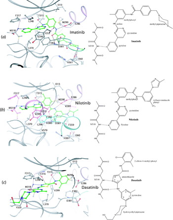
Imatinib has been one of the most successful drugs in the treatment of a cancer. Because CML is driven by a single defect in the early stages of the disease, targeting this defect has been highly effective. Imatinib (Druker et al. Reference Druker, Tamura, Buchdunger, Ohno, Segal, Fanning, Zimmermann and Lydon1996) is based on a phenylamino-pyrimidine scaffold (Fig. 3), which was initially discovered in a programme for PKC inhibitors. The introduction of the methylphenyl group ortho to the pyrimidinyl-amino group switched the specificity towards ABL and PDGFR. The pyridine ring targets the adenine ATP site. The hydrophilic N-methylpiperazine was added to the phenylamide to improve solubility and oral bioavailability (reviewed by Fabbro et al. Reference Fabbro, Ruetz, Buchdunger, Cowan-Jacob, Fendrich, Liebetanz, Mestan, O'Reilly, Traxler, Chaudhuri, Fretz, Zimmermann, Meyer, Caravatti, Furet and Manley2002).
Details of the ABL tyrosine kinase and complexes with imatinib (Glivec), nilotinib (Tasigna) and dasatinib (Sprycel). (left) The secondary structure elements of the kinase are shown in magenta for the glycine-rich loop (residues 249–254), the C-helix (residues 277–291) and the hinge region (residues 316–319); in cyan for the activation segment (residues 381–409); and in grey for the remainder of the molecule. (a) ABL in complex with imatinib. The kinase is in the inactive conformation with the C-helix displaced outward (top right) and the activation segment in an extreme conformation with the phenylalanine (Phe382) of the DFG motif pointing to the ATP site. Side chains are coloured grey, and not all contact residues are shown to keep the diagram simple. Hydrogen bonds include the following: pyridine-N with hinge region back bone Met318 NH, the anilino-NH with the side chain of Thr315, the amide NH to the side chain of Glu286 from the C-helix, the amide-carbonyl with the backbone of NH Asp381 and the protonated methylpiperazine to the back bone carbonyl oxygen of Ile360 and His361. The hydrophobic groups of Leu248, Leu370, Met318 and Phe317 surround the pyridine ring. See text for further details. From PDB ID 1IEP. (b) ABL in complex with nilotinib. The conformation of the kinase is similar to that with imatinib. The diagram has a greater depth than in (a) to show the glycine-rich loop folding over the inhibitor with Tyr253 stacking. Residues in contact with the trifluoromethyl and imidazole group are also shown. Residues are coloured according to the kinase region (glycine loop, C-helix and hinge in magenta; activation segment in cyan). From PDB ID 3CS9. (c) ABL in complex with dasatinib. The kinase is in the active conformation with the C-helix rotated in, the activation segment in the active conformation and Phe382 of the DFG motif tucked into a hydrophobic pocket underneath the C-helix. Three hydrogen bonds are identified between dasatinib and ABL. A pair of hydrogen bonds is formed in the hinge region of the ATP-binding site (i.e. between the 3-nitrogen of the aminothiazole ring of dasatinib and the amide nitrogen of Met318 and between the 2-amino hydrogen of dasatinib with the carbonyl oxygen of Met318). A hydrogen bond is also formed between the side chain hydroxyl oxygen of Thr315 and the amide nitrogen of dasatinib. From PDB ID 2GQC. See text for further details.

The structural results from John Kuriyan's laboratory (Schindler et al. Reference Schindler, Bornmann, Pellicena, Miller, Clarkson and Kuriyan2000; Nagar et al. Reference Nagar, Bornmann, Pellicena, Schindler, Veach, Miller, Clarkson and Kuriyan2002) revealed that imatinib bound to an inactive conformation of ABL in which the C-helix is displaced and the DFG (Asp381, Phe382, Gly383) motif at the start of the activation segment has an extreme inactive conformation. The bound structure is shown in Fig. 3a. There is one hydrogen bond from the pyridine to the hinge region. The remainder of the molecule binds to a pocket lined by the inactive conformation of the DFG loop. The imatinib methylphenyl group extends into a hydrophobic pocket guarded by the small gatekeeper residue Thr315, whereas the amide group contacts the C-helix and the DFG motif. The details of the interactions are described in the figure legend. The pyrimidine ring contacts the hydrophobic side chains of Val299 and Phe382 (the phenylalanine of the DFG motif). The glycine-rich loop bends towards imatinib to form close interactions with the kinase inhibitor, including an edge-to-face aromatic interaction between Tyr253 and the pyridine and pyrimidine rings (Fig. 3a). Imatinib binds preferentially to the inactive conformation. Assays indicate the K i of imatinib for inactive ABL is 37 nM and the K i for the activated conformation is 7 μM (Schindler et al. Reference Schindler, Bornmann, Pellicena, Miller, Clarkson and Kuriyan2000).
The structural studies on the binding of imatinib to c-KIT revealed an interesting variation for the mechanism of inhibition. In the structure of KIT autoinhibited by its juxtamembrane region, the autoinhibitory region inserts into the cleft between the N-terminal and C-terminal kinase lobes and flips the DFG motif to its off state. The activation segment folds back over the catalytic site. To bind imatinib, the autoinhibitory region binding is disrupted by the phenyl and piperazinyl rings of the inhibitor (Mol et al. Reference Mol, Dougan, Schneider, Skene, Kraus, Scheibe, Snell, Zou, Sang and Wilson2004). Imatinib is bound to KIT in a very similar manner to that observed for ABL making similar hydrogen bonds. The authors comment that it is ironic that to achieve the inhibited inactive state, imatinib actually disrupts the natural mechanism that has evolved to maintain the kinase in the inactive state through autoinhibition.
4.2.1 Resistance
Despite the tremendous success of imatinib when administered in the early stages of the disease, many advanced-stage patients develop drug resistance and relapse. Imatinib resistance arises principally from point mutations in the kinase domain of ABL. The structure of ABL in complex with imatinib significantly contributed to understanding mechanism and to the design of new drugs to combat resistance. Most of the resistance mutations occur in regions that interact directly with imatinib or at residues that are crucial in allowing the kinase to adopt the unique inactive conformation (Azam et al. Reference Azam, Latek and Daley2003). For example, the mutation of the gatekeeper threonine to isoleucine (T315I) results in loss of potency, and this can be understood from the structure by the larger isoleucine residue restricting access to the hydrophobic pocket, producing a steric clash with the methylphenyl ring.
Several second-generation inhibitors have been developed that have increased potency and an ability to inhibit many of the imatinib-resistant forms of ABL. Nilotinib (tasigna; AMN107) (Fig. 3b) is an analogue of imatinib with an IC50~30 nM in cells, corresponding to a 15-fold higher potency than imatinib. Nilotinib has good clinical efficacy in imatinib-resistant patients and is a well-tolerated drug (le Coutre et al. Reference Le Coutre, Ottmann, Giles, Kim, Cortes, Gattermann, Apperley, Larson, Abruzzese, O'Brien, Kuliczkowski, Hochhaus, Mahon, Saglio, Gobbi, Kwong, Baccarani, Hughes, Martinelli, Radich, Zheng, Shou and Kantarjian2008). Structural results (Weisberg et al. Reference Weisberg, Manley, Breitenstein, Bruggen, Cowan-Jacob, Ray, Huntly, Fabbro, Fendrich, Hall-Meyers, Kung, Mestan, Daley, Callahan, Catley, Cavazza, Azam, Neuberg, Wright, Gilliland and Griffin2005) with one of the more common resistance mutations M351T showed that nilotinib bound to the inactive conformation of ABL as observed for imatinib. The greater affinity of nilotinib compared with imatinib appears to come from a better topological fit of nilotinib to the kinase. The contacts for the pyridine, pyrimidine, methylanilino and amide groups are the same as for imatinib (Fig. 3b). The trifluoromethyl group binds in a hydrophobic pocket, and residues in contact include Leu298, Val299, Phe359 and those preceding the DFG motif (Val379, Ala380 and Asp381). The imidazole contacts Leu285, Glu286 and Val289 from the C-helix. For nilotinib, the trifluoromethyl/imidazole-substituted phenyl group contributes significantly to the potency, so that the relative contributions from the pyridinyl and pyrimidinyl groups are less for nilotinib than they are for imatinib. Hence, mutations such as F317L from the hinge region and E255K/V in the glycine-rich loop, which contact the pyridinyl and pyrimidinyl groups, have less effect on the overall affinity of nilotinib than imatinib. It is not clear why the M351T mutant (in the C-terminal lobe of the kinase at a site distant from inhibitor binding) is resistant to imatinib but not to nilotinib. It has been suggested (Weisberg et al. Reference Weisberg, Manley, Breitenstein, Bruggen, Cowan-Jacob, Ray, Huntly, Fabbro, Fendrich, Hall-Meyers, Kung, Mestan, Daley, Callahan, Catley, Cavazza, Azam, Neuberg, Wright, Gilliland and Griffin2005) that because imatinib makes directional hydrogen bonds to the carbonyl oxygen of Ile360 and His361 in the C-terminal lobe, it is more sensitive to mutations in the C-terminal region than nilotinib where the imidazole moiety of nilotinib contacts the N-terminal lobe. Analytical structural studies have identified chains of communication through individual conserved residues in the protein kinase core that could explain why perturbations at sites, which are remote from binding sites, can influence activities at binding sites (Kornev et al. Reference Kornev, Haste, Taylor and Eyck2006). Although nilotinib overcomes resistance of many mutant ABLs, the mutant with the T315I mutation of the gatekeeper residue remains resistant because of the loss of a hydrogen bond and introduction of a steric clash, as in the case of imatinib.
4.2.2 Dasatinib binds to the active conformation of ABL
Dasatinib has been developed as a dual SRC/ABL inhibitor. It also affects a wide array of kinases (Bantscheff et al. Reference Bantscheff, Eberhard, Abraham, Bastuck, Boesche, Hobson, Mathieson, Perrin, Raida, Rau, Reader, Sweetman, Bauer, Bouwmeester, Hopf, Kruse, Neubauer, Ramsden, Rick, Kuster and Drewes2007) and does show some side effects that are not seen with imatinib and nilotinib. However, it has clinical efficacy and is generally well tolerated. It is effective against most mutant ABL mutations that occur in the glycine-rich loop and the activation segment with the exception of the T315I mutation. Dasatinib (Fig. 3c) is more than 300-fold more potent than imatinib in cells expressing BCR-ABL. Structural results provided an explanation for these properties (Tokarski et al. Reference Tokarski, Newitt, Chang, Cheng, Wittekind, Kiefer, Kish, Lee, Borzillerri, Lombardo, Xie, Zhang and Klei2006). Dasatinib binds to ABL in its active conformation (Fig. 3c). Dasatinib binds at the ATP site with the aminothiazole moiety of the molecule occupying the site normally bound by the adenine group of ATP. The 2-chloro-6-methylphenyl ring of dasatinib is orthogonal to the thiazole carboxamide group and probes into the hydrophobic pocket near Thr315. The involvement of this pocket guarded by Thr315 explains why the mutant T315I ABL is resistant to dasatinib. Details of the hydrogen bonds are given in the figure legend. Dasatinib also participates in two heteroaromatic CH…O=C interactions with the hinge region backbone (i.e. the C4 thiazole carbon with the carbonyl oxygen of Glu316 and the C5 pyrimidine carbon with the carbonyl oxygen of Met318). These types of interactions have also been observed with other kinase inhibitor complexes (Pierce et al. Reference Pierce, Sandretto and Bemis2002). The N-2-hydroxyethylpiperazine group of dasatinib, for which there is no imatinib counterpart, extends out towards a solvent-exposed region.
The most striking difference between the ABL structures complexed with imatinib or nilotinib in the inactive conformation and that complexed with dasatinib in the active conformation is the conformation of the activation segment. The activation segment in the imatinib-bound state folds back towards the ATP-binding site, forming interactions with both the glycine-rich loop and the inhibitor in the inactive kinase conformation. Phe382 of the DFG motif in the inactive imatinib-bound state points towards the ATP-binding site, contacts the pyrimidine ring of imatinib and forms an edge-to-face interaction with Tyr253 of the glycine-rich loop (Fig. 3a). In contrast, in the active dasatinib-bound form, Phe382 is buried in a hydrophobic pocket, as is typically found with activated kinases (Fig. 3c). Although the central cores of dasatinib and imatinib overlap, the remainder of the structures interacts with different parts of the protein. Specifically, dasatinib does not protrude towards the hydrophobic pocket occupied by Phe382, as does the methylphenyl branch of imatinib. The latter observation shows why the activation loop is able to exist in the active conformation in the presence of bound dasatinib but not in the presence of bound imatinib. The observation that ABL conformational requirements are less stringent for dasatinib than for imatinib and that dasatinib is not involved in critical interactions with many of the mutated residues further explains the positive activity profile of dasatinib. NMR studies have shown that the conformational states selected (inactive by imatinib and nilotinib and active by dasatinib) are also found in solution and are not an artefact of crystallisation (Vajpai et al. Reference Vajpai, Strauss, Fendrich, Cowan-Jacob, Manley, Grzesiek and Jahnke2008).
4.3 EGFR kinase inhibitors: gefitinib (Iressa), erlotinib (Tarceva) and lapatinib (Tykerb)
The deregulation of the EGFR by overexpression, frame deletions or activating somatic mutations is prevalent in many cancers including non-small cell lung cancer (NSCLC), head and neck, breast, glioblastomas, pancreatic, colon and several other cancers with poor clinical outcome (Nicholson et al. Reference Nicholson, Gee and Harper2001). There are four members of the EGFR family: EGFR (Her1, ErbB1), ErbB2 (Her2), ErbB3 (Her3) and ErbB4 (Her4). These are capable of sharing ligands, such as EGF and TGF-α, forming heterodimers and initiating different signal cascades. The EGFR family members are transmembrane proteins with an extracellular ligand-binding domain, and an intracellular kinase domain connected by a transmembrane region. Following the kinase domain, a C-terminal tail contains autophosphorylation sites that recruit signalling molecules. Approximately 20–25% of breast cancer patients have an overexpressed form of Her2. Treatment with trastuzumab (Herceptin), a monoclonal antibody directed against the Her2 extracellular domain, has had a favourable outcome for many but not all patients. Her2 overexpressing breast cancer cells can be identified by immunochemistry and fluorescence in situ hybridisation, allowing selection of patients that are likely to respond. In a form of glioblastomas, mutations occur through frame deletions (most commonly deletion of residues 6–273) in the extracellular domain that remove the ligand-binding domain leading to a constitutively active kinase, analogous to a retroviral oncogene product. Because of the importance of the EGFR system in the development and progression of numerous tumours, it has become an important target for drug discovery. There are three small-molecule EGFR tyrosine kinase inhibitors that are in clinical use: gefitinib (Iressa) (Barker et al. Reference Barker, Gibson, Grundy, Godfrey, Barlow, Healy, Woodburn, Ashton, Curry, Scarlett, Henthorn and Richards2001), erlotinib (Tarceva) (Moyer et al. Reference Moyer, Barbacci, Iwata, Arnold, Boman, Cunningham, Diorio, Doty, Morin, Moyer, Neveu, Pollack, Pustilnik, Reynolds, Sloan, Theleman and Miller1997) and lapatinib (Tykerb) (Rusnak et al. Reference Rusnak, Affleck, Cockerill, Stubberfield, Harris, Page, Smith, Guntrip, Carter, Shaw, Jowett, Stables, Topley, Wood, Brignola, Kadwell, Reep, Mullin, Alligood, Keith, Crosby, Murray, Knight, Gilmer and Lackey2001) (Table 1). All are based on a 4-anilinoquinazoline scaffold and target the ATP site. Typically, these compounds have IC50 values of a few nM against the kinase in vitro and of ~20–30 nM in cells.
Gefitinib and erlotinib have been approved for NSCLC. Treatment of the general, non-selected NSCLC population with these inhibitors resulted in a modest response rate of 10–18% and a 2-month median survival advantage over placebo, and there were even some doubts over survival benefits (Thatcher et al. Reference Thatcher, Chang, Parikh, Rodrigues Pereira, Ciuleanu, Von Pawel, Thongprasert, Tan, Pemberton, Archer and Carroll2005). However, the discovery in a subset of patients with somatic mutations in the EGFR kinase domain, which resulted in an activated kinase (Lynch et al. Reference Lynch, Bell, Sordella, Gurubhagavatula, Okimoto, Brannigan, Harris, Haserlat, Supko, Haluska, Louis, Christiani, Settleman and Haber2004; Paez et al. Reference Paez, Janne, Lee, Tracy, Greulich, Gabriel, Herman, Kaye, Lindeman, Boggon, Naoki, Sasaki, Fujii, Eck, Sellers, Johnson and Meyerson2004; Pao et al. Reference Pao, Miller, Zakowski, Doherty, Politi, Sarkaria, Singh, Heelan, Rusch, Fulton, Mardis, Kupfer, Wilson, Kris and Varmus2004) and which was highly sensitive to the inhibitors, has transformed understanding and the development of patient-specific treatment strategies. In trials with a small number of these patients, response rates were higher than 65% and median survival was 20–30 months (reviewed by Sequist & Lynch, Reference Sequist and Lynch2008; Sequist et al. Reference Sequist, Martins, Spigel, Grunberg, Spira, Janne, Joshi, McCollum, Evans, Muzikansky, Kuhlmann, Han, Goldberg, Settleman, Iafrate, Engelman, Haber, Johnson and Lynch2008). Erlotinib has also been approved for metastatic pancreatic cancer where EGFR is commonly overexpressed. Although the benefits are modest (a 6% increase in survival rates), there is expectation that a better understanding of the defect and combination therapy should improve treatment (Laurent-Puig & Taieb, Reference Laurent-Puig and Taieb2008).
Structural results have shed light on the mechanisms of binding of the drugs to the kinase, on the mechanisms of activating mutation, and on the significance of resistance mutations. In contrast to many protein kinases, EGFR does not require phosphorylation on the activation segment for activity. Activation in response to EGF requires ligand-induced receptor dimerisation (Schlessinger, Reference Schlessinger2000). A detailed analysis of active and inactive EGFR kinase crystal structures, together with biochemical and mutational studies, led to the discovery that the association of the N-terminal lobe of one kinase and the C-terminal lobe of another kinase in an asymmetric heterodimer was responsible for the dimerisation-promoted activation (Zhang et al. Reference Zhang, Gureasko, Shen, Cole and Kuriyan2006). In the active kinase structure, the asymmetric association of the two kinases through the interaction of the C-terminal lobe of one kinase with the N-terminal lobe of the other kinase mimics an association that is observed for the cyclin subunit activating the cyclin-dependent protein kinase CDK2. Association results in the correct orientation of the C-helix and the adoption of the activation segment in the active conformation for the kinase molecule in which the N-terminal lobe is used in the association.
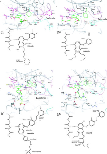
To date, there are more than 20 structures of the EGFR tyrosine kinase domain (reviewed by Kumar et al. Reference Kumar, Petri, Halmos and Boggon2008). Figure 4 illustrates the essential features of the structures of the wild-type EGFR kinase with gefitinib (Yun et al. Reference Yun, Boggon, Li, Woo, Greulich, Meyerson and Eck2007), with erlotinib (Stamos et al. Reference Stamos, Sliwkowski and Eigenbrot2002; Wood et al. Reference Wood, Truesdale, McDonald, Yuan, Hassell, Dickerson, Ellis, Pennisi, Horne, Lackey, Alligood, Rusnak, Gilmer and Shewchuk2004) (both drug complexes have the kinase in the active conformation) and with lapatinib (Wood et al. Reference Wood, Truesdale, McDonald, Yuan, Hassell, Dickerson, Ellis, Pennisi, Horne, Lackey, Alligood, Rusnak, Gilmer and Shewchuk2004) where the kinase is in the inactive state. In the active conformation (Fig. 4a, b), which is generated by a lattice kinase/kinase contact (Stamos et al. Reference Stamos, Sliwkowski and Eigenbrot2002), EGFR exhibits the common kinase fold in which the C-helix is in the right conformation to promote the interaction between the conserved glutamate and lysine (Glu762 and Lys745, using the sequence numbering that includes the 24-amino-acid signal sequence). This interaction locates the triphosphate moiety of ATP. The activation segment adopts a conformation similar to that seen in phosphorylated receptors such as the insulin receptor but with the position of the segment stabilised not by a phosphotyrosine but by many favourable van der Waals and hydrogen bond interactions. The carboxylate group of Glu848 assumes a position similar to the position of the phosphoryl group in those kinases (such as CDK2) that are activated by phosphorylation. The DFG motif (comprising Asp855, Phe856 and Gly857) has the active conformation, as shown in Fig. 4a and b.
Details of the EGFR kinase in complex with gefitinib (Iressa), erlotinib (Tarceva), lapatinib (Tykerb) and HKI272. The 3D structures are shown above the chemical structures. The colour code is glycine-rich loop (residues 719–724), C-helix (residues 756–767) and hinge (residues 791–794) in magenta, activation segment (residues 855–884) in cyan and the remainder of the kinase in grey. (a) EGFR in complex with gefitinib. The kinase is in the active conformation with the C-helix rotated in so that a hydrogen bond is made between Glu762 and Lys745. Phe723 and Leu858 are shown for reference (see text) From PDB ID 2ITY. (b) EGFR in complex with erlotinib. As with gefitinib the kinase is in the active conformation. From PDB ID 1M17. (c) EGFR in complex with lapatinib. The kinase is in the inactive conformation in which the glycine loop covers the inhibitor, the C-helix swung out, the DFG motif has Phe856 directed in to the ATP site and the activation adopts a helical conformation at Leu858. Residue Leu858 contacts a number of non-polar groups including Phe723. In the activated mutant L858R, the larger and positively charged arginine would destabilise the inactive conformation and favour the active conformation. From PDB ID 1XKK. (d) EGFR T790M mutant in complex with HKI272. The kinase is in the inactive conformation. There is a covalent bond between Cys797 and the inhibitor. From PDB ID 2JIV. See text for further details.

Gefitinib binds to the active conformation of the EGFR kinase at the ATP binding site in a conformation similar to that observed for quinazolines binding to CDK2 and p38 MAP kinases (Yun et al. Reference Yun, Boggon, Li, Woo, Greulich, Meyerson and Eck2007) (Fig. 4a). The N1 of the quinazoline accepts a hydrogen bond from the main chain NH of Met793 (hinge region), whereas the N3 atom contacts the gatekeeper Thr790 side chain through a water molecule. The 3-chloro-4-fluoro aniline substituent binds in the gatekeeper hydrophobic pocket forming a 42° angle with the quinazoline. The 6-propylmorpholino group extends into solvent consistent with structure activity data that this group provides only a modest enhancement of binding affinity. The group was added to improve pharmacokinetic properties. Gefitinib was not the most potent inhibitor discovered in the series, but it was chosen because it achieves high and sustained blood levels in vivo over a 24-h period.
The structure of wild-type EGFR kinase with erlotinib (Fig. 4b), the first EGFR kinase structure to be published (Stamos et al. Reference Stamos, Sliwkowski and Eigenbrot2002), is similar to that of the kinase with gefitinib. The quinazoline and anilino groups make similar interactions, whereas the acetylene moiety is located in the pocket behind the ATP site, which is only accessible if the gatekeeper (Thr790) residue is small. The other groups attached to the quinazoline are directed to solvent but make some non-polar contacts with residues Leu844, Phe795 and Gly796.
In contrast to the above structures with erlotinib and gefitinib, the complex of EGFR kinase with lapatinib showed the kinase in an inactive conformation with a significantly different structure that resembles the structures observed for inactive SRC or inactive CDK2 (Wood et al. Reference Wood, Truesdale, McDonald, Yuan, Hassell, Dickerson, Ellis, Pennisi, Horne, Lackey, Alligood, Rusnak, Gilmer and Shewchuk2004) (Fig. 4c). In comparison with the erlotinib complex, the N-terminal and C-terminal lobes of the kinase are rotated by about 12° and the kinase is in the closed conformation. The ATP pocket is enlarged in the inactive conformation by rotation and translation of the C-helix, which is necessary to accommodate the 3-fluorobenzyloxy group and explains why lapatinib can only bind to an inactive conformation. In the crystal structure, the quinazoline moiety makes similar contacts to those observed for gefitinib and erlotinib, whereas the large 3′-chloro-4′-[(3-fluorobenzyl)oxy]aniline group goes into the gatekeeper pocket making mostly hydrophobic contacts. The glycine-rich loop is folded over the inhibitor. The methylsulfonylethylaminomethylfuryl group points into solvent and does not make significant contacts with the protein. The activation segment (residues 855–884) has an inactive conformation with a short helical segment from residues 858 to 862.
The inhibitors exhibit different specificities for EGFR and ErbB2. Erlotinib and gefitinib have inhibition constants K i for the EGFR kinase of 0·4 and 0·7 nM, respectively, but are nearly 2000-fold less potent against ErbB2. In contrast, lapatinib has K i of 3·0 nM for EGFR and K i of 13 nM for ErbB2, i.e. it is an effective inhibitor against both EGFR and ErbB2 (Wood et al. Reference Wood, Truesdale, McDonald, Yuan, Hassell, Dickerson, Ellis, Pennisi, Horne, Lackey, Alligood, Rusnak, Gilmer and Shewchuk2004). There are dramatic differences in the off-rates for inhibitor dissociation: for gefitinib and erlotinib the off-rate is 30 min, whereas for lapatinib, it is very slow (half-life 300 min). The substantial differences between the active and inactive EGFR kinase conformation and the notion that to release the inhibitor would require conformational changes provide a possible explanation for the slow off-rate of lapatinib. ErbB2 kinase domain has 88% sequence identity with EGFR kinase. Sequence differences around the inhibitor binding sites do not explain why erlotinib and gefitinib inhibit EGFR but not ErbB2 while lapatinib inhibits both enzymes with similar potency. Wood et al. (Reference Wood, Truesdale, McDonald, Yuan, Hassell, Dickerson, Ellis, Pennisi, Horne, Lackey, Alligood, Rusnak, Gilmer and Shewchuk2004) suggest that the differences between active and inactive structures of EGFR allow inhibitors to target different forms of the enzyme, active or inactive. The lack of inhibition of ErbB2 by erlotinib may reflect the inability of ErbB2 to adopt the precise active conformation as observed for the apo and EGFR/erlotinib structures.
4.3.1 Activating mutations
How do activating mutations in EGFR lead to greater drug sensitivity? A number of mutations have been identified that affect the catalytic site (Chan et al. Reference Chan, Gullick and Hill2006; Shigematsu & Gazdar, Reference Shigematsu and Gazdar2006). These include an in-frame deletion of exon 19, residues 747–749 (sequence LRE), at the region before the C-helix that accounts for ~44% of the activating mutations; a single nucleotide substitution in which Leu858 in the activation segment is substituted by arginine that accounts for ~41% of activating mutations (achieved by just a single base change); a Gly719 to serine, alanine or cysteine substitution in the glycine-rich loop that accounts for a further 4% of activating mutations. Any one of these mutations leads to an active kinase that is transforming when present in cells. The L858R mutant is 50-fold more active, 10- to 100-fold more sensitive to gefitinib and erlotinib, and binds gefitinib 20 times more tightly than the wild-type kinase. In a comprehensive analysis with gefitinib, Yun et al. (Reference Yun, Boggon, Li, Woo, Greulich, Meyerson and Eck2007) showed that the L858R and G719S EGFR mutants adopt very similar active conformations to that observed for the wild-type kinase. In the L858R mutant, the arginine points into the solvent. The structural results show that this mutation destabilises the inactive conformation of EGFR kinase. In the inactive conformation, the C-helix is shifted and rotated outward, allowing the start of the activation segment to take a helical turn. Leu858 is in this helical turn and makes non-polar contacts with nearby residues including Phe723 from the glycine-rich loop (Fig. 4c) as recognised by Zhang et al. (Reference Zhang, Gureasko, Shen, Cole and Kuriyan2006). Substitution with the large and positively charged arginine is not compatible with the inactive conformation but is with the active conformation. The lower affinity for ATP and the higher affinity for gefitinib and erlotinib in the L858R mutant combine to block growth of tumours that are ‘addicted’ to the activated EGFR for survival and explain the greater efficacy of the inhibitors in treatment of NSCLC patients carrying this mutation.
Lapatinib, which binds preferentially to the inactive state, would be less effective against the activating mutant EGFR kinases that occur in NSCLC patients. Instead lapatinib, with its ability to inhibit both EGFR and ErbB2 (Her2), has had success in treatment of Her2-positive breast cancer for patients with trastuzumab resistance. It has been approved for use in combination with capecitabine (trade name Xeloda; a prodrug that is converted to 5-fluorouracil in the tumour and inhibits DNA synthesis). In a phase III study, favourable tumour responses were seen in ~25% of patients (Geyer et al. Reference Geyer, Forster, Lindquist, Chan, Romieu, Pienkowski, Jagiello-Gruszfeld, Crown, Chan, Kaufman, Skarlos, Campone, Davidson, Berger, Oliva, Rubin, Stein and Cameron2006). It has been suggested that treatment could be improved and made more cost effective with different dose administration regimens (Gomez et al. Reference Gomez, Doval, Chavez, Ang, Aziz, Nag, Ng, Franco, Chow, Arbushites, Casey, Berger, Stein and Sledge2008). The preclinical background for anti-EGFR drugs in combination with cytotoxic agents has been reviewed (Milano et al. Reference Milano, Spano and Leyland-Jones2008) in an attempt to understand why in some cases there are synergistic effects while in others there are not. Currently, a large randomised phase III trial is in progress to assess the dual benefits of lapatinib (Tykerb) and trastuzumab (Herceptin) separately, sequentially or together for treatment of Her2-positive breast cancer patients.
4.3.2 Resistance
Development of resistance to the EGFR kinase inhibitors has become a major clinical problem. Patients initially responsive to gefitinib and erlotinib become resistant through second site mutations; most notable are mutations in the gatekeeper residue Thr790 for ~50% of patients. In ABL, the equivalent T315L mutation sterically blocks the binding of imatinib and hence confers resistance to the drug. In EGFR kinase, structural and biochemical studies have shown the mechanism is more complex (Yun et al. Reference Yun, Mengwasser, Toms, Woo, Greulich, Wong, Meyerson and Eck2008). Binding studies showed that the T790M mutant bound gefitinib with a K d=2·4 nM, almost as tightly as the most sensitive L858R mutant. Crystal structures of the T790M mutant alone and in complex with two inhibitors showed that the gatekeeper mutation did not block binding of inhibitors in either the active or the inactive states. The two inhibitors were AEE788 (a compound that is similar to gefitinib and erlotinib, which binds to the active conformation and which is also in phase II clinical trials for glioblastoma) and HKI-272 (neratinib) (an irreversible inhibitor, which reacts with a cysteine Cys797 at the ATP site, binds to the inactive conformation and for which the T790M EGFR mutant is not resistant). The inhibitor AEE788 bound to the mutant T790M in a similar fashion as to active wild-type EGFR with a small change in the Met790 side chain, which adopts a different rotamer conformation to allow access of the phenylethylamine group to the gatekeeper pocket. The irreversible inhibitor HKI-272 bound to the inactive conformation in which the C-helix was swung out to accommodate the bulky aniline group, similar to the change needed for lapatinib binding (Fig. 4d). HKI-272 forms a covalent bond with Cys797, rendering the binding irreversible (Yun et al. Reference Yun, Mengwasser, Toms, Woo, Greulich, Wong, Meyerson and Eck2008).
The explanation for resistance lies in the relative values of ATP affinity (as measured by K m) and gefitinib affinity (as measured by K d) (Yun et al. Reference Yun, Mengwasser, Toms, Woo, Greulich, Wong, Meyerson and Eck2008). Kinetic studies showed that although the L858R mutant has a lowered affinity for ATP (K m 148 μM), allowing the drug (K d 2·4 nM) to compete successfully for the ATP site, the T790M mutant and the double-mutant L858R/T790M have almost the same ATP affinity as the wild type (K m 5·9 and 8·4 μM) with K d of 4·6 and 10·9 nM, respectively, for gefitinib. Analysis of abilities of the drug to compete with cellular concentrations of ATP showed that whereas the lowered affinity of L858R mutant for ATP allows gefitinib to compete successfully, the wild-type-like affinity of the T790M or T790M/L858R mutants for ATP leads to a decrease in efficacy of the drug. (The structural explanation for the change in ATP affinity for the double L858/T790M mutant is not readily apparent.) The balance between drug and ATP affinities is crucial in determining a favourable outcome for ATP site-directed inhibitors when in competition with cellular concentrations of ATP. Thus, a ratio K d (drug)/K m (ATP) of 1·6×10−5 appears favourable, but a ratio of 78×10−5 is not. In contrast, the irreversible inhibitor HKI-272 is able to overcome the T790M resistance because once covalently bound it is no longer in competition with ATP. HKI-272 is progressing in clinical trials as one of second-generation EGFR inhibitors (reviewed by Wissner & Mansour, Reference Wissner and Mansour2008).
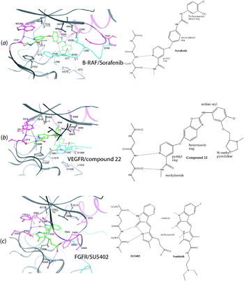
4.4 B-RAF and VEGFR inhibitors: sorafenib (Nexavar) and sunitinib (Sutent)
The protein kinase RAF is part of the RAS signalling pathway, a pathway that is frequently deregulated in cancer. The GTPase, RAS, is a key player in the transmission of signals from the receptor tyrosine kinases to the nucleus that promote transcription for survival and growth. RAS is localised in the membrane by its C-terminal farnesyl group (and for H-RAS and N-RAS additional palmitoylation). The protein SOS, a GDP/GTP exchange factor (GEF) for RAS, is recruited to the cell membrane through recognition of the receptor autophosphorylation sites and its proximity activates RAS. RAS-GTP promotes RAF membrane recruitment and activation. The serine/threonine kinase RAF initiates a protein kinase cascade comprising the MEK and the extracellular signal-regulated kinase (ERK) to achieve activation of transcription factors. Unwanted cell growth can occur through upstream defects in unregulated activation of the tyrosine kinase receptor, or most frequently, in RAS itself, leading to an oncogenic form of RAS that is constitutively active, or in RAF. There are three members of the RAF family, A-RAF, B-RAF and C-RAF, with slightly different regulatory properties. B-RAF, which has a higher basal kinase activity than the other members of the family, is mutated in 70% of malignant melanomas and at lower frequencies in several other cancers (Davies et al. Reference Davies, Bignell, Cox, Stephens, Edkins, Clegg, Teague, Woffendin, Garnett, Bottomley, Davis, Dicks, Ewing, Floyd, Gray, Hall, Hawes, Hughes, Kosmidou, Menzies, Mould, Parker, Stevens, Watt, Hooper, Wilson, Jayatilake, Gusterson, Cooper, Shipley, Hargrave, Pritchard-Jones, Maitland, Chenevix-Trench, Riggins, Bigner, Palmieri, Cossu, Flanagan, Nicholson, Ho, Leung, Yuen, Weber, Seigler, Darrow, Paterson, Marais, Marshall, Wooster, Stratton and Futreal2002a). Significantly, RAS and B-RAF mutations are restricted to the same tumour types, usually in a mutually exclusive fashion, suggesting that these genes are on the same oncogenic pathway. Thus, B-RAF has become a prime target for kinase inhibitors for therapy.
Sorafenib (Nexavar, BAY 439006) is a multikinase inhibitor initially developed to inhibit B-RAF but it also targets VEGFR2, VEGFR3, PDGFRβ, Flt3 and KIT (Wilhelm et al. Reference Wilhelm, Carter, Tang, Wilkie, McNabola, Rong, Chen, Zhang, Vincent, McHugh, Cao, Shujath, Gawlak, Eveleigh, Rowley, Liu, Adnane, Lynch, Auclair, Taylor, Gedrich, Voznesensky, Riedl, Post, Bollag and Trail2004). The structure of B-RAF in complex with sorafenib provided an explanation for oncogenic mutations and the mechanism of sorafenib inhibition (Wan et al. Reference Wan, Garnett, Roe, Lee, Niculescu-Duvaz, Good, Jones, Marshall, Springer, Barford and Marais2004). The inhibitor is buried deep within the crevasse between the N-terminal and C-terminal lobes of the kinase structure with the kinase in the inactive conformation (Fig. 5a). The pyridyl ring occupies the adenine part of the ATP site, interacting with three aromatic residues including Trp530 from the hinge, Phe582 at the end of the catalytic loop and Phe594 of the DFG motif, which also contacts the central phenyl ring. The nitrogen atom of the pyridyl ring accepts a hydrogen bond from the main chain nitrogen of hinge region Cys531 and the methylamide side group donates a hydrogen bond to the Cys531 main chain carbonyl. The central ring contacts Thr528, the gatekeeper residue, but there is no hydrogen bond. The urea group makes three hydrogen bonds, one from each of its NH groups to the carboxylate side chain of Glu500 (C-helix) and one from the carbonyl oxygen to the main chain nitrogen of Asp593 of the DFG motif, in a similar fashion to the hydrogen bonds observed with imatinib and ABL (Figs 3a and 5a). The lipophilic trifluoromethylphenyl ring inserts into the hydrophobic pocket between the αC and αE helices and the start of the activation segment and occupies a position that would be anticipated for Phe594 of the DFG motif in the active conformation. The flipped inactive conformation of the DFG motif orients the activation segment towards the glycine-rich loop in which the side chain of Val599 contacts Phe467. There is considerable similarity between the inactive conformation of B-RAF and that of ABL with respect to the orientation of the C-helix, the N and C lobes and the DFG motif. The interactions of their inhibitors, sorafenib and imatinib, with their respective kinases are also similar.
B-RAF and VEGFR inhibitors. (a) Sorafenib in complex with B-RAF. The colour code is as in Fig. 2. Sorafenib binds in a similar way to imatinib binding to ABL. The kinase is in the inactive conformation. From PDB ID 1UWH. (b) Compound 22 in complex with VEGFR kinase domain. Compound 22 is a derivative of sorafenib and binds to the inactive conformation. From PDB ID 2QU6. (c) SU5402 in complex with FGFR kinase domain. The glycine-rich loop is well ordered and Phe489 caps the inhibitor-binding site. SU5402 is similar to sunitinib, which is in clinical use. From PDB ID 1FGI. See text for further details.

In the inactive conformation of B-RAF, there is an active-like interaction between the C-helix Glu500 and Lys482 (Fig. 5a), the interaction that is important for aligning the triphosphate of ATP. Only a change in position of the DFG motif and activation segment is required to trip the kinase to adopt the active conformation. Significantly, almost all RAF activating mutants are located in the activation segment, within or adjacent to the DFG motif or in the glycine-rich loop (Wan et al. Reference Wan, Garnett, Roe, Lee, Niculescu-Duvaz, Good, Jones, Marshall, Springer, Barford and Marais2004). The mutation Val599 to Glu is frequently found in cancer and accounts for 95% of mutations at this site. (In recent literature, this residue is referred to as Val600. Here we keep the numbering used in the crystal structure coordinates.) It is generated by a single base change possibly explaining why it occurs at high frequency. The structural results show that the mutation of Val599 to a larger and charged residue such as glutamic acid would destabilise the activation segment/glycine-rich interaction between residue 599 and Phe467, which appears to be important in maintaining the DFG motif in the inactive conformation (Fig. 5a). This destabilisation of the inactive conformation by mutation is very similar to that discussed above for the frequent EGFR mutation L858R. The same regions of the kinase are involved. The structure provides a clear explanation for the effects of almost all of the mutants in their ability to destabilise the inactive conformation of B-RAF. The observation that mutations favour the active conformation also implies that sorafenib would be less effective with these mutants, and this has been confirmed experimentally. The V599E B-RAF is ~2-fold less sensitive to sorafenib than wild-type B-RAF (IC50 38 and 22 nM, respectively).
Oncogenic B-RAF stimulates constitutive MEK-ERK signalling in melanoma cells, and this signalling is required for proliferation. However, sorafenib as a B-RAF inhibitor has not been successful for the treatment of melanomas, although there may be some benefits in combination with dacarbazine, a triazene DNA alkylating agent commonly used for treatment of malignant melanoma (Lacouture et al. Reference Lacouture, Desai, Soltani, Petronic-Rosic, Laumann, Ratain and Stadler2006). The reasons are not entirely clear but may be attributed to two possible factors. First, with an IC50 of 38 nM for the activated mutant B-RAF, sorafenib is not a powerful enough inhibitor. Second, downstream of RAF, there are several feedback pathways that act to regulate and compensate for upstream perturbations. For example, the ERK phosphatases that serve to inactivate ERK may become down-regulated in response to RAF inhibition. Other unexpected responses of this pathway to inhibitors have been noted (Hall-Jackson et al. Reference Hall-Jackson, Eyers, Cohen, Goedert, Boyle, Hewitt, Plant and Hedge1999). However, sorafenib is in clinical use for renal cell carcinoma as an inhibitor of VEGFR kinase activity.
To obtain nourishment and to spread, tumour cells need to form new blood vessels by the process of angiogenesis. The members of the split kinase-domain family of tyrosine kinase receptors promote angiogenesis and metastasis. Receptors, such as VEGFR and PDGFR, respond to raised levels of the appropriate growth factors. Their kinases have an interruption to their kinase domain of ~65–97 residues, termed the kinase insertion domain between αD and αE helices that has a role in signalling but not in kinase activity. The most common form of renal carcinoma, clear cell renal cell carcinoma that represents ~80% of renal carcinomas, is triggered by mutations in the tumour suppressor Von Hippel–Lindau (VHL) gene. As a result of VHL inactivation, the hypoxia inducible factors (HIFs) are not degraded, and they continue to induce transcription of a number of genes including VEGF, PDGF and other growth factors (Gnarra et al. Reference Gnarra, Zhou, Merrill, Wagner, Krumm, Papavassiliou, Oldfield, Klausner and Linehan1996). Up-regulation of the VEGFR and PDGFR receptors by their respective ligands leads to vascularised tumours and tumour progression. There are three forms of VEGFR: VEGFR1 (Flt1), VEGFR2 (KDR) and VEGFR3 (Flt4), of which VEGFR2 is of primary importance in mitosis and survival of endothelial cells. Thus, inhibition of tumour growth and angiogenesis through targeting the receptor kinases including VEGFR, PDGFR, the stem cell factor receptor (KIT) and FMS-like tyrosine kinase 3 (Flt3) has become an important route for therapy, especially for treatment of kidney cancers.
Metastatic renal cell carcinoma is resistant to conventional chemotherapy, and the major form of treatment has been cytokine-based immunotherapy with response rates of 10–20%. There are now four compounds approved for clinical use that give hope for improved treatment: bevacizumab (Avastin), an anti-VEGF monoclonal antibody; sorafenib (Nexavar) and sunitinib (Sutent), VEGFR inhibitors; and temsirolimus, an inhibitor of mTOR (discussed later) (reviewed by Costa & Drabkin, Reference Costa and Drabkin2007; Samlowski et al. Reference Samlowski, Wong and Vogelzang2008). Although these compounds have provided a significant improvement in quality of life, delayed progression and extended survival, the average progression-free survival is less than 1 year, and further evaluation of treatment sequences, combinations and first- and second-line schedules are underway to improve the cure rate (Rini et al. Reference Rini, Rathmell and Godley2008). The end points for successful cancer treatment are often difficult to quantify. One end point that is increasingly being used is progression-free survival – the time until a cancer worsens, rather than overall survival, which can be complicated by other factors. Sorafenib and sunitinib were approved in early 2006 for treatment of renal cell carcinoma and for GISTs (sunitinib).
Among potent VEGFR inhibitors (summarised by La et al. Reference La, Belzile, Bready, Coxon, Demelfi, Doerr, Estrada, Flynn, Flynn, Graceffa, Harriman, Larrow, Long, Martin, Morrison, Patel, Roveto, Wang, Weiss, Whittington, Teffera, Zhao, Polverino and Harmange2008), there are no structures in the public domain of sorafenib with VEGFR, but there is a structure of a promising related benzoxazole compound (compound 22; Fig. 5b) that has been developed as a specific VEGFR2 inhibitor based on docking of sorafenib to the VEGFR ATP site. Compound 22 has K i 3 nM, cellular activity IC50 15 nM, is highly selective and has good pharmacokinetic properties (Potashman et al. Reference Potashman, Bready, Coxon, Demelfi, Dipietro, Doerr, Elbaum, Estrada, Gallant, Germain, Gu, Harmange, Kaufman, Kendall, Kim, Kumar, Long, Neervannan, Patel, Polverino, Rose, Plas, Whittington, Zanon and Zhao2007). It was based on cyclisation of the urea group of sorafenib into an oxazole ring to enhance conformational rigidity while retaining hydrogen bonds. The crystal structure (Fig. 5b) showed that compound 22 bound to the inactive conformation of VEGFR2 in a mode very similar to that of sorafenib binding to B-RAF. The nitrogen of the pyridine and the methylamide NH form hydrogen bonds with the hinge region Cys919 main chain atoms as for sorafenib binding to B-RAF. The benzoxazole core is oriented in the hydrophobic pocket flanked by Val899 and Phe1047 (from the DFG motif). The endocyclic nitrogen of the aminobenzoxazole forms hydrogen bond with the NH of Asp1046 (from the DFG motif) and the exocyclic NH contacts the side chain of Glu885. The aniline aryl ring occupies the hydrophobic pocket in a similar way to the trifluoromethylphenyl group of sorafenib with B-RAF. The N-methylpyrrolidine side chain is directed to a solvent-exposed portion of the protein.
Sunitinib (Sutent, SU112488) (Fig. 5c) was developed as an agent against tumour cells dependent for proliferation and survival on signalling through the receptors PDGFR, KIT, FLT3 and VEGFR (Mendel et al. Reference Mendel, Laird, Xin, Louie, Christensen, Li, Schreck, Abrams, Ngai, Lee, Murray, Carver, Chan, Moss, Haznedar, Sukbuntherng, Blake, Sun, Tang, Miller, Shirazian, McMahon and Cherrington2003). In vitro, sunitinib exhibits K i for VEGFR2 (KDR) and PDGFR of 9 nM. In cells, sunitinib inhibited VEGF-induced proliferation with IC50 value of 40 nM. Following successful clinical trials (Motzer et al. Reference Motzer, Michaelson, Redman, Hudes, Wilding, Figlin, Ginsberg, Kim, Baum, Deprimo, Li, Bello, Theuer, George and Rini2006, Reference Motzer, Michaelson, Rosenberg, Bukowski, Curti, George, Hudes, Redman, Margolin and Wilding2007) where median response rates were 33%, progression-free survival was 9 months and median survival was 24 months, sunitinib was approved for renal cell carcinoma therapy in 2007. Sunitinib is partly metabolised to SU12662 in vivo after N-demethylation by cytochrome P450, but this does not affect its efficacy. There are no structures in the public domain of sunitinib with VEGFR or PDGFR kinase domains, but there is a structure of a similar compound SU5402 with the kinase domain of the FGFR1 kinase domain (Mohammadi et al. Reference Mohammadi, McMahon, Sun, Tang, Hirth, Yeh, Hubbard and Schlessinger1997) (Fig. 5c). The compound SU5402, which exhibits IC50 of 10–20 μM in cells, binds at that ATP site with the oxindole making two hydrogen bonds to the hinge region; the nitrogen to the carbonyl oxygen of Glu562 and the oxygen to the main chain nitrogen of Ala564. The NH of the pyrrole forms hydrogen bond to the O2 oxindole in an intramolecular interaction and is also within hydrogen bonding distance of the carbonyl oxygen of Ala564. Hydrophobic groups (Leu484, Phe489 (glycine-rich loop), Ala512, Leu561 and Leu630) line the pocket that accommodates the oxindole and the pyrrole rings. The methyl group of the pyrrole contacts Gly567 and the carboxyethyl group forms hydrogen bond to the side chain of Asn568. The phenylalanine Phe489 from the glycine-rich loop caps the hydrophobic pocket in which the oxindole binds and is stabilised by an extensive network of oxygen aromatic interactions. SU5402 is also an inhibitor of VEGFR, which has conserved Asn equivalent to Asn568 that contacts the carboxy ethyl group.
4.5 Temsirolimus (Torisel, CCI-779) and mTOR inhibition
Temsirolimus (cell cycle inhibitor CCI-779), a soluble ester of rapamycin (sirolimus), has recently been approved as an anti-cancer drug for renal cell carcinoma (May 2007) based on a phase III trial that showed an overall survival advantage over interferon α monotherapy (Hudes et al. Reference Hudes, Carducci, Tomczak, Dutcher, Figlin, Kapoor, Staroslawska, Sosman, McDermott, Bodrogi, Kovacevic, Lesovoy, Schmidt-Wolf, Barbarash, Gokmen, O'Toole, Lustgarten, Moore and Motzer2007). Temsirolimus has also shown promise in treatment of other cancers (reviewed by Rini, Reference Rini2008). Rapamycin (Fig. 6d), first isolated from a soil bacterium found on Rapa Nui (Easter Island), is a 31-membered macrolide natural product. Like the related compound FK506, rapamycin has potent immunosuppressive properties. However, its anti-cancer properties are exerted through the inhibition of mammalian target of rapamycin (mTOR) kinase function.
Rapamycin in the complex of FKBP12/rapamycin/FRB domain of mTOR. (a) Schematic diagram showing secondary structures of the two proteins. FKBP12 is in blue (right) and FRB in pink (left). Rapamycin (carbon atoms in green) is sandwiched between the two proteins. (b) Details of the interaction from the same view as in (a). The FKBP12 interactions (in blue) are extensive and include van der Waals interactions to a number of aromatic residues (Y26, F36, F46, W59, Y82, Y85 and F99) and hydrogen bonds from rapamycin to D37 and Y82 side chains and Q53, E54 and I56 main chain atoms. The contacts from rapamycin to the FRB domain (pink) are less extensive and include the aromatic residues F2039, W2101, Y2105 and F2108. (c) Space filling representation of the FKBP12/rapamycin/FRB complex in the same orientation and with the same colour code as (a). Rapamycin is almost entirely buried. Only the C40, C41, O13 and O2 atoms are exposed. (d) Chemical structure of rapamycin with ring carbon atoms numbered. The extra atoms substituted at the O13 position in temsirolimus are shown. The view is similar to that of (b). (e) View of space filling representations of the FKBP12/rapamycin/FRB structure rotated 90° from (c) to show the rapamycin surface docked against the FRB domain (left) and rotated 90° in the opposite direction to show the surface of rapamycin docked against FKBP12 (right). From PDB ID 2FAP.

mTOR is a massive 289-kDa serine/threonine kinase, which promotes cell growth when conditions are favourable and which is down-regulated in response to environmental stress. It is a member of the phosphatidylinositol kinase-related kinase family and is regulated through the PI3K and Akt/PKB pathway (reviewed by Wullschleger et al. Reference Wullschleger, Loewith and Hall2006). mTOR phosphorylates the p70 S6 kinase leading to enhanced translation of certain ribosomal proteins and elongation factors and the production of HIF1α, which regulates the transcription of genes that stimulate growth including VEGF. mTOR also phosphorylates the 4E-binding protein 1, promoting dissociation of this complex and allowing the eukaryotic initiation factor 4 subunit E to increase the translation of mRNAs that encode cell cycle regulators such as c-myc and cyclin D. The kinase domain is located at the C-terminal region. Immediately N-terminal to the kinase domain is the 100-amino-acid FKBP12-rapamycin binding domain (FRB) (residues 2015–2114). Further upstream are FAT domains and HEAT repeats. Both mTOR and PI3K are targets for anti-cancer drugs. There are several selective small-molecule mTOR kinase inhibitors in preclinical trials and for PI3K in clinical trials (Marone et al. Reference Marone, Cmiljanovic, Giese and Wymann2008; Yuan & Cantley, Reference Yuan and Cantley2008). Some inhibitors have dual specificity and have an ATP competitive mechanism.
Rapamycin (like FK506) forms a tight binary complex with the small immunophilin 12-kDa protein FKBP12 (FK506 binding protein) (K D=0·2 nM) and inhibits the peptidyl-prolyl isomerase activity of FKBP12. Rapamycin also binds to mTOR FRB domain using a part of its binding surface that is distinct from the FK506 binding site. Rapamycin and FK506 have different biological functions based on their ability to target other proteins. Immunosuppressive activity is due to the ability of FKBP12/FK506 (but not the FKBP12/rapamycin complex) to inhibit the phosphatase calcineurin. In contrast, the FKBP12/rapamycin complex (but not the FKBP12/FK506 complex) inhibits mTOR through binding to the FRB domain. The FRB domain appears to regulate mTOR signal transduction, although the exact mechanism by which rapamycin inhibits mTOR kinase function is not understood. In the absence of rapamycin, FKBP12 and FRB do not interact. Rapamycin binds to FRB with low affinity (K D=26 μM), but the FKBP/rapamycin complex binds 2000-fold higher affinity (K D=12 nM) (Banaszynski et al. Reference Banaszynski, Liu and Wandless2005). Rapamycin acts as an adaptor to mediate the association of two proteins. In vitro temsirolimus and rapamycin inhibit mTOR at nanomolar concentrations, but growth inhibitory properties in cells are not apparent until much higher concentrations (micromolar) are reached. Recently, the effects at high concentration in the biphasic response have been shown to be due to an FKBP12-independent suppression of mTOR kinase activity that leads to significant reduction in protein synthesis and cell growth (Shor et al. Reference Shor, Zhang, Toral-Barza, Lucas, Abraham, Gibbons and Yu2008).
Structural studies have characterised the FKBP12/rapamycin interactions (Bierer et al. Reference Bierer, Mattila, Standaert, Herzenberg, Burakoff, Crabtree and Schreiber1990; Van Duyne et al. Reference Van Duyne, Standaert, Karplus, Schreiber and Clardy1991) and the FKBP/rapamycin/FRB ternary complex (Choi et al. Reference Choi, Chen, Schreiber and Clardy1996; Liang et al. Reference Liang, Choi and Clardy1999). In the ternary complex FKBP12/rapamycin/FRB, rapamycin uses two different faces to interact with FKBP12 and FRB (Fig. 6a). FKBP12 comprises a 5-stranded twisted β-sheet wrapped around a single α-helix. Rapamycin binds in a hydrophobic cleft formed between the β-sheet and the α-helix (Fig. 6a, b). Three loops surround and contribute to binding. In addition to hydrophobic interactions, there are several hydrogen bonds. A total of 52% of the molecular surface area of rapamycin is buried. The FRB domain forms a four-helix bundle (Fig. 6a). The rapamycin triene arm (atoms C17–C24) (Fig. 6d) binds at a hydrophobic crevice containing conserved aromatic residues at the interface of helices 1 and 4. The crevice is relatively shallow. The interactions between FRB and rapamycin are all hydrophobic. A total of 44% of the molecular surface area of rapamycin is buried on association with FRB. In the FKBP12/rapamycin/FRB complex, rapamycin has 92% of its molecular surface area buried (Fig. 6c, e). Only the O13, C40 and C41 atoms are exposed. This is the region where additional groups have been incorporated in temsirolimus. Although these would disrupt the hydrogen bond of rapamycin to Gln53 main chain (Fig. 6b), the additional atoms could be readily accommodated in the solvent-exposed region.
The conformation of rapamycin appears rigid. The structures of free rapamycin, rapamycin in a binary complex with FKBP12 and rapamycin in the ternary complex are all similar. The planarity of the triene arm is maintained in all structures. What then is the origin of the observation that rapamycin by itself binds with low affinity to the FRB domain but that rapamycin/FKBP12 binds with high affinity to FRB? A small change in the orientation of rapamycin relative to FKBP12 was observed between the binary and ternary complexes. This results in a shift in the triene arm of rapamycin that allows the methyl group on C22 to contact Phe2108. This may provide a part of the explanation, but a full structural explanation is not clear at present.
An intriguing insight into the significance of the FRB domain in mTOR signalling has come from the observation that phosphatidic acid (PA) is required for mTOR to phosphorylate S6 kinase (Fang et al. Reference Fang, Vilella-Bach, Bachmann, Flanigan and Chen2001). PA is generated by the action of phospholipase D, and phospholipase D has been implicated in survival signals in cancer. PA was reported to bind mTOR FRB domain in a mode that was competitive with rapamycin/FKBP12 leading to a model in which rapamycin inhibited the localisation of mTOR to the plasma membrane and prevented access to other proteins such as raptor or substrates (Chen & Fang, Reference Chen and Fang2002; Foster, Reference Foster2007). Recent NMR structural data have confirmed that the PA binding site on the FRB domain does indeed overlap with the rapamycin binding site with Arg2109, one of the seven residues critical for binding (Veverka et al. Reference Veverka, Crabbe, Bird, Lennie, Muskett, Taylor and Carr2008) (Fig. 6b). The remarkable properties of rapamycin to interfere with protein or protein/lipid targeting suggest that drug-like compounds might be discovered that exploit other facets of kinase mechanisms apart from the ATP binding site.
4.6 CDK inhibitors in phase II: seliciclib (roscovitine), PD0332991 and alvocidib (flavopiridol)
The CDKs have been prime targets for inhibition in cancer therapy because of their role in control of the cell cycle and their up-regulation through overexpression of cyclins or down-regulation of tumour suppressor proteins in cancer (reviewed by Malumbres et al. Reference Malumbres, Pevarello, Barbacid and Bischoff2008). For example, breast cancer that is driven by the ErbB2 oncogene is dependent on CDK4/cyclin D1 function and mice lacking cyclin D1 are resistant to ErbB2-driven mammary neoplasia (Yu et al. Reference Yu, Geng and Sicinski2001; Landis et al. Reference Landis, Pawlyk, Li, Sicinski and Hinds2006). The observation that small interfering RNA knockdown of CDK2 failed to halt proliferation of osteosarcomas and Rb-negative cervical cancer cells and that CDK2−/− mice are viable (Berthet et al. Reference Berthet, Aleem, Coppola, Tessarollo and Kaldis2003; Ortega et al. Reference Ortega, Prieto, Odajima, Martin, Dubus, Sotillo, Barbero, Malumbres and Barbacid2003; Tetsu & McCormick, Reference Tetsu and McCormick2003) suggested that CDK2 might not be a good target for inhibition. However, most CDK2 inhibitors also target CDK1 and the joint down-regulation of the CDKs are likely to have beneficial effects. CDK2 has received much attention in CDK inhibitor design, partly because of ease of expression of the kinase and the early availability of a structure in 1993 (reviewed by Davies et al. Reference Davies, Pratt, Endicott, Johnson and Noble2002b). More than 133 CDK2/inhibitor complexes have been determined (Marsden & Knapp, Reference Marsden and Knapp2008). There are several compounds in phase I clinical trials, including an interesting compound derived from fragment-based discovery methods, that have good pharmacokinetic and pharmacodynamic properties (Wyatt et al. Reference Wyatt, Woodhead, Berdini, Boulstridge, Carr, Cross, Davis, Devine, Early, Feltell, Lewis, McMenamin, Navarro, O'Brien, O'Reilly, Reule, Saxty, Seavers, Smith, Squires, Trewartha, Walker and Woolford2008). Recent structural results have also shed light on the binding geometry of specific inhibitors to CDK6 and CDK9. Thus, within a family of closely related kinases, it has been possible to achieve specificity. The structural basis for this is examined below.
Seliciclib (R-roscovitine, CYC-202) is in phase II clinical trials for NSCLC and nasopharyngeal cancer. It targets CDKs, has good oral bioavailability and is well tolerated but suffers from a short half-life and weak potency in cells. Second-generation compounds such as CR8 with an N 6-biaryl substituent have improved properties (Bettayeb et al. Reference Bettayeb, Oumata, Echalier, Ferandin, Endicott, Galons and Meijer2008). CR8 inhibits CDK2/cyclin A with IC50 72 nM, whereas roscovitine has an IC50 of 210 nM. Both also inhibit CDK7 and CDK9, but not CDK4 or CDK6. CR8 has a 50-fold higher potency than roscovitine in cells, suggesting that this compound should have improved anti-tumour potential (Bettayeb et al. Reference Bettayeb, Oumata, Echalier, Ferandin, Endicott, Galons and Meijer2008). Roscovitine and CR8 bind to active CDK2/cyclin A in similar binding modes (Fig. 7a). The purine ring occupies the adenine ATP site and makes two hydrogen bonds to the hinge region to Leu83 main chain atoms. Both inhibitors make non-polar contacts to Ile10 and Leu134 in the adenine-binding pocket. The phenyl group contacts Phe82. The pyridine ring of CR8 points towards the solvent but is accommodated in a hydrophobic pocket of the alkyl portion of Glu8, Ile10 and Phe82 side chains. The structural reasons for the enhanced biological activity of CR8 over roscovitine are not clear, but the explanation could lie in the improved cell permeability and intracellular stability of CR8.
CDK inhibitor structures. Each CDK is in complex with its cyclin (not shown) and is in the active conformation. The C-helix glutamate and β3 lysine which form an ion pair to create the ATP triphosphate binding site and the phenylalanine of the DFG motif are shown for reference although they do not contact the inhibitors. (a) CDK2 in complex with the roscovitine analogue CR8. The phenyl pyridine occupies a hydrophobic pocket formed by Phe82 and the aryl part of Glu8 and extends towards the solvent. From PDB ID 3DDP (b) CDK6 in complex with PD0332991. There are three hydrogen bonds to the hinge region. The 2-aminopyridine and piperazine rings extend into the pocket lined by Thr107 (equivalent to Lys89 in CDK2). The gatekeeper residue Phe98 is shifted slightly from the position of the gatekeeper (Phe80) in CDK2. From PDB ID 2EUF. (c) The CDK9 complex with flavopiridol. The chlorophenyl ring is in the ‘CDK2 Lys89’ pocket but in CDK9 the equivalent residue to CDK2 Lys89 is Gly112. The small glycine creates a larger pocket allowing the chlorophenyl ring to be docked. The secondary structure of CDK9 from the complex of CDK9/cylin T1/ATP is shown in pale green. On binding flavopiridol there is an induced conformation change. Phe30 from the glycine loop folds down and contacts the inhibitor with an edge to face interaction with the piperidinyl ring. Leu51 takes the place of Phe30. From PDB ID 3BLR. For further details see text.

The compound PD0332991, a potent and selective CDK4 and CDK6 inhibitor, is in phase II clinical trials for multiple myeloma. PD0332991 is an orally active inhibitor that has been shown to induce G1 cell cycle arrest in primary human myeloma cells and to suppress tumour growth in xenograft models (Fry et al. Reference Fry, Harvey, Keller, Elliott, Meade, Trachet, Albassam, Zheng, Leopold, Pryer and Toogood2004). Progression through G1 phase of the cell cycle requires phosphorylation of the tumour suppressor protein pRb by CDK4 or CDK6 in association with their D-type cyclins. Phosphorylation of pRb results in the release the transcription factor E2F, which then stimulates the expression of downstream activators for the G1/S phase transition such as cyclin E and cyclin A that are important for DNA replication. PD0332991 has shown promise in combination with bortezomib, a proteasome inhibitor widely used in treatment of myeloma. It was shown that the tumour suppression by the cell-cycle CDK4 inhibitor combined with the cytotoxic killing by bortezomib gave an improved outcome in a mouse model of myeloma (Menu et al. Reference Menu, Garcia, Huang, Di Liberto, Toogood, Chen, Vanderkerken and Chen-Kiang2008).
PD0332991 is based on a pyridol[2,3-d]pyrimidin-7-one scaffold including a 2-aminopyridine side chain at the C2 position, which provides selectivity for CDK4 and CDK6. PD0332991 has IC50 for CDK4/cyclin D of 11 nM and is not active against CDK2/cyclin A (IC50>5 μM) (Toogood et al. Reference Toogood, Harvey, Repine, Sheehan, Vanderwel, Zhou, Keller, McNamara, Sherry, Zhu, Brodfuehrer, Choi, Barvian and Fry2005). A crystal structure of PD0332991 in complex with CDK6, activated by binding the herpes viral cyclin, showed binding at the ATP site (Lu & Schulze-Gahmen, Reference Lu and Schulze-Gahmen2006) (Fig. 7b). The N3 and N2-H groups form hydrogen bond to the main chain atoms of Val101 in the hinge region, as expected for a CDK inhibitor. There are further hydrogen bonds between the aminopyridine side chain and the Asp102 carbonyl oxygen and between the C6-acetyl group and the peptide amide of Asp163, the aspartate of the DFG motif. The C5-methyl and the C6-acetyl groups fill the pocket in front of the gatekeeper residue Phe98. This pocket appears slightly larger in CDK6 in contrast to CDK2 partly because of a shift in orientation of the main chain in the hinge region. Modelling suggests that binding of PD033291 to CDK2 would lead to overcrowding with the gatekeeper residue of CDK2, Phe80, providing a partial explanation for specificity. The piperazinylpyridine substituent in the C2 position points away from the ATP site into a pocket, which in CDK2 is lined by Lys89. In CDK6 the equivalent residue is a threonine, Thr107 (Thr102 in CDK4). The piperazine interacts with Thr107 and Asp104 side chains, but the interactions are just outside hydrogen binding distances. Although modelling suggests that CDK2 Lys89 would interfere with the interaction, the flexibility of the lysine side chain would allow it to move away but with an associated energy penalty that may decrease binding affinity.
Flavopiridol (alvocidib) is a derivative of the flavonoid, rohitukine, obtained from the bark of an indigenous tree used in Ayurvedic medicine, Dysoxylum binectiferum, which grows in India, Nepal, Ceylon and China. Flavopiridol is a broad-specificity CDK inhibitor with a distinct preference for CDK9. K i values for CDK9/CycT (3 nM) are ~10-fold lower than those for CDK1, CDK2 and CDK4 (40–70 nM) and for CDK7 (300 nM) (Chao et al. Reference Chao, Fujinaga, Marion, Taube, Sausville, Senderowicz, Peterlin and Price2000). CDK9/cyclin T (P-TEFb, positive transcription elongation factor b) regulates the elongation phase of transcription, where it phosphorylates the C-terminal domain of RNA polymerase II and inhibitors of transcriptional elongation. Flavopiridol administration has profound effects on transcription and results in decreased levels of labile mRNAs including those for early transcription factors, cytokines, cell cycle regulators and kinases, as well as those proteins that mediate the anti-apoptotic response such as Mcl1 and XIAP. Flavopiridol is currently in phase II clinical trials as a CDK9 inhibitor for a number of cancers (Shapiro, Reference Shapiro2006), with promising results for the treatment of B-cell chronic lymphocytic leukaemia (CLL) (Byrd et al. Reference Byrd, Lin, Dalton, Wu, Phelps, Fischer, Moran, Blum, Rovin, Brooker-McEldowney, Broering, Schaaf, Johnson, Lucas, Heerema, Lozanski, Young, Suarez, Colevas and Grever2007). CLL is an uncommon cancer (about 8000 cases in the United States in 2004) with a significant risk ratio for first- or second-degree relatives that is associated with epigenetic silencing and/or germline mutations in death-associated protein kinase 1, a positive mediator of apoptosis (Raval et al. Reference Raval, Tanner, Byrd, Angerman, Perko, Chen, Hackanson, Grever, Lucas, Matkovic, Lin, Kipps, Murray, Weisenburger, Sanger, Lynch, Watson, Jansen, Yoshinaga, Rosenquist, De Jong, Coggill, Beck, Lynch, De La Chapelle and Plass2007). CLL cells are refractory to apoptosis. Flavopiridol appears to promote apoptosis by inhibiting transcription of anti-apoptotic proteins. Flavopiridol has also shown promise in combination therapy with docetaxel (Fornier et al. Reference Fornier, Rathkopf, Shah, Patil, O'Reilly, Tse, Hudis, Lefkowitz, Kelsen and Schwartz2007) or with irinotecan (Shah et al. Reference Shah, Kortmansky, Motwani, Drobnjak, Gonen, Yi, Weyerbacher, Cordon-Cardo, Lefkowitz, Brenner, O'Reilly, Saltz, Tong, Kelsen and Schwartz2005) where information based on known mode of action of the inhibitors was used to decide the order in which the drugs were administered. In human patients, flavopiridol is bound to serum (only ~5–8% flavopiridol is in the free state compared with 63–100% for bovine serum). This has been partly addressed to achieve pharmacological active concentrations by using a bolus injection for 30 min of 30 mg/m2 (plasma concentration achieved 1·5–2·2 μM) followed by 4-h infusion of 30 mg/m2 (plasma concentration 0·9–1·5 μM) administered over 4- to 6-week period (Byrd et al. Reference Byrd, Lin, Dalton, Wu, Phelps, Fischer, Moran, Blum, Rovin, Brooker-McEldowney, Broering, Schaaf, Johnson, Lucas, Heerema, Lozanski, Young, Suarez, Colevas and Grever2007). Serum binding probably through human serum albumin or α1 acid glycoprotein may have beneficial effects in promoting drug solubility, transport and preventing too rapid elimination, but too tight binding means that high concentrations are required to achieve the desired free concentrations in cells.
Recent structural studies have shown that flavopiridol binds to CDK9/cyclinT1 at the ATP binding pocket (Baumli et al. Reference Baumli, Lolli, Lowe, Troiani, Rusconi, Bullock, Debreczeni, Knapp and Johnson2008) in a similar fashion to the binding of deschloro-flavopiridol to inactive monomeric CDK2 (De Azevedo et al. Reference De Azevedo, Mueller-Dieckmann, Schulze-Gahmen, Worland, Sausville and Kim1996) (Fig. 7c). Flavopiridol is almost entirely buried in CDK9 with only 8% of its molecular surface area exposed. There is one hydrogen bond from the flavopiridol O4 oxygen to hinge residue Cys106 NH. The piperidinyl group N1, which is assumed to be protonated, contacts Asn154 side chain, whereas the O3 hydroxyl and the N1 contact Asp167 side chain (the ‘DFG’ aspartate). The chlorophenyl ring is oriented ~30° to the flavone ring to accommodate the chlorine, which makes a long contact to the main chain oxygen of Ile25 (4·2 Å) and an internal contact to the oxygen of the adjacent ring (2·8 Å). The pocket that accommodates the chlorophenyl ring is more open in CDK9. Gly112 takes the place of CDK2 Lys89 and CDK6 Thr107. This creates a less crowded and a more favourable electrostatic environment than in CDK2, thus providing a partial explanation for the higher affinity for CDK9 than for CDK2.
A further explanation for specificity is found in the conformational changes induced on binding flavopiridol to CDK9. The glycine-rich loop between β1 and β2 folds over the active site bringing Ile25 and Val33 into van der Waals contact with the inhibitor (Fig. 7c). Phe30 in the glycine-rich loop rotates and makes additional van der Waals contacts (edge to face) with the piperidinyl group of the inhibitor (similar to the contact between imatinib and ABL Tyr253 (Nagar et al. Reference Nagar, Bornmann, Pellicena, Schindler, Veach, Miller, Clarkson and Kuriyan2002; Schindler et al. Reference Schindler, Bornmann, Pellicena, Miller, Clarkson and Kuriyan2000). The β3/αC loop changes its conformation so that Leu51 occupies the space of Phe30 in the native structure and locks the glycine-rich loop in its closed conformation. The new position of the glycine-rich loop would exclude ATP binding. These conformational changes are unlikely to be accomplished by CDK2 because of sequence changes. The residue equivalent to CDK9 Leu51 is an arginine, Arg35, in CDK2.
5. Concluding remarks
The ATP binding site of protein kinases has proved to be remarkably adaptable for the design of specific drugs, although for many inhibitors specificity is now recognised to be broader than was first envisaged. Despite the conserved nature of the adenine-binding pocket, there are sufficient adjacent pockets that can be targeted by both polar and non-polar groups. It is often said that to design an inhibitor is easy, but to design a drug is challenging. In addition to specific targeting of just a few kinases, the drug must fulfill requirements of pharmacokinetic and pharmacodynamics properties, ease of delivery, metabolism in the host, lack of toxicity and elimination mechanisms. Often such properties can be designed into a compound at an early stage and structure can be enormously informative on where and where not to add groups to confer desired properties of solubility, stability and specificity. Such modifications can also be used to govern the likely route of administration of a drug either orally or intravenously (Lipinski, Reference Lipinski2000).
In a new slant on the protein kinase inhibitor story, the rapamycin action on mTOR has shown that it is possible to have a drug that apparently interferes with protein docking interactions. The basis for the inhibition of the FKB12/rapamycin complex on mTOR is not completely understood but appears likely to involve kinase sub-cellular location and substrate recognition.
A feature that has emerged with prominence from the structural studies are the benefits or otherwise of targeting the active or the inactive conformation of the kinase, especially in connection with drug-resistant mutations. With the EGFR kinase, mutations that favour the active conformation result in a kinase that is more susceptible to those inhibitors (e.g. gefitinib and erlotinib) that target the active conformation. Activating mutations, such as in B-RAF V599E or in EGFR L858R, can be understood in terms of structure where the mutations destabilise the inactive kinase conformation. This results in a conformation that is insensitive to sorafenib (B-RAF) or lapatinib (EGFR), where both drugs bind preferentially to the inactive conformations of their kinases.
Mutations in the gatekeeper residue have resulted in resistance phenotypes for both ABL and EGFR, but the explanations are different. In ABL, mutation of the threonine gatekeeper to isoleucine results in steric blocking of drug binding in both inactive and active states, whereas for EGFR, mutation of the gatekeeper threonine to methionine still allows inhibitor binding but changes the relative ratios of ATP and drug affinities, resulting in a less effective drug. An emerging theme in the effectiveness of a drug in vivo has been the ability to compete with cellular concentrations of ATP (typically about 1–10 mM). For most kinases the K m for ATP is of the order of 10 μM. As the work with the EGFR inhibitor gefitinib, Yun et al. (Reference Yun, Boggon, Li, Woo, Greulich, Meyerson and Eck2007) has shown it is the balance between ATP affinity and drug affinity that determines the efficacy of the drug. For EGFR, such problems have been overcome with a compound, HKI-272, that binds covalently to a cysteine residue. The success of a drug in patients depends on complex interactions among a number of factors. Drug potency is critically important for ensuring that the drug targets the appropriate kinase. Some mutations can cause intermediate sensitivity rather than outright resistance. High potency allows scope to target some drug resistance mutants without compromising the side effect profiles. For the EGFR inhibitors where patients with activating mutations have responded well, it is still not clear what EGFR-related profile in a patient gives the best survival. Is it solely mutation or overexpression or EGFR status in the context of other deficits? The concept of identifying patients most likely to respond to one or another drug has someway to go before becoming a reality.
There are a large number of kinase inhibitors (at least eight) in phase III trials (Gill et al. Reference Gill, Verdonk, Boyle and Taylor2007) (http://clinicaltrials.gov). Some of these target similar protein kinases to those discussed above but are in trials for different disease indications. For example, there are a variety of VEGFR inhibitors that also target other kinases and that are in trials for NSCLC, breast, pancreatic, thyroid cancers and glioblastomas; or there is a staurosporine-like compound CEP-701 that targets Flt3, TrkA and JAK2 for acute lymphoblast leukaemia; or a PKCβ, GSK3, AKT inhibitor LY-317615 that is in phase III for glioma and non-Hodgkin lymphoma cancers. What is regarded as ‘success’ in cancer treatment, for example, prolongation of life by a few months, is often painfully modest. Nevertheless, the increasing awareness of the basic biology that regulates tumour progression, the defects that can occur which change a normal cell to a tumour cell, and the structural understanding of how to target these defects is beginning to deliver new drugs to supplement those already in clinical use either in monotherapy or in combination with existing drugs.
6. Acknowledgments
I am grateful to David Barford, Jeremy Davis, Mathias Gautel, Roger Griffin and Karen Zinkewich-Peotti for their helpful comments and information.