Introduction
Trichinellosis is a prevalent zoonotic disease observed worldwide. It affects a variety of mammals, birds and reptiles. Trichinella spiralis (T. spiralis) has infected millions of individuals globally. Recent trichinellosis outbreaks have been documented in various countries, highlighting its significance as a public health concern (WHO 2021). Humans may acquire T. spiralis larvae by consuming raw or inadequately cooked meats, such as pork and bear meat (Wang et al. Reference Wang, Liu, Sun, Song, Jiang, Zhang and Cui2017). After digestion of the infected meat in the stomach, the larvae are released and migrate to the small intestine, where they infiltrate epithelial cells. Active migration occurs through the epithelial layer of the gut, typically at the junction of the crypt and villus. They develop into adults after four molts (Pozio Reference Pozio2007). The larvae necessitate approximately 17 days to infiltrate the striated muscles. The host fibroblasts secrete collagen fibers that form the capsule wall to evade the humoral and cellular immune responses. These muscle larvae can survive throughout the host’s lifespan (Gottstein et al. Reference Gottstein, Pozio and Nöckler2009; Ren et al. Reference Ren, Liu, Wang and Cui2013).
The enteric epithelium constitutes the primary physical barrier against Trichinella infection and functions as a crucial interface between the parasite and the host (Hu et al. Reference Hu, Shi, Zhang, Miao, Zhang and Jiang2016; Hu et al. Reference Hu, Zeng, Yang, Yue, Dan Liu, Long, Zhang, Jiang, Cui and Wang2020). During the enteric phase of T. spiralis infection, adult T. spiralis establishes an intra-multicellular niche composed of several epithelial cells (Lawrence et al. Reference Lawrence, Paterson, Wei, Liew, Garside and Kennedy2000; Li et al. Reference Li, Seth, Gray, Bayston, Mahida and Wakelin1998). The intestinal phase of trichinellosis is critical, as it determines the disease’s course and outcome (Abd-Rabou et al. Reference Abd-Rabou, El-Hamed and Faramawy2021). Inhibiting T. spiralis at this stage would prevent progression to the muscle phase (Zhang et al. Reference Zhang, Yuan Sun, Bai, Wen Yue, Yue, Song, Cui and Wang2020).
Multiple anthelmintic medications, such as albendazole and mebendazole, are used to eliminate adult worms and migratory and encysted larvae. Current anthelmintic drugs exhibit several limitations, mainly due to their reduced efficacy against encysted larvae living in muscle tissues. This may be attributed to the low bioavailability of drug particles, which restricts their ability to penetrate tissues, as well as the resistance developed from repeated use. Consequently, a key aim in addressing this parasite is to develop novel drug formulations that exhibit improved bioavailability characteristics (Fahmy et al. Reference Fahmy, Zalat and Rabei2021). Innovative compositions, such as nanoforms, have significantly improved the materials’ intrinsic effects (Klaine et al. Reference Klaine, Alvarez, Batley, Fernandes, Handy, Lyon, Mahendra, McLaughlin and Lead2008). SLNs have emerged as promising agents in the rapidly evolving field of nanotechnology. SLNs play a vital role as drug carriers (Akanda et al. Reference Akanda, Mithu and Douroumis2023). SLNs integrate the advantages of polymeric nanoparticles, emulsions and liposomes. SLNs address the limitations associated with conventional drug delivery methods by combining the biological compatibility of lipid carriers with the stability of a solid matrix (Bahari and Hamishehkar Reference Bahari and Hamishehkar2016). SLNs do not exhibit common disadvantages, such as uncontrolled particle development or unanticipated drug expulsion from the lipid core (Ghasemiyeh and Mohammadi-Samani Reference Ghasemiyeh and Mohammadi-Samani2018; Mukherjee et al. Reference Mukherjee, Ray and Thakur2009).
Numerous parasitic diseases, such as gnathostomiasis, myiasis, strongyloidiasis, scabies and lice infestations, can be treated with ivermectin (Failoc-Rojas et al. Reference Failoc-Rojas, Silva-Díaz, Maguiña, Rodriguez-Morales, Díaz-Velez, Apolaya-Segura and Valladares-Garrido2023). Ivermectin has recently gained popularity due to substantial evidence supporting its potential use against severe acute respiratory syndrome coronavirus 2 (SARS-CoV-2), the pathogen responsible for COVID-19 (Castañeda-Marín et al. Reference Castañeda-Marín, Gonzalez-Gonzalez, Grau-Bocanegra and Caballero-Alvarado2021). Ivermectin functions in helminths by selectively binding to glutamate-sensitive chloride channels. The medication prevents the parasite’s central nervous system from operating normally, leading to paralysis and, ultimately, the death of the parasite (Fox Reference Fox2006).
This study aims to assess the SLNs for the oral administration of IVM against the enteric phase of experimental trichinellosis. Parasitological, biochemical, histological, histochemical and immunohistochemical parameters were utilized to evaluate IVM-SLN efficacy against the intestinal phase of T. spiralis.
Materials and methods
Experimental animals
The study included 35 parasite-free male Swiss albino mice weighing 15–20 grams and aged 8 weeks. The mice were obtained from Theodor Bilharz Research Institute (TBRI) in Giza, Egypt, and maintained following institutional and national standards.
Parasites and infection
Larvae of T. spiralis utilized for infecting mice were sourced from TBRI and perpetuated through multiple passages in mice. Five weeks post-infection, infected mice were sacrificed and skinned. Muscles infected with T. spiralis were excised and treated with a solution containing 1% pepsin and 1% concentrated hydrochloric acid in distilled water. The mixture was incubated at 37°C for one hour with continuous stirring. Liberated larvae were collected via sedimentation, washed twice in phosphate-buffered saline (PBS; pH 7.4) and resuspended in PBS (Dunn and Wright Reference Dunn and Wright1985). Viable larvae, identified by active motility and coiled morphology, were quantified using McMaster counting chambers following the International Quality Assurance for Diagnostic Laboratories. (Gajadhar et al. Reference Gajadhar, Noeckler, Boireau, Rossi, Scandrett and Gamble2019). Each mouse was orally infected with 200–250 larvae of T. spiralis (Shoheib et al. Reference Shoheib, Abdin, Shamloula and El–Segaey2006).
Experimental design
The current study includes seven groups of 5 mice each – Group 1: normal healthy control; Group 2: positive control (infected mice without treatment); Group 3: infected and treated with albendazole; Group 4: infected and treated with IVM: Group 5: infected and treated with SLNs; Group 6, infected and treated with IVM-SLNs; and Group 7, infected and treated with albendazole and IVM-SLNs.
Drug therapy
On the third day post-infection, albendazole was administered orally for three consecutive days at a dosage of 50 mg/kg. Alzental suspension (EIPICO, Egypt; containing 20 mg/ml) was utilized (Attia et al. Reference Attia, Mahmoud, Farrag, Makboul, Mohamed and Ibraheim2015). Ivermectin (IVM) was administered as a single dose of 0.25 mg/kg of body weight on the fifth day post-infection (dpi). Ivermectin was purchased in 6 mg tablets branded as ‘Iverzine®’, manufactured by Unipharma (Universal Pharmaceutical Industries, Al Obour City, Cairo, Egypt). IVM was dissolved in distilled water. SLNs were given as a single oral dosage of 1.25 mg/mouse on the fifth dpi (Hassan et al. Reference Hassan, El-Rahman, El-Hamed, Fattah, Harb, Mohamed and Sarhan2019). IVM-SLNs were given on the fifth dpi at a dose of 0.25 mg/kg body weight. Mice treated with albendazole and IVM-SLNs combination received the total dose of both drugs on the same days.
Assessment of samples
Mice were euthanized using cervical dislocation while under anesthetic on the seventh dpi. The small intestine was extracted, cleaned and longitudinally incised. A segment of the mid-intestinal region, approximately 1 cm in length, was immersed in 10% formalin for subsequent histological, histochemical and immunohistochemical analyses. The residual portion was utilized to count T. spiralis adults. Blood samples were collected from the orbital plexus on the seventh dpi to assess renal and liver biomarkers, including serum levels of ALT (alanine aminotransferase), AST (aspartate aminotransferase), total protein, albumin, globulin, urea and creatinine.
Preparation of IVM-SLNs
The lipid phase consisted of IVM, lipid and lipophilic surfactant. The components were precisely measured using a Shimadzu electronic balance (AUX 220, Shimadzu Corporation, Kyoto, Japan) and then dissolved in a mixture of chloroform and methanol in a ratio of 2:1. The solution was transferred to a rotary evaporator (IKA RV 10, Staufen, Germany) which was operated under a pressure of 300 mbar and a temperature of 50°C. The heated aqueous phase, which included a hydrophilic surfactant, was subsequently adjusted to the temperature of the molten lipid phase. An Ultra-Turrax T25 homogenizer was utilized (IKA Werke GmbH & Co., Staufen, Germany). The temperature was maintained at 5°C above the lipid’s melting point to prevent recrystallization. An ultrasonic homogenizer (UP200S, Hielscher Ultrasonics GmbH, Teltow, Germany) was employed to sonicate the hot oil in water. The heated nanoemulsion was quickly transferred into 200 mL of cold water to produce SLNs containing IVM. Cool centrifugation was then performed using a C-24 BL centrifuge (REMI Ins. Ltd., Mumbai, India). The SLNs were subsequently washed three times and immersed in purified water. Lyophilization was subsequently conducted using a Lyophilizer (Decibel, India) (Venkateswarlu and Manjunath Reference Venkateswarlu and Manjunath2004).
Characterization of IVM-SLNs
The morphology of the IVM-SLNs was evaluated using transmission electron microscopy (TEM) (JEM-2100, JEOL, Tokyo, Japan). A drop of the produced solution was applied to 300-mesh formvar and carbon-coated copper grids (EMS, USA), which were subsequently air-dried. The dried samples were then examined using TEM, and images were captured at an appropriate magnification. The Zetasizer (Malvern Instruments, UK) was utilized to verify that the produced particles were within the nanometer size range, utilizing the dynamic light scattering technique (DLS) (Figure 1).
(a) Transmission electron micrograph of IVM-SLNs revealing their spherical shape (bar =500nm). (b) Size distribution of IVM-SLNs. Most of them ranged from 60 to 80 nm.

Parasitological assessment
Isolation and counting of adult worms
On the 7th dpi, the small intestine was excised, rinsed with physiological saline, longitudinally incised, segmented into smaller portions and incubated in saline at 37°C for 3-4 hours. The adult worms were allowed to sediment for 30 minutes following the shaking and rinsing of the samples with saline. Following removing the supernatant, the sediment was transferred to a Petri dish containing three to five drops of physiological saline. The adult worms were subsequently enumerated and examined under a dissecting microscope (Figure 2) (Basyoni and El-Sabaa Reference Basyoni and El-Sabaa2013). The treatment efficacy was assessed by calculating the percentage reduction in the mean number of adult worms retrieved from the treated mice relative to the number obtained from the control-infected mice that received no therapy. This calculation was performed using the provided equation:
(a) T. spiralis female with larvae in the uterus. (b) T. spiralis male with its posterior claspers.

Reduction percentage = [(A – B)/A] × 100. A denotes the number of T. spiralis adults in control-infected mice, and B denotes the number of adults in treated mice.
Biochemical assessment
Blood samples were collected from the orbital plexus on the seventh dpi. The specimens were collected in Eppendorf tubes and allowed to coagulate at ambient temperature for 20 minutes. The samples were subsequently centrifuged at 1500 RPM. The sera collected were utilized to quantify the levels of liver and kidney biomarkers, including ALT, AST, total protein, albumin, globulin, urea and creatinine. The biomarkers were examined utilizing Spinreact kits (Spinreact S.A., Girona, Spain) and a Chem-7 chemical analyzer (Erba Molecular, Cambridgeshire, UK) following the manufacturer’s instructions. The ALT (Cat. No. 1001170) and AST (Cat. No. 41270) were measured using the kinetic UV technique and measured at a wavelength of 340 nm following the International Federation of Clinical Chemistry (IFCC) protocol (Bergmeyer et al. Reference Bergmeyer, Horder and Rej1986). The Biuret method was utilized to examine the total protein content (Cat. No. 1001290) (Gornall et al. Reference Gornall, Bardawill and David1949). The albumin concentration (Cat. No. 1001020) was analyzed using the bromocresol green method, which involves measuring observations at a wavelength of 605 nm (Doumas et al. Reference Doumas, Watson and Biggs1971). Subsequently, the concentration of globulin was calculated by subtracting albumin from total protein, and the albumin/globulin (A/G) ratio was determined. The urea serum concentrations (Cat. No. 1001332) were measured using the enzymatic colorimetric test developed by Fawcett and Scott in 1960 (Fawcett and Scott Reference Fawcett and Scott1960). The serum concentration of creatinine (Cat. No. 1001110) was measured using the Jaffé reaction method, which employs a colorimetric test (Toora and Rajagopal Reference Toora and Rajagopal2002). The absorbance of the resultant color was measured at a wavelength of 492 nm.
Histopathological study
The intestinal tissue samples were fixed in 10% formalin for 24 hours and washed in water for 12 hours. Samples were then dehydrated using ascending concentrations of ethanol (70%, 90% and 100% for 120 minutes, 90 minutes and one hour, respectively, totaling two cycles). The samples were washed by immersion in a solution of equal parts ethanol and xylene for one hour, followed by immersion in pure xylene for one and a half hours. Subsequently, the samples were immersed in paraffin wax. The paraffin sections, measuring 4–5 μm in thickness, were stained with hematoxylin and eosin (Kiernan Reference Kiernan2015). The degree of inflammatory cell infiltration in the core area of the intestinal villi and submucosa was evaluated. An analysis was performed on five histological slices for each animal. The mean score of ten low-power fields (100X) was then computed. A score of +1 indicates a mild reaction, +2 denotes a moderate reaction and +3 represents an intense reaction (Elgendy et al. Reference Elgendy, Othman, Hasby Saad, Soliman and Mwafy2020).
Histochemical assessment
Apoptotic changes dependent on the hydrolysis of nuclear DNA were evaluated using Feulgen staining. To prevent over-destaining, tissues were fixed without strong acids. DNA usually manifests as bright red color. Apoptotic cells were classified by nuclear DNA staining intensity: faint red (severe apoptosis), pale red (moderate), less than bright red (mild) and bright red (normal nuclei) (Chieco and Derenzini Reference Chieco and Derenzini1999).
Immunohistochemical assessment
Intestinal tissue sections (5 μm) underwent deparaffinization in xylene, rehydration in graded ethanol and endogenous peroxidase blocking with 3% hydrogen peroxide to suppress endogenous peroxidase activity. The sections were subsequently rinsed in phosphate-buffered saline (PBS) and immersed in citrate buffer (pH 6.0) for antigen retrieval. They were then incubated in a 1% biotin-streptavidin (BSA) solution diluted in PBS to prevent ambiguous background stains. After that, sections were incubated with rabbit polyclonal anti-COX-2 antibody (cat. no. ab15191, Abcam®, Cambridge, UK.) and subsequently with HRP-conjugated secondary antibody. The slides were then rinsed in PBS and incubated with a secondary antibody, conjugated to horseradish peroxidase, at room temperature. After washing in PBS, 3–3′-diaminobenzidine-tetrahydrochloride (DAB) chromogen was applied to the sections (Bancroft and Gamble Reference Bancroft and Gamble2002). After rinsing with distilled water, the sections were counterstained for 2 minutes with Mayer’s hematoxylin. Subsequently, rinsing with running water was performed, followed by drying, clearing and mounting using di-n-butyl-phthalate-polystyrene-xylene (DPX). A negative control was prepared using the same procedure, except for the primary antibody (Buchwalow and Böcker Reference Buchwalow and Böcker2010).
The immunohistochemistry score (IHS) for COX-2 was calculated by combining the quantity score, representing the percentage of positively stained cells, with the staining intensity score. The quantity score ranges from 0 to 4: 0 indicates no immunostaining; 1 signifies 1–10% of cells stained; 2 denotes 11–50% positivity; 3 represents 51–80% positivity; and 4 reflects ≥81% positivity. The staining intensity was classified as follows: 0 (negative), 1 (weak), 2 (moderate) and 3 (strong). The raw data were converted into the IHS by multiplying the quantity score (0–4) by the staining intensity score (0–3). Thus, the score can vary from 0 to 12. An IHS of 9–12 indicated strong immunoreactivity; 5–8, moderate; 1–4, weak; and 0, negative. The average score was calculated by examining ten high-power fields (400X) from each section (Gou et al. Reference Gou, Chen, Zhu, Jiang, Yang, Cao and Hou2011).
Statistical analysis
The statistical analysis was conducted utilizing GraphPad Prism 8 software (GraphPad Software, California, USA). The results were expressed as the mean ± standard deviation (SD). A one-way analysis of variance (ANOVA) was employed to assess the statistical differences between groups, followed by Tukey’s post hoc test for multiple group comparisons. The chi-squared test was employed to analyze qualitative data. Statistical significance was determined at a P-value of < 0.05, while high significance was set at P < 0.01.
Results
Parasitological findings
T. spiralis adult worm count
All treatment groups demonstrated significant reductions in the mean number of adult worms compared to G2 (infected control). The most significant reduction was observed in mice receiving a combination of albendazole and IVM-SLNs, followed by those treated with albendazole and IVM-SLNs, with reductions of 96.75%, 92.86% and 83.44%, respectively (Table 1).
Mean adult T. spiralis count among the studied groups

SD: standard deviation; P: probability; R: reduction in adult count; *Significant difference; **Highly significant difference.
P1: G3 vs. G2, P2: G4 vs. G2, P3: G5 vs G2, P4: G6 vs. G2, P5: G7 vs. G2, P6: G4 vs. G3, P7: G5 vs. G3, P8: G6 vs. G3, P9: G7 vs. G3.
Biochemical findings
Mice infected with T. spiralis demonstrated a statistically significant elevation in serum levels of ALT, AST, urea and creatinine compared to the control negative mice (P<0.001). In contrast, there was a significant reduction in serum albumin and total protein levels. The administered medications significantly decreased the abnormal serum levels of ALT, AST, urea and creatinine while increasing albumin and total proteins toward normal values. The most significant improvement was observed in G7 (Table 2).
Liver and kidney function tests seven days after T. spiralis infection

F: one-way ANOVA test, highly significant (P < 0.001).
Histopathological findings
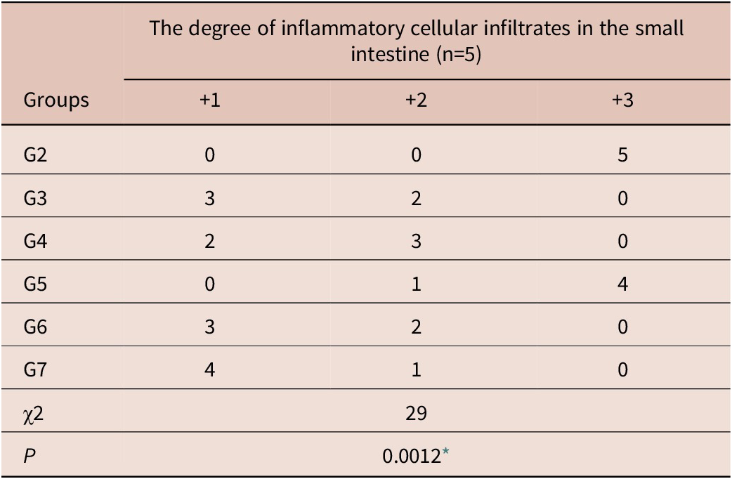
Histological analysis revealed that compared to the intestine in the negative control group, the positive control group (G2) exhibited significant infiltration of inflammatory cells, primarily concentrated in the core of the villi and extending into the submucosa. The inflammatory infiltrates were predominantly composed of eosinophils and neutrophils. Villi flattening and villous tips sloughing were also observed. Intraluminal cut sections of adult T. spiralis were observed. All treated groups exhibited significant histopathological changes and reduced inflammation compared to G2, except in G5 (treated with SLNs). The group administered a combination of albendazole and IVM-SLNs exhibited a notable decrease in the degree of inflammation and elongation of the villi. Mice treated with albendazole showed mild inflammation at the center of the intestinal villi. The ivermectin-treated group demonstrated moderate inflammation (Figure 3). Table 3 illustrates the degree of inflammatory cellular infiltrates in the small intestine among the studied groups.
Histopathological examination of the intestinal sections 7 dpi. (a) Normal intestinal mucosa with intact villi (V). The underlying crypts are rich in goblet cells (blue arrows) (200X); (b) Section in the small intestine of infected nontreated mice with marked inflammatory infiltrate formed of eosinophils, lymphocytes, plasma cells and macrophages (green star) (200X); (c) Cross section of adult T. spiralis was detected in the intestinal lumen of control positive group (blue arrow). Intestinal mucosa shows marked inflammatory infiltrate (green star) (400X); (d) albendazole treated group revealing intact villi (V) with mild lymphocytic infiltration (green stars) (200X); (e) IVM treated group revealing intestinal mucosa with villous regenerative changes including hyperplasia and dark vesicular nuclei (V). Villous infiltration with moderate lymphocytic infiltration is observed (green star) (200X); (f) SLNs treated group showing marked inflammatory infiltrate in villous core (green star) (200X); (g) IVM-SLNs treated group revealing intestinal villi (V) infiltrated by mild to moderate lymphocytic infiltrate (green star). Cut section of T. spiralis adult can be seen (blue arrows) (200X); (h) IVM-SLNs and albendazole combination group revealing intact intestinal villi (V) with minimal inflammatory cell infiltration (green star) indicating best improvement. Paneth cell regeneration can be observed deep in the crypts. (P). Goblet cells increased in size and number (blue arrows) (200X).

The degree of inflammatory cellular infiltrates in the small intestine of the studied groups

⁎ Significant (P < 0.05).
Histochemical findings
Feulgen staining of normal enterocytes showed bright red coloration. The control positive group showed enterocytes’ DNA damage, which displayed a very faint red stain, indicating severe apoptosis. Group 7 enterocytes showed bright red coloration (no dysregulated apoptosis), indicating that the combination of albendazole and IVM-SLNs minimized trichinellosis-induced apoptosis. Pale red staining of enterocyte DNA was observed in groups 4 and 6, suggesting moderate apoptosis. Group 3, administered albendazole, exhibited less bright red coloration indicative of mild apoptosis (Figure 4).
Intestinal sections stained by Feulgen stain (400X). (a) Normal nuclei of enterocytes showed bright red coloration (blue arrows) without histochemical changes; (b) The positive control group exhibited damage of DNA, which stained very faint red, indicating significant apoptosis (blue arrows); (c) albendazole treated group showed less bright red coloration indicating mild apoptosis (blue arrows); (d) Pale red staining of enterocyte DNA was observed in group 4 treated with IVM indicating moderate apoptosis (blue arrows); (e) SLNs treated group showed severe apoptosis (blue arrows); (f) IVM-SLNs treated group showed moderate apoptosis (blue arrows); (g) Group 7 enterocytes showed bright red coloration (no dysregulated apoptosis) (blue arrows), which indicated that the combination of albendazole and IVM-SLNs minimized apoptosis induced by trichinellosis.

Immunohistochemical
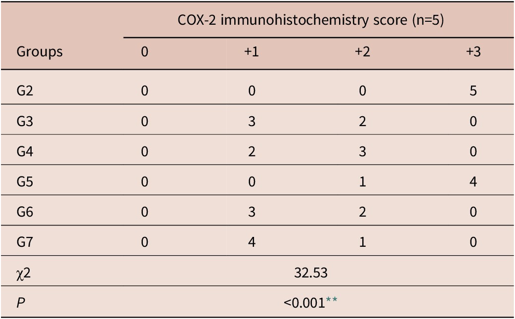
The infected control group demonstrated significant (+3) COX-2 expression. Pronounced COX-2 immunoreactivity was primarily observed in the inflammatory infiltrate of the villous core. In the treatment groups, COX-2 expression decreased to weak levels in the albendazole-treated group (+1) and moderate levels in the ivermectin-treated groups (+2) compared to the infected control group. The decrease in COX-2 expression was more pronounced in the IVM-SLNs and albendazole combination group compared to the other treated groups (Figure 5). The expression of COX-2 in the studied groups is presented in Table 4.
Immunohistochemical expression of COX-2 in small intestine of studied groups. (a) normal healthy control; (b) The positive control group showing pronounced COX-2 immunoreactivity (+3), predominantly identified in the inflammatory infiltrate of the villous core; (c) albendazole-treated group showing weak COX-2 expression (+1); (d) IVM treated group showing moderate cytoplasmic immunohistochemical expression of COX-2 (+2); (e) SLNs treated group showing strong COX-2 cytoplasmic expression in the intestinal mucosa and the inflammatory cells; (f) IVM-SLNs treated group showed moderate immunoreactivity; (g) group 7 exhibiting a significant reduction in COX-2 expression (400X).

The expression of COX-2 in the studied groups

** Highly significant (P < 0.001).
Discussion
Trichinellosis is a significant and extensively prevalent parasitic disease. It can elicit an inflammatory response and apoptosis in the affected tissues, potentially resulting in lethality if it disseminates to critical organs such as the lungs, heart or central nervous system (CNS) (Piekarska et al. Reference Piekarska, Miśta, Houszka, Króliczewska, Zawadzki and Gorczykowski2011; Xu et al. Reference Xu, Hou, Zhu, Liu, Shi, Li, Li, Cai, Zhou and Qiu2019). This study aimed to identify more effective medications for trichinellosis, addressing significant issues such as drug resistance and low water solubility associated with existing treatments. The nanoscale dimensions (10–100 nm) and chemical properties of nanoparticles render them effective drug carriers (Norouzi Reference Norouzi2017).
SLNs are a colloidal delivery system consisting of solid lipids at ambient temperature, stabilized by a coating of either a single surfactant or a combination of surfactants (Mishra et al. Reference Mishra, Bansal, Verma, Yadav, Thakur, Sudhakar and Rosenholm2018; Souto and Müller Reference Souto and Müller2007; Wissing et al. Reference Wissing, Kayser and Müller2004). Particle diameters typically range from 50 to 500 nm. SLNs were introduced in 1991 as an alternative drug delivery technology to conventional colloidal carriers, such as oil-in-water emulsions and liposomes. SLNs are significant as they consist of spherical lipid particles with a small diameter in the nanometer range, a high specific surface area and advantageous zeta potential. They comprise crystallized lipid components combined with emulsifiers and surfactants utilized for medication encapsulation (Eldem et al. Reference Eldem, Speiser and Hincal1991; Pardeike et al. Reference Pardeike, Hommoss and Müller2009).
SLNs integrate the advantages of liposomes, emulsions and polymeric nanoparticles. They offer both the solid matrix’s stability and lipid carriers’ biological compatibility, circumventing the constraints associated with such drug delivery techniques (Bahari and Hamishehkar Reference Bahari and Hamishehkar2016). Their solid matrix plays a crucial role in protecting the encapsulated drug against instability and facilitating controlled drug release. SLNs can maintain stable nano-suspensions for prolonged durations compared to liposomal delivery systems. SLNs formulations demonstrate good tolerance and are acknowledged for their safety profile. The low cytotoxicity of SLNs facilitates its extensive use in cutaneous, oral, pulmonary and parenteral delivery (Dolatabadi et al. Reference Dolatabadi, Hamishehkar and Valizadeh2015; Kakkar et al. Reference Kakkar, Singh, Singla and Kaur2011). SLNs do not display disadvantages, such as erratic particle growth or unforeseen drug release from the lipid core, which are recognized as prevalent occurrences (Mehnert and Mäder Reference Mehnert and Mäder2001). This research aimed to assess the effects of oral IVM-SLNs, independently and in combination with albendazole, on the enteric phase of experimental trichinellosis.
The current study demonstrated a significant reduction in the mean adult count among the studied groups following the application of different treatment protocols. Monotherapies with albendazole, IVM and IVM-SLNs reduced intestinal worm counts by 92.86%, 74.35% and 83.44%, respectively. Group 7 demonstrated the highest reduction percentage. These findings are consistent with prior research indicating a significant effect of IVM on T. spiralis adults (Basyoni and El-Sabaa Reference Basyoni and El-Sabaa2013; Elmehy et al. Reference Elmehy, Hasby Saad, El Maghraby, Arafa, Soliman, Elkaliny and Elgendy2021; Fadil et al. Reference Fadil, Mahmoud, El-Ahl, Abd-Elaal, El-Shafaey, Badr, Elesawy, A, Hamed, Abdel-Shafi, Reda, Elsayed and Abdeltawab2022; Soliman et al. Reference Soliman, Taher and Mahmoud2011). Recent studies have also indicated comparable efficacy of IVM monotherapy against T. spiralis adults, with observed decrease percentages of 73.3%, 72.4 and 68.6% (El Saftawy et al. Reference El Saftawy, Aboulhoda, Hassan, Ismail, Alghamdi, Hussein and Amin2024; Hassan et al. Reference Hassan, El-Sayed, Zekry, Ahmed, Abd-Elhamid, Salama, Taha, Mahmoud, Mohammed and Amin2024; Moawad et al. Reference Moawad, Elgendy, Mohamed, Mousa, Moustafa and Saleh2024). In contrast, Elmehy et al. (Reference Elmehy, Gamea, El-Guindy, Tahoon, Elkholy and Zoghroban2024) found that a single IVM subcutaneous injection only resulted in a 16.7% reduction percentage.
The efficacy of ivermectin against trichinellosis was attributed to its ability to alter GABA-gated chloride channels in nematode parasites rather than the afflicted host (Bai and Ogbourne Reference Bai and Ogbourne2016). The highest efficacy is observed during the intestinal phase, wherein IVM induces paralysis in T. spiralis adults within the small intestine lumen (González Canga et al. Reference González Canga, Sahagún Prieto, José Diez Liébana, Martínez, Vega and Vieitez2009). Ivermectin induced topographic changes, leading to the degeneration and destruction of adult populations, resulting in decreased larval populations (El-Azzouni Reference El-Azzouni1997).
Albendazole exerts its effect by specifically binding to the β-tubulin monomer of the parasite, thereby inhibiting microtubule polymerization (Yu and Qi Reference Yu and Qi2015). Furthermore, albendazole causes various metabolic changes in the target worm, such as the inhibition of mitochondrial fumarate reductase, glucose transport reduction and oxidative phosphorylation uncoupling (Shoheib et al. Reference Shoheib, Abdin, Shamloula and El–Segaey2006). Our findings are consistent with prior studies indicating a notable reduction in T. spiralis adult count in mice treated with albendazole (Nada et al. Reference Nada, Mohammad, Moad, Shafey, Al-Ghandour, Nagwa and Moawad2018; Nassef et al. Reference Nassef, Moharm, Atia, Brakat, Abou Hussien and Mohamed2018).
Alanine aminotransferase (ALT), a cytosolic enzyme, almost exclusively indicates hepatocellular damage. Elevated serum levels indicate the extent of hepatocellular injury (Kaneko et al. Reference Kaneko, Harvey and Bruss1997). Aspartate aminotransferase (AST) is present in various cells; however, it is a marker for assessing hepatic and muscular damage, given its increased activity in these tissues. Urea, commonly referred to as blood urea nitrogen (BUN) in blood assessments, is a byproduct of protein metabolism (Wang et al. Reference Wang, Gao, Hong, Hou, Liu, Li, Yang and Zhang2023). Creatinine, a byproduct of creatine and phosphocreatine metabolism, functions as a marker for renal function (Price and Finney Reference Price and Finney2000).
This study found that the levels of ALT, AST (liver enzymes), urea and creatinine (renal parameters) were significantly elevated in the infected control group compared to the normal healthy mice. Additionally, total protein and albumin levels showed a significant decrease while globulin levels increased, as illustrated in Table 2. Increased levels of ALT and AST indicate potential hepatic damage, possibly due to drug detoxification processes or metabolites released from parasitic stages (Abuelenain et al. Reference Abuelenain, Fahmy, Elshennawy, Fahmy, Ali, Hammam and Abdel-Aziz2021). Our results align with those obtained by Basyoni and El-Sabaa (Reference Basyoni and El-Sabaa2013) and Swilam et al. (Reference Swilam, Nasreldin, Abd-Elrahman and nasr2022). The histopathological findings of the liver and kidney in infected mice supported these findings. Swilam et al. (Reference Swilam, Nasreldin, Abd-Elrahman and nasr2022) reported that the liver showed vacuolar degeneration and lymphoid cell reaction in the portal area. Moreover, the kidney showed blood vessel congestion. Acute hepatitis was observed in rats infected with T. spiralis, along with several metabolic alterations (Farid et al. Reference Farid, Fath, Mido, Nonaka and Horii2019).
All plasma proteins, except immunoglobulins (antibodies), are synthesized and secreted by the liver. Plasma proteins represent various macromolecules with varying structures and functions, essential for maintaining plasma oncotic pressure (Potter Reference Potter, Enna and Bylund2007). Albumin is the primary protein in plasma, making up approximately 60% of serum proteins. It is essential for regulating body fluid distribution, maintaining acid-base balance and binding vital components in the bloodstream (Sheinenzon et al. Reference Sheinenzon, Shehadeh, Michelis, Shaoul and Ronen2021). The total globulin fraction is calculated by subtracting albumin from the total protein. Reduced serum albumin and globulin levels may arise from reduced hepatic production or augmented loss due to renal pathology (Busher Reference Busher1990).
The elevated levels of globulins observed in our experiment may be ascribed to a compensatory mechanism aimed to restore osmotic pressure, which decreases due to lower albumin levels caused by impaired glomerular function in conjunction with heightened serum antibodies in response to the parasite or its metabolic byproducts (Basyoni and El-Sabaa Reference Basyoni and El-Sabaa2013).
All treated groups demonstrated improvements in the assessed biochemical parameters toward normal levels. A significant reduction in ALT, AST, globulin, urea and creatinine levels was noted. Moreover, a significant increase in total serum proteins and albumin levels was recorded in all treated groups compared to the positive control group. Comparable outcomes were reported by (Basyoni and El-Sabaa Reference Basyoni and El-Sabaa2013) and (Swilam et al. Reference Swilam, Nasreldin, Abd-Elrahman and nasr2022). In contrast, (Arise and Malomo Reference Arise and Malomo2008) reported a significant increase in serum AST activity following the administration of ivermectin, both independently and in combination with albendazole. The administration of albendazole did not significantly impact serum AST activity.
The initial response of the host to infection is inflammation. Inflammation plays a critical role in maintaining homeostasis and facilitating tissue repair (Kunnumakkara et al. Reference Kunnumakkara, Sailo, Banik, Harsha, Prasad, Gupta, Bharti and Aggarwal2018; Munn Reference Munn2017). However, uncontrolled inflammation is detrimental (de Sá Coutinho et al. Reference de Sá Coutinho, Pacheco, Frozza and Bernardi2018). Histopathological analysis of the positive control group demonstrated significant inflammation in the intestinal tissues. The shedding of intestinal villi was also detected. The adult T. spiralis secretes excretory-secretory proteins that facilitate intestinal penetration and primarily contribute to the observed degenerative changes. The results are consistent with those reported by Abou Rayia et al. (Reference Abou Rayia, Saad, Ashour and Oreiby2017), Elguindy et al. (Reference Elguindy, Ashour, Shamloula and Assad2019), Nassef et al. (Reference Nassef, Moharm, Atia, Brakat, Abou Hussien and Mohamed2018) and Shalaby et al. (Reference Shalaby, Moghazy, Shalaby and Nasr2010). The administered drugs resulted in the improvement of intestinal degenerative changes. A significant reduction in inflammatory cellular infiltrate and a lower inflammatory score were noted compared to the positive control group. The combination group (G7) exhibited restoration of normal villous architecture. The significant decrease in adult worm populations and the inhibition of intestinal epithelial penetration by the infective larvae account for these improvements. The IVM-treated group revealed villous regenerative changes and moderate lymphocytic infiltration. Recent studies have demonstrated comparable efficacy of IVM monotherapy (Elmehy et al. Reference Elmehy, Gamea, El-Guindy, Tahoon, Elkholy and Zoghroban2024; El Saftawy et al. Reference El Saftawy, Aboulhoda, Hassan, Ismail, Alghamdi, Hussein and Amin2024). Moawad et al. (Reference Moawad, Elgendy, Mohamed, Mousa, Moustafa and Saleh2024) reported that the IVM-treated group demonstrated irregular, slightly short villi with moderate inflammatory infiltrate. Hassan et al. (Reference Hassan, El-Sayed, Zekry, Ahmed, Abd-Elhamid, Salama, Taha, Mahmoud, Mohammed and Amin2024) noted the presence of cut sections of T. spiralis adults in the IVM-treated group, as well as the shedding of some villous tips.
The most favorable outcomes were observed in mice treated with IVM-SLNs and albendazole combination. The morphology of intestinal sections was comparable to that of the negative control group. Regeneration of Paneth cells was observed in the deeper regions of the crypts. They inhibit parasitic penetration (Bevins and Salzman Reference Bevins and Salzman2011). Paneth cells are the primary components of innate immunity in GIT. Their secretory granules encompass diverse components essential in immunological and inflammatory responses. Consequently, Paneth cells contribute to maintaining the integrity of the intestinal mucosal barrier (Adolph et al. Reference Adolph, Mayr, Grabherr and Tilg2018). Goblet cells exhibited an increase in both size and number. Mucus secretion contributes to intestinal defense against parasitic invasion. Goblet cells and their secretions augment the host immune response, thus impeding helminth colonization in the intestinal mucosa and facilitating worm expulsion from the gut (Marillier et al. Reference Marillier, Michels, Smith, Fick, Leeto, Dewals, Horsnell and Brombacher2008). The enhanced efficacy of the IVM-SLNs and albendazole combination over other treatment groups is due to improved drug bioavailability and the mitigation of challenges associated with the oral administration of IVM, which is marked by a slow and inconsistent dissolution rate (Elmehy et al. Reference Elmehy, Hasby Saad, El Maghraby, Arafa, Soliman, Elkaliny and Elgendy2021).
Cyclooxygenase has two isoforms: COX-1 and COX-2. It catalyzes the production of prostaglandins and thromboxane from arachidonic acid. The expression of the COX-2 isoform varies in response to inflammatory situations and malignancies (Matsumoto et al. Reference Matsumoto, Ferino, Monteleone and Ribeiro2009). Selective COX-2 inhibitors can overcome apoptosis and attenuate intestinal muscle hypercontractility that persists for six weeks after recovery from Trichinella infection despite the absence of observable mucosal inflammation (Barbara et al. Reference Barbara, Vallance and Collins1997; Barbara et al. Reference Barbara, De Giorgio, Deng, Vallance, Blennerhassett and Collins2001; Liu et al. Reference Liu, Qu and Yan2015).
Significant COX-2 immunoreactivity was observed in the positive control group, primarily within the inflammatory cells. The results are consistent with those reported by Akiho et al. (Reference Akiho, Deng, Blennerhassett, Kanbayashi and Collins2005) and Serna et al. (Reference Serna, Porras and Vergara2006). Consistent with prior studies, COX-2 expression diminished in the treatment groups compared to the positive control group. Zhang et al. (Reference Zhang, Song, Xiong, Ci, Li, Yu, Zhang and Deng2009) revealed that IVM reduced LPS-induced COX-2 in a concentration-dependent manner. IVM significantly reduced COX-2 expression and demonstrated potential anti-inflammatory effects in the treatment of acetic acid-induced colitis in rats (Aryannejad et al. Reference Aryannejad, Tabary, Noroozi, Mashinchi, Iranshahi, Tavangar, Mohammad Jafari, Rashidian and Dehpour2022).
Apoptosis, also known as programmed cell death, is a systematic process comprising an initiator phase followed by an effector phase, which leads to cell death through the degradation of various cellular components (Osborne Reference Osborne1996). Parasite-induced apoptosis can be initiated via active mediators secreted by the parasite or indirectly through inflammatory mediators (Lundy et al. Reference Lundy, Lerman and Boros2001; Tato et al. Reference Tato, Fernández, Solano, Borgonio, Garrido, Sepúlveda and Molinari2004). Gastrointestinal helminth infection, such as T. spiralis, triggers a sequence of alterations in intestine structure, cellular composition and physiological function (Yu et al. Reference Yu, Deng, Lu, Zhang, Jia, Huang and Qi2014). Apoptosis plays a crucial role in the pathophysiology of trichinellosis (Karmanska et al. Reference Karmanska, Houszka and Piekarska2000). Apoptosis was detected in the intestines of infected mice by the third dpi (Yu et al. Reference Yu, Deng, Lu, Zhang, Jia, Huang and Qi2014).
The control positive group showed DNA damage. It stained very faint red, indicative of significant apoptosis. Minimal apoptotic changes were observed, as indicated by intense red staining in the group administered albendazole and IVM-SLNs (G7), followed by the albendazole group (G3). G5 exhibited marked apoptosis, comparable to the infected control. This finding suggested a minimal effect of SLNs when administered independently on the apoptotic alterations induced by trichinellosis.
Conclusion
This study demonstrated that a biocompatible and safe delivery system utilizing IVM-SLNs markedly enhanced the adulticidal efficacy of a single oral dose of IVM in T. spiralis-infected mice. IVM-SLNs exhibited significant anti-inflammatory and anti-apoptotic properties, successfully restoring hepatic and renal function parameters to near-normal levels. Consequently, they may serve as alternatives or adjuncts to current nematicidal agents. However, comprehensive pharmacokinetic and toxicological investigations are essential for optimizing dosing regimens and ensuring safety. Further research is needed to develop an economical and precise drug delivery system for trichinellosis.
Abbreviations
- SLNs
-
solid lipid nanoparticles
- IVM
-
Ivermectin
- IVM-SLNs
-
ivermectin-loaded solid lipid nanoparticles
- COX-2
-
Cyclooxygenase-2
- T. spiralis
-
Trichinella spiralis
- dpi
-
day post-infection
Financial support
The current work has not received any grant funding.
Competing interests
No conflict of interest.
Ethical standard
The ethics committee of the Faculty of Medicine, Zagazig University, approved the protocol of this study (Approval number: ZU IACUC/3/F/91/2023).