Introduction
Therapeutic radiographers are an essential part of a cancer patient’s journey and play a vital and changing role in the delivery of radiotherapy services. 1 Multiple professional, guidance documents have been written 2–5 highlighting the changing practice of radiographers and the requirement to develop the scope of practice to meet service needs and deliver patient-centred care. As a result, radiographers have seized opportunities to develop diverse and expanded roles at both advanced practice and consultant levels building upon the four-tier structure. 4 Roles include technical specialists in imaging/pre-treatment, dosimetry specialists, tumour site and treatment specialists all providing high-quality treatment and care.
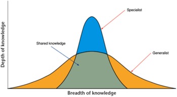
The breast advanced practitioner is involved within the multidisciplinary team meetings (MDT) discussing the management of breast cancer patients and has responsibility for virtual simulation CT planning of all breast patients undergoing radiotherapy, on-treatment review clinics, follow-up clinics, undertaking informed consent, service development and research. Specialist roles therefore require additional specific knowledge and skills, and Figure 1 clearly demonstrates this difference in both depth and breadth of knowledge.
NHS Education for Scotland Four Pillars of Practice: The difference in depth and breadth of knowledge between generalists and specialists.

Breast cancer patients are identified either through the breast screening programme or by referral into specialist breast clinics via their GP. The patients undergo a process of triple assessment, which involves clinical examination, mammography/ultrasound and cytology to detect the presence of invasive cancers and ductal carcinoma in situ (DCIS). Once a cancer diagnosis has been identified, they then proceed to staging investigations to rule out metastatic disease. Approximately 6% of all patients presenting with breast cancer will have metastatic disease at presentation. 6
The most common sites for distant metastases include the brain, lungs, liver, bones and skin. Reference Wang, Zhu and Liu7 However, routine CT staging for asymptomatic patients with early-stage disease (T1 or T2) is not recommended in early breast cancer, 8 so patients usually only undergo routine blood tests and a chest X-ray especially if lymph node involvement has been ruled out.
Computed tomography (CT) based treatment planning for breast cancer is standard practice in radiotherapy centres as it allows better visualisation of the breast tissue and underlying structures such as heart and lung. Reference Cameron9 Studies have demonstrated an increased uptake in the detection of pulmonary nodules as a result of these planning CT scans; Reference Krishna, Varghese and Gopurathingal10 however, the presence of incidental nodules should not alter the definitive treatment for breast cancer. Reference Lee, Lim and Lalvani11,Reference Callister, Baldwin and Akram12 The detection of pulmonary nodules is common and in populations at high risk of lung cancer (such as smokers), and nodules are detected in 20–50% of individuals. Reference Krishna, Varghese and Gopurathingal10,Reference MacMahon, Naidich and Goo13
The radiotherapy CT planning scans are non-contrast and not routinely reported by radiologists and are therefore not considered wholly diagnostic. The CT scan is used to assist with visualisation of the extent of breast tissue plus nodes, to create treatment fields appropriate for the stage of disease, for example breast alone when no nodes are involved or breast plus lymphatics for nodal disease. The Breast Advanced Practitioner is therefore viewing the CT scans closely throughout the thoracic and upper abdominal areas and abnormalities may be visualised.
Due to the quantity and frequency of abnormalities identified by the Breast Advanced Practitioner, a new process was created, to allow the planning CT scan to be uploaded directly to the national picture archiving and communication systems (PACS) radiology system. This involved the hospital IT staff creating a link to the PACS system and required clinicians to send a Trak Patient Management System request to the radiology teams to ask for a review of the radiotherapy scan including pertinent information such as smoking status.
This paper is designed to highlight the advanced practice role in evaluating CT planning scans identifying abnormalities, ensuring they are uploaded for formal radiology review and analysing the patient management outcomes. This process has involved collaboration with the multi-disciplinary team including Oncologists, IT staff and radiology.
Methods
Any breast cancer patient who had an abnormality identified on the CT planning scan was included in the audit, except patients presenting with obviously benign liver cysts. Key information recorded included patient age, their smoking status (if a lung abnormality) and a description of the abnormality, e.g. lung nodule, liver lesion. The Breast Advanced Practitioner ensured that all patients had their planning CT scan uploaded to the national PACS radiology system for radiology review. A timely turnaround of a maximum of 48 hours allows the patients to be appointed to either further formal CT imaging or direct referral to appropriate multi-disciplinary team meetings (MDT’s).
All patients with suspected abnormalities (excluding benign liver cysts) had their outcomes recorded in an Excel spreadsheet once a Radiologist had reviewed the CT scan. All radiological outcomes were analysed to identify what further treatment the patient required or if their treatment plan changed as a result of the identification of an abnormality.
A retrospective audit was therefore undertaken in all patients who were found to have an incidental CT abnormality in the period May 2016 – June 2021 The total number of planning scans (normal and abnormal) viewed by the advanced practitioner was not recorded as it was a retrospective audit of only the abnormal scans. However, for reference the cancer centre treats in excess of 900 breast patients per annum and the radiographer led planning is shared between three radiographers. The key outcomes of the audit were recording the radiology opinion and the subsequent patient management as a result of finding the incidental abnormalities.
Results
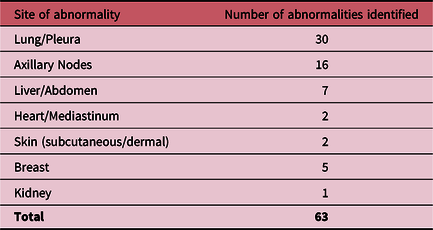
The Breast Advanced Practitioner identified sixty-three significant abnormalities during the 5-year audit (Table 1) and thirty seven were found to be malignant. The majority of these were lung abnormalities (n=30), and these were further analysed and demonstrated twelve new lung primaries, six lung metastases, five indeterminate lung nodules, three pleural metastases, three infections and one primary lymphoma.
Site and number of abnormalities identified on planning CT scans

Seven patients with a new lung primary diagnosis went on to have further treatment with surgery alone, surgery plus chemoradiation or stereotactic ablative radiotherapy and had their breast radiotherapy cancelled to allow their lung cancer to be treated aggressively first. Those with lung metastases or incurable lung cancer had their radical breast radiotherapy cancelled to allow the patients to progress onto palliative chemotherapy. Indeterminate nodules required six-month follow-up CT scans to exclude malignancy.
Six patients presenting with abnormal axillary lymphadenopathy were found to have reactive lymph nodes post axillary ultrasound and biopsy. However, ten had further axillary surgery performed or their radiotherapy altered to include the nodal lymphatic’s region.
Liver metastases were identified in five patients, which unfortunately changed their prognosis to palliative. Benign cystic liver lesions are commonly identified on CT scans and were excluded. A thymoma was identified in one patient, measuring 10 cm x 7 cm, however, the patient was completely asymptomatic, and this was successfully excised requiring no further treatment. The remaining breast abnormalities were found to benign (both intra- and contra-lateral breasts). The kidney and heart abnormalities were found to be benign cystic lesions.
Discussion
Radiographers have a duty of care to their patients at the pre-treatment stage to appraise image information for clinical manifestation and technical accuracy, taking appropriate action as required. 14 There is no expectation, however, that their role is equivalent to a reporting radiographer or radiologist as only formal training and assessment will permit such specialist knowledge.
The advanced practice radiographer has extensive experience within breast cancer, however, has not undertaken any formal training in CT evaluation and has just gained experience in-house through evaluating images and updating anatomical knowledge. This may be a limitation of the study as there may have been more abnormalities on the CT scans that were not observed by the radiographer and further training in evaluating CT scans may be beneficial. This will be of particular importance to the other members of the advanced practice team who have less experience in identifying incidental abnormalities.
Recent research Reference Nguyen, Dietz and Singh15 has reported a small number (n=4) of incidental findings of significance on radiotherapy planning CT scans by a radiation oncologist. This study over a 2-year period reviewed a smaller number of scans of all cancer types. The authors comment that there is uncertainty whether other departments are routinely scrutinising CT planning scans for abnormalities and recommend implementing a formal quality assurance process to interpret radiotherapy scans. It is therefore encouraging that a quality assurance process was created and has been utilised within this department since 2016. The process permits a timely review of the radiology usually within 48 hours, avoiding delays in commencing radiotherapy or referral for other treatment options.
Service development and evaluation is part of the breast advanced practice role, and this audit has demonstrated a positive development of sharing imaging across different specialties to enhance patient-centred care. It has required collaborative working with different stakeholder groups to achieve changes in practice, which benefit patients.
The identification of sixty-three abnormalities during routine CT breast planning has benefited seven patients to enable them to have curative treatment for previously undiagnosed and asymptomatic primary cancers. This has improved their prognosis as they had early-stage curable lung cancers or a lymphoma.
Two of the patients had their breast cancer diagnosed through a routine mammogram from the breast screening programme and then went onto have lung cancers diagnosed as a result of the radiotherapy CT planning scan. Therefore, both patients presented with asymptomatic breast and simultaneous asymptomatic lung cancers, and this really highlights the benefits of cancer screening programmes. The earlier cancers are picked up and treated can significantly impact on long term survival, Reference In, Kwon and Oh16 and both of these patients were fortunate to have curable breast and lung cancers diagnosed. The absence of symptoms of lung cancer at diagnosis is a favourable prognostic factor as they often present as early Stage 1 or 2 disease, with 54% asymptomatic versus 14% symptomatic presentation. Reference Polanco, Pinilla and Gracia-Lavedan17 This study also demonstrated a 3-year overall survival (OS) rate of 63.6% versus 30.3% for symptomatic cancers and this is supported by another study, Reference Kocher, Lunger and Seeber18 which confirmed a median OS benefit of 38.9 months versus 16.1 months. However in contrast, recent evidence has suggested Reference Farjah, Monsell and Gould19 that intensive diagnostic evaluation of pulmonary nodules is associated with greater procedural complications and unnecessary radiation exposure with little benefit. Certainly, in the retrospective audit five patients with indeterminate nodules were advised to have a six-month follow-up CT scan to determine any changes and to exclude a small lung malignancy.
Sadly, for those in whom metastatic disease was identified, this meant an alteration in treatment plan often including the use of palliative chemotherapy. However, the identification of the metastases at an early stage will have allowed the patients’ to commence treatment earlier than normal, which may offer a slight benefit in terms of length of survival.
Conclusion
This retrospective audit has permitted the introduction of a new pathway to allow a timely review of the radiotherapy planning CT scans, ensuring the best management for patients with incidental, asymptomatic abnormalities. Radiotherapy departments should consider collaborating with radiology colleagues to set up a local process to review CT planning scans for radiotherapy to aid identification of potentially significant abnormalities. Radiographers have a duty of care to ensure that any abnormalities identified on CT planning scans are investigated thoroughly, as the early diagnosis of new primary or metastatic cancers has a potential impact in terms of prognosis and improvement in survival.
Conflicts of Interest
The author declares that there is no conflict of interest.