
Introduction
The production of tambaqui (Colossoma macropomum, Cuvier Reference Cuvier1816) in the northern region of Brazil has been affected by problems of a sanitary nature due to the development and spread of acanthocephalosis on fish farms. Even 12 years after the registration of the first case, the occurrences continue to increase due to the lack of specific legislation, a contingency plan and good management practices (Castro et al., Reference Castro, Jerônimo, Silva, Santos, Ramos and Andrade-Porto2020). Acanthocephalosis is a disease caused by the endoparasite Neoechinorhynchus buttnerae (Golvan, Reference Golvan1956), which infects the intestine of tambaqui and its hybrids, and is considered the disease that has the greatest impact and importance in the production of this fish (Valladão et al., Reference Valladão, Gallani, Jerônimo and Seixas2019). In tambaqui fish farming, acanthocephalosis can cause economic impacts of up to 200% on fish growth and, as such, directly affects producer income. This can result in a difference of more than 1000% between infected and uninfected farms (Silva-Gomes et al., Reference Silva-Gomes, Gomes, Viana-Silva, Braga-Oliveira, Bernardino and Costa2017).
Neoechinorhynchus buttnerae has an indirect life cycle, with an ostracod, Cypridopsis vidua as the intermediate host and a vertebrate as the definitive host (tambaqui). The development period of the immature stages of the larva is 29 days, when the manifestation of three stages occurs (acanthor, acanthella and cystacanth). The cystacanth is considered the infecting phase because it has well-defined hooks in the proboscis, similar to those found in adult specimens (Lourenço et al., Reference Lourenço, Morey and Malta2018). Ostracods are naturally present in the aquatic environment and are part of the diet of the definitive host, which ingests the intermediate host and thus the parasite continues its development. The egg is one of the fundamental parts of the life cycle of N. buttnerae, but to date there are no studies that provide more detailed knowledge of how embryonic development occurs, from copulation to its elimination in the environment. In a study conducted by Wharton (Reference Wharton1983), it was reported that an important function of the eggshell is the control of permeability and the maintenance of a microenvironment within the egg that allows embryonic and organism development. In acanthocephalans, information about the origin and chemical composition of their shell is either insufficient or scarce.
Studies of the embryonic development of the egg and the life cycle of a pathogen are relevant, since they allow the correct form of intervention that will reduce the proliferation of the disease. In the present study, stereological quantification was applied in an evaluation of the adult parasite to show the detailed proportion of its internal components, especially the proportion of eggs and ovarian balls. Stereology is the gold standard for morphological quantification (Gundersen et al., Reference Gundersen, Bagger, Bendtsen, Evans, Korbo, Marcussen, Møller, Nielsen, Nyengaard, Pakkenberg, SØRensen, Vesterby and West1988a; Howard and Reed, Reference Howard and Reed2005). Few studies have applied this tool to expand the knowledge regarding the biology and life cycle of parasites, though stereology has been accurate and precise in the most varied fields of biology (Felix and McGuire, Reference Felix and McGuire1981; Mandarim-de-Lacerda, Reference Mandarim-de-Lacerda2003; Hartlev et al., Reference Hartlev, Klose-Jensen, Thomsen, Nyengaard, Boel, Laursen, Laurberg, Nielsen, Steengaard-Pedersen and Hauge2018). Thus, the aim of the present study was to present a detailed description of the stages of embryonic development of N. buttnerae eggs, as well as a quantitative evaluation of the body of adult females of the parasite.
Materials and methods
Fish sampling and acclimation
Specimens of tambaqui (C. macropomum) naturally parasitized by N. buttnerae were obtained from commercial farms. The fish (n = 20) were acclimated for 24 h in four 120 L aquariums with constant aeration, at the density of five fish per aquarium in the Aquatic Animal Health Laboratory of the Faculty of Agricultural Sciences of the Federal University of Amazonas (LASAA/FCA/UFAM). This study was approved by the Ethics Committee for the Use of Animals (CEUA/UFAM), under protocol No 017/2017, and access to the genetic heritage of the animals involved in this research was approved under the register number AB1F0FA from the Genetic Heritage Management Council (CGEN), Ministry of the Environment (MMA).
Necropsy of the fish and processing of parasites
The fish were measured and then euthanized according to the recommendations of The National Council for the Control of Animal Experimentation (CONCEA, 2013). The gastrointestinal tract of the fish was removed and the intestine was excised, conditioned in a petri dish with distilled water, dissected, and analysed under a stereoscopic microscope (Feldmann, Germany) for the presence of acanthocephalan parasites. The parasites were transferred to Petri dishes with distilled water and cooled in a refrigerator for 3 h so that the proboscis remained everted (Thatcher, Reference Thatcher2006). Subsequently, the parasites were quantified to obtain the abundance index. The anatomical study of the acanthocephalans was carried out according to Amin (Reference Amin1987). The specimens were fixed in 2.5% glutaraldehyde solution buffered with 0.1 m sodium phosphate buffer for 24 h and stored in the refrigerator. After fixation, the specimens were processed for light microscopy in order to describe the embryonic development and stereology.
Histological processing
The samples were dehydrated in increasing the concentrations of ethanol (70 and 96%) for 6 h and pre-infiltrated in ethanol 96%+hydroxyethyl methylcrylate plastic historesin (50:50 v/v) (Technovit 7100, Külzer-Heraues, Germany) overnight. The next day, the samples were infiltrated in pure historesin for 2 h. For inclusion, the parasites were placed in parallel in a teflon Histobloc mould (Külzer-Heraues, Germany) and covered with the same historesin plus polymerizing solution (15:1 v/v). The moulds were kept in an oven at 37°C for 24 h. After total polymerization and formation of the resin, the blocks were sectioned in a microtome (RM 2345, Leica, Germany). The sections were later stained with toluidine blue 0.5% and basic fuchsin. All procedures adopted for histological processing according to Kiernan (Reference Kiernan1999).
Description of stages and morphometry of eggs
The 1.5 μ m histological sections stained with toluidine blue and haematoxylin-eosin were used to elaborate the drawings of the eggs of the parasite, in a clear chamber coupled to a microscope with phase contrast (BH-2, Olympus, Tokyo, Japan and Axioscope 2 plus, Zeiss, Jena, Germany) with a 100x lens, and subsequently digitized. The samples were analysed, photographed and recorded using the software Bel Photonics Microimage Analyser 2.3 (BEL Engineering s.r.l., Italy). Egg measurements (length and width) were recorded in micrometric units (μ m).
Stereology of the parasite
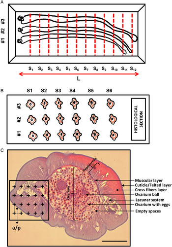
For stereology, a single inclusion block was created containing five parasites (Fig. 1A). After inclusion, the block was observed under a stereomicroscope (EZ4D Digital System, Leica, Germany) for the identification and marking of the extremities of the parasites aligned inside it. The total length of the parasites (L) was determined and this value was divided into 8–12 equidistant sections (L = S1 + S2 + … + S12) and marked on the block (Fig. 1A). The markings served as a guide for obtaining serial cross-sections made on a microtome (RM 2145, Leica, Germany) (Fig. 1B). The images were obtained through a photomicroscope (DM 500, Leica, Switzerland) at a magnification of 100. Then, the images were analysed using the program Imod version 4.7/stereology module (Kremer et al., Reference Kremer, Mastronarde and McIntosh1996), where a test counting system containing points was superimposed on the images (Fig. 1C). Each time a point coincided with the desired structure, this was counted. The parasite was composed of the body wall formed by the cuticle and felted layer; hypodermis formed by cross-fibre layer, muscular layer, lacunar system with eggs and lacunar system without eggs; ovarium formed by ovarium with eggs, empty spaces and ovarian balls. The total volume of the parasite was the sum of the volume of these components. The applied technique is based on the Cavalieri principle (Cavalieri, Reference Cavalieri1635), which determines the volume of any structure, regardless of its shape (Gundersen et al., Reference Gundersen, Bendtsen, Korbo, Marcussen, Moller, Nilsen, Nyengaard, Pakkenberg, Sorensen, Vesterby and West1988b; Howard and Reed, Reference Howard and Reed2005). The volume was calculated using the following equation: $V{\boldsymbol \;}( {{\rm m}{\rm m}^3} ) = {\boldsymbol \;}\sum\nolimits_{i = 1}^m {Pi{\boldsymbol \;} \times T{\boldsymbol \;} \times a/p} {\boldsymbol \;}$ Where V is the absolute volume, $\sum\nolimits_{i = 1}^m {Pi}$
 is the total number of points over each specific structure, T (1000 μ m) is the distance between each section, and a/p is the area represented by each point (576 μ m2). A coefficient of error of 5% was considered acceptable (Gundersen and Østerby, Reference Gundersen and Østerby1981; Gundersen et al., Reference Gundersen, Bendtsen, Korbo, Marcussen, Moller, Nilsen, Nyengaard, Pakkenberg, Sorensen, Vesterby and West1988b).
(A) Representation of the process of inclusion of parasites in a single block of plastic resin. For better visualization and clarification, only three parasites are shown (#1, #2 and # 3), however, five parasites were analysed in this study and included in a single block. The red lines indicate the markings on the block for obtaining equidistant serial sections (S1 + S2 + ….. + S12). L, total length of parasites in the block. (B) Final arrangement of sections in histological slide. (C) Stained and overlapping histological section with a counting system for determining the total volume of the parasite and its components. Crosses indicate points that touch the different structures of the parasite. Bar = 100 μ m.

Statistical analysis
The Prism program (GraphPad Software, Inc., CA, USA) was used for the descriptive statistics and graphs of this study. All values were presented as mean ± standard deviation (5% confidence limit). For multivariate analyses (PCA – principal component analysis), the PAST-Paleontological Statistics Program, version 4.01 was used (Hammer et al., Reference Hammer, Harper and Ryan2001). The stereological data obtained using light microscopy were evaluated for each animal and the variance estimator was determined using the coefficient of error (CE) for each parameter (Gundersen and Østerby, Reference Gundersen and Østerby1981). The accuracy of the Cavalieri principle was determined according to Cruz-Orive (Reference Cruz-Orive1999):

where $CE( \sum\nolimits_{i = 1}^n {Pi} )$ is the coefficient of error for determining the volume; $B/\sqrt A$
 is the variance of cross-sectional areas (shape coefficient) and depends on the complexity of the forms of the structure; n is the number of sections evaluated; and $\sum\nolimits_{i = 1}^n {Pi}$
 is the number of points counted on the sections.
Results
The parasitological analysis revealed that of the 20 fish examined, 100% were parasitized with the acanthocephalan N. buttnerae, with a parasitic index of average abundance of 43.9. There were a total of 878 parasites, of which 410 were males and 468 were females, 46.70 and 53.30%, respectively. The sexual ratio of female to male in this study was one female to one male (1:1), with a tendency to two females to one male (2:1). No clinical signs or pathological changes were observed in the fish analysed.
Description of the development stages of the eggs
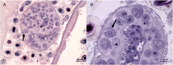
After copulation, the female's body cavity is filled with developing eggs. We observe that as the number of embryos increases, the number of ovarian balls is reduced (Fig. 2A and B). Females that failed to copulate contain only one or two ovarian balls.
(A) Ovarian ball with oocytes in different stages of development (arrow), with the central region composed of clustered nuclei in the centre (oogonial syncytium) (*) HE staining. (B) Ovarian ball with oocyte migrating to peripheral region of the ball (arrow) to detach and continue its development in the female body cavity (toluidine blue and HE staining).

Ovarian ball: the ovarian ball of the species N. buttnerae is a circular structure that is loose in the pseudocelomatic cavity of the female, and composed of a mass of hundreds of nuclei clustered in the centre (oogonial syncytium) (Fig. 2A). The multinucleated central mass (ovarian ball), after successive cell divisions, generates the large and mature oocytes (Fig. 2A and B) that migrate to the peripheral region of the ovarian ball and where they will be fertilized and subsequently detached forming the zygote, which is released to continue their development in the pseudocelomatic cavity of the female.
When the oocytes are fertilized and form the zygote, it is possible to identify 13 embryonic stages (Table 1), which are divided into three phases of embryonic development (Nicholas and Hynes, Reference Nicholas and Hynes1963; Nicholas, Reference Nicholas1967; Schmidt, Reference Schmidt1973), as described below:
Description of the egg stages of Neoechinorhynchus buttnerae, with respective measurements (mean ± standard deviation) of length (L) and width (W)

Phase 1: cell division
After fertilization, successive cell divisions (mitosis/cleavage of macromeres) occur, causing several stages of development until reaching the second stage that consists of the formation of the internal nuclear mass. In this first step, the eggs are on average 25.77 ± 3.20 μ m long and 14.75 ± 0.64 μ m wide.
Phase 2: formation of the internal nuclear mass
In the species N. buttnerae, the process that will cause the formation of the internal nuclear mass, which in the next step will correspond to the body of the larva, results from the process that promotes invagination in the tissues of the embryo forming the germ layer, which corresponds to gastrulation in other animals. In this step, the eggs are on average 29.48 ± 1.52 μ m long and 16.73 ± 0.84 μ m wide.
Phase 3: formation of the larval acanthor
As the embryo completes its development inside the cavity of the female, it becomes lined with a denser and thicker layer. It is also possible to observe the formation of the larva. In this phase, the eggs are on average 32.86 ± 2.02 μ m long and 26.35 ± 2.02 μ m wide.
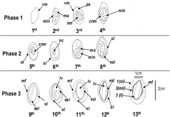
In the cell division phase, four stages of egg development were identified, in the second phase, four stages were also identified, and in the third phase, five stages, among them the final stage (mature). As such, in total, there are 13 stages of eggs, which are divided into three phases of development. Descriptive drawings of the stages corresponding to each phase and stage of egg development, with their respective images and morphological analysis, are presented in Fig. 3.
Representation of the 13 stages of development of the egg of Neoechinorhynchus buttnerae. Phase I: cell division, phase II: formation of the internal nuclear mass, phase III: acanthor organs. ol, outer layer; nc, nuclear condensation; esl, embryonic surface layer; ps, perivitelline space; lv, larva; ma, macromeres; mf, multiple filament; min, tiny nuclei; nm, nuclear mass; cnm, central nuclear mass; vm, vitelline membrane; pb, polar body; 1 (ol), outer layers; 2 (esl), embryonic surface layer; 3 (il), inner layer.

Morphometric analysis of eggs
Morphometric data are presented in Table 1. We observed that the dimensions of the egg increase in proportion to the embryonic development, and show a relationship between length and width of the eggs according to the stages (Fig. 4).
Linear regression between length and width of eggs according to the stages. *Significant difference (P < 0.0001) in the elevation of the two lines.

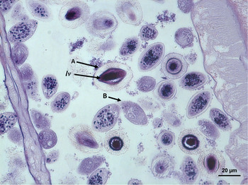
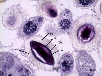
Through the results of the process of identifying the stages of egg development, two stages of development (initial and final) were emphasized. In the initial stage, the egg is smaller and has a thin layer in development. However, in the final stage of formation, it is possible to visualize the layers of the egg and the presence of the well-developed larva (Figs 5 and 6). The larva is covered in an apparently rigid and thick shell, maintaining the fusiform and elongated shape of the egg. It is possible to observe three layers: an outer layer (1) thicker, transparent and with multiple filaments; an intermediate layer (2) that has a darker colour; and the inner layer (3) with an appearance in the form of ripples that covers the larva (Fig. 6).
Eggs of Neoechinorhynchus buttnerae at different stages of development. (A) Egg specimen in the mature (final) stage with visible larva (lv = Larva). (B) Egg specimen in the immature (initial) stage.

Layers of the egg of Neoechinorhynchus buttnerae in the final stage of development. (1) Thicker outer layer, transparent and with multiple filaments – mf, intermediate layer (2) that has a darker colour and inner layer (3) with a covering in the form of ripples over the larva.

In the initial stage, the egg layers are in the process of formation, and are covered only by a thin vitelline membrane, which is difficult to measure (Table 2, Fig. 5). In the final stage, measurements of the larva (acanthor) and the outer layer were recorded, since it was thicker and more visible (Table 2, Fig. 6). It was not possible to measure the remaining layers because they were extremely thin and difficult to visualize.
Morphometric data of eggs of Neoechinorhynchus buttnerae at the initial (immature) and final (mature) stage

s.d., standard deviation; L, length; W, width.
Stereology of the parasite
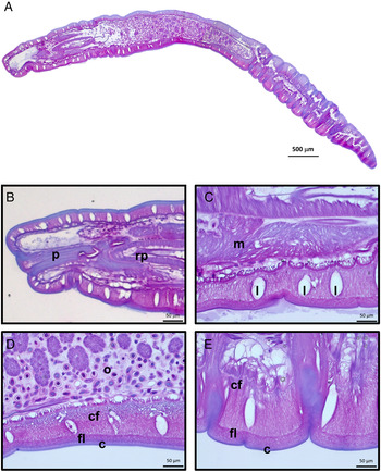
The internal anatomy of N. buttnerae is made up of a cuticle, followed by a thin layer of fibres (felted layer) that separates the animal from the external environment. A thick layer of fibres with diagonal arrangement relative to the central axis of the animal occupies a large proportion of the body (cross fibres). This layer is interrupted in several regions by the presence of fluid-filled gaps (lacunar system). The presence of eggs inside the gaps was observed in three animals that were analysed. A double layer of muscles was seen enveloping the ovary. This layer was formed by circular (external) and longitudinal (internal) muscle bundles, which were not individualized in the present study. The ovary is always filled with eggs at different stages of development and ovarian balls. Reduced empty spaces were observed in the ovary (Fig. 7).
(A) Longitudinal section of the parasite revealing its internal organization. (B) Praesoma revealing the proboscis and its receptacle. (C) Praesoma evidencing muscular layer and thick cross fibres with three evident spaces. (D) Metasoma evidencing the ovary, cross fibres and cuticle/felted layer. (E) Caudal region revealing thick cross fibres and cuticle/felted layer. p, proboscis; receptacle (rp); o, ovary; cf, cross fibres; m, muscular layer; l, lacunar; fl, felted layer; c, cuticle.

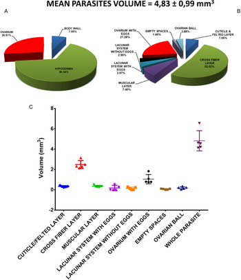
The quantitative results obtained for N. buttnerae are expressed in Fig. 8. The mean total volume of parasites was 4.83 ± 0.99 mm3. The hypodermis comprised 66% of the volume of the animal, with cross fibres contributing 52%, the muscle layer 7% and the spaces with and without eggs 4 and 3%, respectively. The ovary comprised 27% of the volume of the animal and most of it contained eggs (21%) and ovarian balls (4%). Few spaces in the ovary were free of eggs (empty spaces, 1%). The respective absolute values were obtained from these data in relation to the Cavalieri volume and are presented in Fig. 8. Table 3 shows the individual absolute values of the components. Animal 2 had the highest absolute volume (6.57 mm3) and animal 3 the lowest volume (4.15 mm3). Overall, the CE for stereological analyses remained below 5%, except for the ovarian ball of animal 1 (5.16%) and the empty spaces of animal 3 (5.28%).
Quantitative analysis in Neoechinorhynchus buttnerae. (A) Percentage of contribution of hypodermis, ovary and body wall (n = 5). (B) Fractionation and quantification of components (%). (C) Absolute volume (mm3) of components. All values as mean ± standard deviation.

Absolute values of the volumes (mm3) of the body components of N. buttnerae and coefficient of error (%) of estimations by point counting

The morphological variables obtained in the present study (eight components in total) were analysed by multivariate analysis (PCA) for a better understanding of their correlations. The result of this analysis is presented in Fig. 9A. The analysis reduced the complexity of the data in two components (PC1 and PC2). In total, these two components explained more than 87% of the data variability (PC1 = 53.09% and PC2 = 34.44%). The animals did not show homogeneity in their distribution (variability between the specimens). Animal 2 remained isolated from the others because it had high factors for muscle layer, cross fibres, ovary with eggs and ovarian ball. Animals 1 and 5 are similar to each other since they present high factors for cuticle/felted layer and empty spaces. Animals 3 and 4 are intermediary in regards to the previously mentioned characteristics. A high positive correlation was observed between muscle, cross fibres and ovary with eggs. There is no correlation between these variables and cuticle/felted layer. Lacunar system without eggs was inversely correlated to ovarian balls. Clustering analysis was used as a pattern recognition technique in order to determine the relationships between individuals (Fig. 9B). Thus, the analysis confirmed the proximity between animals 3 and 4 and between animals 1 and 5, in addition to isolating animal 2 from the others.
Principal component analysis (PCA) (A) and clustering analysis (B) of the stereological data of Neoechinorhynchus buttnerae.

Discussion
Neoechinorhynchus buttnerae presents sexual dimorphism with anatomical characteristics that are specific to the male and female sex (Golvan, Reference Golvan1956), and are able to perform sexual reproduction. In the present study, the proportion of females (468) was higher for males (410), corresponding to 53.30 and 46.70%, respectively. Research related to sexual ratio in acanthocephalans has shown that females are more abundant than males (Poulin and Morand, Reference Poulin and Morand2000; Sasal et al., Reference Sasal, Jobet, Faliex and Morand2000; Violante-González et al., Reference Violante-González, Villalba-Vásquez, Monks, García-Ibáñez, Rojas-Herrera and Flores-Garza2016) and in some species this may be related to the fact that females remain in the host longer than the male, consequently they have greater representativeness after copulation. Crompton and Walters (Reference Crompton and Walters1972) analysed Moniliformis dubius infection and observed that males and females are present in equal numbers during the first 5 weeks of infection, and after this period, females showed greater survival rates than males. In brown trout, Salmo trutta, males of Echinorhynchus truttae do not survive in the final host, and disappear earlier than females (Awachie, Reference Awachie1966) and males of Polymorphus minutus survived for a shorter time than females in domestic ducks (Crompton and Whitfield, Reference Crompton and Whitfield1968). Sasal et al. (Reference Sasal, Jobet, Faliex and Morand2000) suggest that the lower number of males may be related to reproductive competition between males to copulate, and that this mortality is generally related after sexual maturity. Therefore, it is believed that the females have a long life due to their reproductive importance and carry out spawning in instalments until all the eggs are mature and released into the environment.
Acanthocephalan females have ovarian balls that are fundamental structures in reproduction (Nicholas, Reference Nicholas1967; Schmidt, Reference Schmidt1973; Crompton et al., Reference Crompton, Arnold and Walters1976). Crompton and Whitfield (Reference Crompton and Whitfield1974) analysed ovarian balls of the species M. dubius and P. minutus and found that they are similar in their general cytological organization, presenting a spheroid shape composed of oogonial syncytium in the inner region. This is surrounded by a zone of developing oocytes and mature oocytes ready for fertilization, and zygotes, and the production and maturation of oocytes and the fertilization process occurs in both species. For species Acanthosentis oligospinus, Anantaraman and Subramoniam (Reference Anantaraman and Subramoniam1975) described that the ovarian balls have numerous well-compacted oocytes with distinct cell walls and oocytes of different and irregular sizes within the ovarian balls, although in many cases the enlarged oocytes occupy the peripheral zone making the outer membrane appear thin and wavy. In our study, fertilized or unfertilized females presented ovarian balls of a circular shape with characteristics common to other species, with a central nuclear mass (oogonial syncytium), immature oocytes in development and internal fertilization, which are structures that are freely dispersed in the cavity of the female. However, the presence of zygote in the ovarian ball was not visualized, as occurs in Centrorhynchus corvi (Parshad and Guraya, Reference Parshad and Guraya1977). This is because after fertilization, the zygote detaches from the ovarian ball to continue its development in the female cavity, and we estimate that this process is immediate and therefore cannot be recorded. Even under the lens of the highest magnification, the presence of zygotes adhered to the ovary ball was not found in the samples.
In the present study, after copulation, the fertilized oocytes of N. buttnerae detach from the ovarian balls and go through three stages of development within the female cavity. During this process, it is possible to verify the 13 stages of development, and such embryonic modifications are similar in the species M. dubius (Nicholas, Reference Nicholas1967), P. minutus (Nicholas and Hynes, Reference Nicholas and Hynes1963) and Mediorhynchus grandis (Schmidt, Reference Schmidt1973). These species have the polar bodies and the condensation of the embryo nuclei in common, and it becomes progressively smaller and denser to form a compact nuclear mass from which it will give rise to the body of the larva and future organs of the acanthor. With regard to cleavage, the species diverge in small differences in the initial cleavage and in the clarity of the division of the nuclear mass.
The condensation of nuclei for the formation of internal nuclear mass is considered by Nicholas (Reference Nicholas1967) to be a characteristic process of the phylum Acanthocephala, and corresponds to gastrulation in other animals. Schmidt (Reference Schmidt1973) cites that about 180–200 condensed nuclei form a nuclear mass of the egg of M. grandi. In the present study, the gastrulation process occurred in the second stage of development of N. buttnerae eggs, in stages 5, 6, 7 and 8, though it was not possible to accurately quantify the number of nuclei in these stages.
In the last stage of development of the egg of N. buttnerae, the layers that compose it are in intense transformation in order to ensure the protection of the larva. According to Nicholas and Hynes (Reference Nicholas and Hynes1963), when development is complete, the acanthor is contained within a triple shell (layers lining the larva). Schmidt and Nickol (Reference Schmidt, Nickol, Crompton and Nickol1985) reported that, at the moment when the embryo develops as described in the last stage, it is surrounded by a series of shells or membranes and the outermost would probably be the fertilization membrane.
The nomenclature and quantity of structures that cover the larva, as well as the shape of the egg, are variable in the classes of the phylum Acanthocephala (Nikishin, Reference Nikishin2001). Diversity in the composition of the egg can be observed between species of the same genus. In the genus Neoechinorhynchus, there are species that present three layers in their composition, such as Neoechinorhynchus rutili (Merritt and Pratt, Reference Merritt and Pratt1964) and Neoechinorhynchus emydis (Hopp, Reference Hopp1954), and species with four layers, such as Neoechinorhynchus iraqensis (Al-Sady, Reference Al-Sady2009), Neoechinorhynchus saginatus (Uglem and Larson, Reference Uglem and Larson1969) and Neoechinorhynchus cristatus (Uglem, Reference Uglem1972).
Lourenço et al. (Reference Lourenço, Morey and Malta2018) reported an ovoid and elliptical shape for N. buttnerae eggs, which are composed of three membranes (thin and transparent outer membrane, fertilization membrane and inner layer covered with refractory granules on the sides). Serra (Reference Serra2019) also described three coverings; however, they diverged from Lourenço et al. (Reference Lourenço, Morey and Malta2018) in the description of the first layer, which they considered thicker. In our study, the characteristics described above were recorded in the 13th stage of the eggs, where they also presented elliptical or fusiform shape, and were composed of three layers, namely the thicker outer shell, corroborating with the description of Serra (Reference Serra2019), with an intermediate layer and another inner layer. These are also present in other species of acanthocephalans of the class Eoacanthocephala, but with distinct names, such as in the parasite N. emydis (outer eggshell, middle eggshell membrane and the inner fertilization membrane) (Hopp, Reference Hopp1954), N. rutili (transparent outer membrane, inner or middle egg membrane and the fertilization membrane) (Merritt and Pratt, Reference Merritt and Pratt1964) and Acanthogyrus oligospinus (outer layer slightly brown, refractory layer almost transparent and inner layer similar to the second) (Anantaraman and Ravindranath, Reference Anantaraman and Ravindranath1976).
The layers that coat the eggs have the purpose of promoting the protection of the larva from any mechanical and chemical actions of the host and the environment in which they happen to be, and present strategies that help in its attractiveness to the host, thus facilitating its ingestion (Wharton, Reference Wharton1983; Nikishin, Reference Nikishin2001). In the case of Pallisentis nagpurensis eggs, for example, the thin membrane lining the eggs in contact with water swells and floats freely, facilitating their ingestion by the host (George and Nadakal, Reference George and Nadakal1973). However, for the eggs of Leptorhynchoides thecatus, the thin outer membrane of the egg is lost in the environment, leaving the fibrillated structure exposed, allowing the eggs to settle in filamentous algae that will serve as food for the intermediate host (amphipods) (Uznanski and Nickol, Reference Uznanski and Nickol1976). For Acanthocephalus dirus, Pfenning and Sparkes (Reference Pfenning and Sparkes2019) report that fibrillated eggs are more likely to adhere to the substrate and hosts exposed to fibrillated eggs have a higher prevalence of infection. In this study, in the final stage, the eggs of N. buttnerae presented multiple filaments and a fibrillated structure in the outer layer, and it is suggested that these characteristics may be related to one of the mechanisms of attraction and/or dispersion and facilitate its ingestion by the intermediate host, the ostracod C. vidua, since the fibrils also increase the likelihood of infection by attaching themselves to the intestine wall. This characteristic is most commonly cited in species that carry out their life cycle with aquatic animals (Nikishin, Reference Nikishin2001).
Marchand (Reference Marchand1984) studied the layers of the lining of the eggs out of the 13 species of acanthocephalans using electron microscopy (five species of the class Eoacanthocephala, six species of the class Palaeacanthocephala, and two species of the class Archiacanthocephala). It was found that the fibrillated structure is present in all species of the class Eoacanthocephala, but it is rarely present in species of the class Palaeacanthocephala, and is absent in the class Archiacanthocephala. In this study, this information was confirmed for the acanthocephalan N. buttnerae, which also belongs to the Eoacanthocephala class, and corroborates the idea that certain morphological characteristics of the fibrillar structure of eggs are related to different mechanisms of transmission to intermediate hosts.
In our study, the morphological analysis revealed that the shape of the egg, the nuclear mass and the growth of the layers of N. buttnerae vary according to the stage of development. Thus, the thickness of each of these layers differs with the phase of evolution of the eggs, and the innermost layers are visualized after the formation of the outer layer. In the initial stage, the internal cavity of the eggs has a cluster of granular cells, which is in the course of development, and this cluster of cells will give rise to acanthor larva. This pattern of development corroborates with the literature, in descriptions of M. dubius (Nicholas, Reference Nicholas1967), P. minutus (Nicholas and Hynes, Reference Nicholas and Hynes1963) and M. grandis (Schmidt, Reference Schmidt1973). Therefore, the description of embryonic development for this species is unprecedented, and the discussion of the results was based on similar works with species of the phylum Acanthocephala. Research such as this was not found for the genus (Neoechinorhynchus) or family (Neochinorhynchidae) to which the species N. buttnerae belongs.
Lourenço et al. (Reference Lourenço, Morey and Malta2018) reported that the intermediate host of the parasite is the ostracod (C. vidua) and 29 days is the length of time for the development of the immature stages of N. buttnerae, which consists of acanthor, acanthella and cystacanth. When describing the life cycle of N. buttnerae, they report that the mature eggs measure 36 μ m long and 26 μ m wide. However, for morphometric analyses, the stages of egg development were not taken into account. Serra (Reference Serra2019) reported egg size as being 31.9 ± 4.6 and 21.0 ± 4.3 μ m. In this study, the morphometry of the egg of N. buttnerae in the final stage (13th) corroborated with the measurements found by the authors, with similar dimensions (36.73 and 26.49 μ m). The other phases could not be compared, since the research of Lourenço et al. (Reference Lourenço, Morey and Malta2018) and Serra (Reference Serra2019) do not differentiate the stages and phases of the egg.
The stereological analysis in this study detected differences in body volume and distribution of anatomical components of N. buttnerae females, which may be related to the host–parasite relationship. According to Parshad and Crompton (Reference Parshad and Crompton1982), such intraspecific variations may be related to age, reproductive status, population structure of parasites and several other host-related factors (species, sex, diet and environment). Nesheim et al. (Reference Nesheim, Crompton, Arnold and Barnard1978) and Parshad et al. (Reference Parshad, Crompton and Nesheim1980) showed that males and females of M. dubius may have body length and mass affected by the quality and quantity of carbohydrates ingested by the host and by the density of parasites present in the intestine.
The lacunar system (with and without eggs) accounted for 6.92% of the parasite's body. This fluid transport system has the function of decreasing the diffusion distance of gases and solutes internally and between the animal and the medium (Miller and Dunagan, Reference Miller and Dunagan1985). The flow is due to the action of the muscular contractile system that represented 7.40% of the volume. According to Miller and Dunagan (Reference Miller and Dunagan1985), the lacunar system plays the role of a hydrostatic skeleton that confers rigidity to the animal due to the internal pressure of the fluid on the walls of the canals. The authors warn that the contraction of the longitudinal and circular muscles during the fixation process of the animal can restrict large areas of the lacunar system, thus underestimating the real dimension of the system. The highly positive correlation between muscle, cross fibres and ovary variables with eggs (PCA analysis) may be related to the structural support necessary for egg maintenance. In addition, the more ovarian balls present, the fewer eggs are found in the lacunar system.
Through linear equations, the analysis of the body volume of the parasite can contribute to a greater understanding of the reproductive characteristics between males and females, transmission strategies, fecundity rate and sexual dimorphism of acanthocephalans (Poulin and Morand, Reference Poulin and Morand2000; Sasal et al., Reference Sasal, Jobet, Faliex and Morand2000; Violante-González et al., Reference Violante-González, Villalba-Vásquez, Monks, García-Ibáñez, Rojas-Herrera and Flores-Garza2016). Poulin and Morand (Reference Poulin and Morand2000) calculated the body volume of males and females of 112 species of acanthocephalans and, in the species of the group Neoechinorhynchus, the volume of females varied from 0.416 mm3 (Neoechinorhynchus limi) to 57.638 mm3 (Neoechinorhynchus tylosuri). In general, females are larger than males and, consequently, they have a larger body volume. Through stereology, it was possible to observe that part of this body volume is dedicated exclusively to the development of eggs and another significantly representative part that is composed basically of a thick fibrous layer that protects the internal structures of the female, which in this case are the developing eggs. Therefore, the embryos of the species N. buttnerae are protected both by the layers of the eggs and by the structures of the female hypodermis.
The volume of the ovarium of N. buttnerae corresponded to 21% of the total volume of the female, which is a representative value and indicates the investment of the animal in reproduction. These data are unprecedented for the species and should serve as a parameter for future studies that seek to solve the problem of acanthocephalosis in the rearing of tambaqui. One approach that could generate positive results would be the use of natural compounds in the control of this parasitosis, based on the effect caused on the volume of eggs present in females. Parshad and Crompton (Reference Parshad and Crompton1982) highlighted that the estimate of the average daily release of eggs/female is 5500 for the species M. dubius with a release period of 106 ± 16 days, 1700–2000 for the species P. minutus in the period of 21 days and 260 000 for Macracanthorhynchus hirudinaceus in the period of 10 months.
The results of the stereology allowed us to conclude that there is heterogeneity of body volume among the specimens used in this study. Differences in body volume may be correlated with the age of the parasite, sex and diet of the host. Histological analyses showed that the eggs are scattered throughout the cavity and lacunar system of the female, with the absence of specific anatomical structures that aggregate them. In addition, it was possible to observe that the development of eggs is asynchronous, and is present at different stages in the female. The description of the embryonic development of the eggs of N. buttnerae allowed us to understand that, despite the large number of eggs produced by the female, not all of them are ready to be released and ingested by the intermediate host, possibly performing a split spawning. This was the first research to present the description of the embryonic development of the N. buttnerae egg, and generates information that contributes greatly to the knowledge of the egg, which is the infecting stage for the intermediate host and, as such, will help in the establishment of future strategies to control acanthocephalosis through the unfeasibility of the hatching of eggs.
In conclusion, the eggs of N. buttnerae are scattered throughout the cavity and lacunar system of the female, with the absence of specific anatomical structures that aggregate them and their development is asynchronous. Despite the large number of eggs produced by the female, not all of them are ready to be released in the environment. The eggs are in different stages of development since the oocytes present in the ovarian balls are also developing and being gradually fertilized as they mature (migrate to the peripheral region of the ovarian ball for fertilization to occur). Therefore, within the female, fertilization and collective maturation of eggs does not occur, which allows it to carry out spawning in instalments as the eggs reach the mature stage. Females have a genital system with a complex egg selection apparatus (uterine bell), which allows the selection of eggs that should be or are expelled into the environment. Thus, eggs that are immature return to the female cavity and the mature ones are released. Our description showed that the shape of the eggs is related to the stage of development. We hope that our results will contribute to a greater knowledge regarding the species and will aid the plans for monitoring acanthocephalosis in fish farming.
Acknowledgements
The authors would like to thank the thematic Laboratory of Optical and Electronic Microscopy of the National Institute for Amazonian Research (LTMOE/INPA) for the technical support in obtaining microscopy images, as well as the Laboratory of Quantitative Microscopy/UFAM for the stereological analyses. We also thank the Institutional Program of Scholarships for Scientific Initiation (PIBIC/UFAM) for the encouragement of academic research via the Call 004/2016/PROPESP.
Author contributions
All authors were responsible for conceiving and designing the study. Mayra Gonçalves: methodology, validation, formal analysis, investigation, writing of original draft; Oscar Costa: methodology, investigation, formal analysis, writing – review and editing; Germán Morey: methodology, investigation, writing; Lucas Dias: methodology, formal analysis; Edsandra Chagas: methodology, investigation, writing – review; Sanny Porto: methodology, investigation, formal analysis, writing – review and editing.
Financial support
This study was supported by the Empresa Brasileira de Pesquisa Agropecuária – Embrapa (MP2 – 02.13.09.003.00.00) and the Federal University of Amazonas through the ‘Projeto Jovens Doutores-PJD’ 041/2016 (PROPESP/UFAM). The Coordination for the Improvement of Higher Education Personnel – Brazil (CAPES) through the National Academic Cooperation Program in the Amazon – PROCAD/Amazônia – Finance Code 001 (88881.200614 / 2018-01).
Conflict of interest
None.
Ethical standards
All applicable institutional and/or national guidelines for the care and use of animals were followed.