Introduction
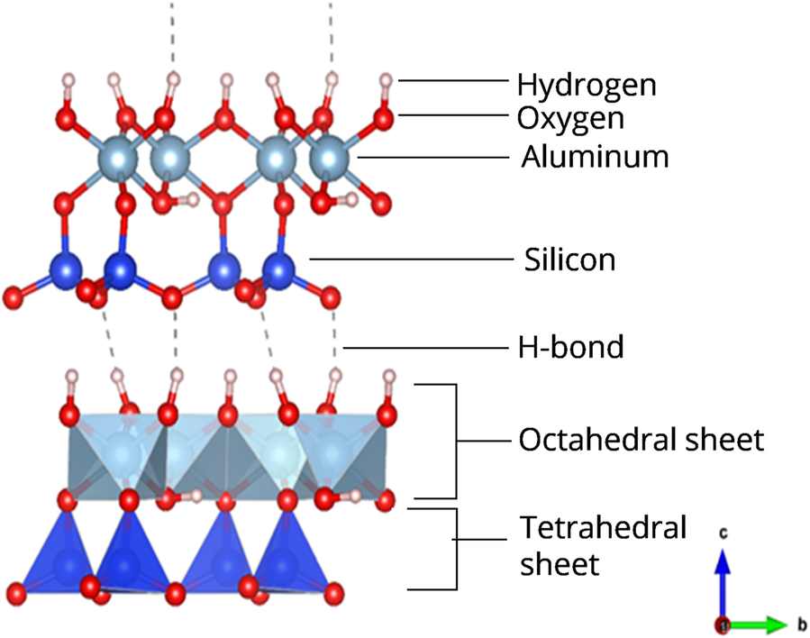
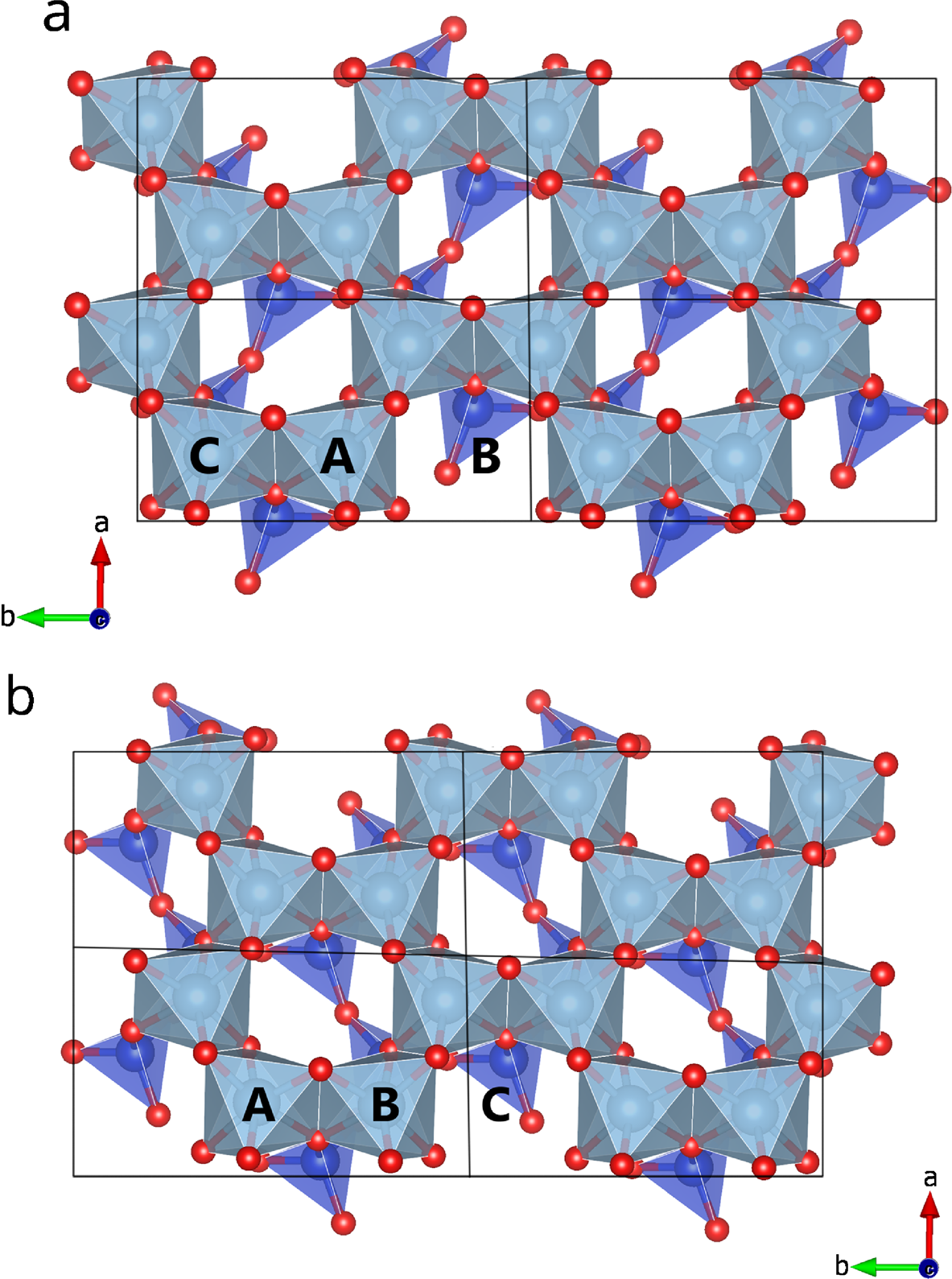
Kaolinite is a 1:1 dioctahedral member of the kaolin subgroup of minerals with an ideal formula of Al2Si2O5(OH)4. Each 1:1 layer consists of a silica tetrahedral sheet and a dioctahedral alumina octahedral sheet (Fig. 1). Successive layers are held together primarily by long hydrogen bonds along the c axis (Giese, Reference Giese1973). Two of the three (A, B, and C) possible octahedral sites in the octahedral sheet are occupied by Al. The layers are designated A, B, or C depending on the position of the unoccupied octahedral site (Fig. 2).
Ball and stick (upper) and polyhedral (lower) representation of the kaolinite structure

2×2×1 (x,y,z) kaolinite layer types: a B and b C with vacant B and C positions, respectively. Based on atomic coordinates by Bish and Von Dreele (Reference Bish and Von Dreele1989)

Structural disorder in kaolinite could either be natural ― through crystal growth, polymorphic transformations (Veblen, Reference Veblen1985), transport processes (White & Dixon, Reference White, Dixon, Dixon and Schulze2002) ― or mechanically induced through milling (Kristof et al., Reference Kristof, Juhasz and Vassanyi1993), sonication (Franco et al., Reference Franco, Pérez-Maqueda and Pérez-Rodríguez2003), etc. An ideal, perfect (defect-free) kaolinite has stacks of only one layer type (CC… stacking sequences) with a layer displacement vector t 1 (≈ –a/3) or t 2 (≈ a/6–b/6). The relationship between t 1 and t 2 is a pseudo-mirror plane passing through the center of the vacant octahedral site of the kaolinite unit cell (Bookin et al., Reference Bookin, Drits, Plançon and Tchoubar1989; Drits & Tchoubar, Reference Drits, Tchoubar, Drits and Tchoubar1990; Giese, Reference Giese and Bailey1988). This pseudo-mirror reflection makes the t 1 and t 2 displacements symmetric about the pseudo-mirror plane. Herein, the layers in a t 2 displacement are denoted as CeCe. Most stacking disorder in kaolinite is introduced into the structure by random interstratification of t 1 and t 2 displacement vectors (Bookin et al., Reference Bookin, Drits, Plançon and Tchoubar1989; Plançon et al., Reference Plançon, Giese, Snyder, Drits and Bookin1989). Results from high-resolution transmission electron microscopy (HRTEM) studies and the modeling of X-ray diffraction (XRD) patterns have revealed that the interstratification of enantiomorphic C layers (i.e. CCe) is the most common source of stacking disorder in kaolinite (Plançon et al., Reference Plançon, Giese, Snyder, Drits and Bookin1989; Kogure et al., Reference Kogure, Elzea-Kogel, Johnston and Bish2010; Kogure, Reference Kogure2011; Sakharov et al., Reference Sakharov, Drits, McCarty and Walker2016). Though present in just small proportions, another form of stacking disorder that has been observed in kaolinites arises from the alternation of C and B (±120° mutual rotation between adjacent layers in ideal structures) layers (Plançon et al., Reference Plançon, Giese, Snyder, Drits and Bookin1989; Kogure et al., Reference Kogure, Elzea-Kogel, Johnston and Bish2010; Kogure, Reference Kogure2011). The regular CB… or BC… stacking sequence with the t 1 displacement is characteristic of dickite — another member of the kaolin subgroup of minerals. Dickite-like sequences in kaolinite have been confirmed in HRTEM studies (Kogure et al., Reference Kogure, Elzea-Kogel, Johnston and Bish2010; Kogure, Reference Kogure2011). Infrared spectra of some kaolinites have shown dickite-like features, such as enhanced 3650 and 3620 cm–1 bands, suggesting that the random stacking of kaolinite and dickite-like layers is one of the sources of disorder in kaolinites (Beauvais & Bertaux, Reference Beauvais and Bertaux2002; Fraser et al., Reference Fraser, Wilson, Roe and Shen2002; Johnston et al., Reference Johnston, Elzea-Kogel, Bish, Kogure and Murray2008; Prost et al., Reference Prost, Dameme, Huard, Driard and Leydecker1989). Another form of minor stacking disorder is due to the layer displacement t 0 ( ≈ –0.3154a – 0.3154b), located along the long diagonal of the oblique layer unit cell that contains the vacant octahedral site (Sakharov et al., Reference Sakharov, Drits, McCarty and Walker2016). To model the XRD patterns of kaolinites successfully, it was necessary to incorporate some dickite-like sequences or displacement vectors (t o ) that have similar effects to introducing B layers (Bookin et al., Reference Bookin, Drits, Plançon and Tchoubar1989; Plançon et al., Reference Plançon, Giese, Snyder, Drits and Bookin1989; Sakharov et al., Reference Sakharov, Drits, McCarty and Walker2016).
Because it is the most common type of disorder in clays (Brindley, Reference Brindley, Brindley and Brown1980; Lombardi et al., Reference Lombardi, Russell and Keller1987), stacking disorder in kaolinites has been studied extensively during the past ~70 years using both qualitative and quantitative approaches. Given that the properties of minerals are a function of chemical and structural blueprints, addressing the implications of order-disorder for the reactivities and applications of kaolinites is critical, but many contrary observations have been reported in the literature.
Structural Disorder and Reactivity of Kaolinites
Few studies have attempted to address the question above by investigating the influence of stacking disorder in kaolinites on the properties: rate and degree of intercalation, rate of dissolution and transformation, and thermal stability.
Intercalation of kaolinites by organic molecules is important for producing nanoparticles for desired agricultural, environmental, and industrial applications (Sugahara et al., Reference Sugahara, Nagayama, Kuroda, Dio and Kata1991; Mahdavi et al., Reference Mahdavi, Abdul and Khanif2014). The ease and extent of intercalation determines the quantity and quality of the nanoparticles recovered as well as their applications. Conclusions drawn about the effect of stacking disorder on the intercalation of kaolinite are often non-definitive or contradictory. In some studies, highly ordered layers were easier to intercalate (Frost et al., Reference Frost, Kristof, Horvath and Kloprogge1999; Deng et al., Reference Deng, White and Dixon2002; Cheng et al., Reference Cheng, Liu, Yang, Du and Frost2010) which suggests a weaker interlayer binding strength, in a domain with the same type of stacking sequence, than that between two domains with different stacking sequences. Others have recorded an opposite trend (Wiewióra & Brindley, Reference Wiewióra and Brindley1969; Cruz-Cumplido et al., Reference Cruz-Cumplido, Sow and Fripiat1982) or concluded that the interlayer binding strength of kaolinite layers was not influenced by structural disorder (Uwins et al., Reference Uwins, Mackinnon and Thompson1991, Reference Uwins, Mackinnon, Thompson and Yago1993).
The dissolution and transformation of kaolinite, particularly in alkaline solution, has long been studied. The dissolution of kaolinite in alkaline solution at elevated temperature is a fast process and is used as a source of Al and Si for the synthesis of a variety of geopolymers, zeolites, and feldspathoids (Panagiotopoulou et al., Reference Panagiotopoulou, Kontori, Perraki and Kakali2007; Reyes et al., Reference Reyes, Williams and Alarcón2013; Xu et al., Reference Xu, Smith, Wingate and De Silva2010; Zhao et al., Reference Zhao, Deng, Harsh, Flury and Boyle2004). The extent of kaolinite dissolution determines the quantity of Al and Si in solution and determines indirectly the properties and applications of the synthesized materials. An experiment lasting for up to 4 weeks concluded that structural disorder did not influence the transformation of kaolinite to sodalite in NaOH (Zhao et al., Reference Zhao, Deng, Harsh, Flury and Boyle2004). In another study, highly disordered kaolinite dissolved twice as quickly as highly ordered kaolinite in oxalic acid but a similar dissolution rate was observed in HNO3 (Sutheimer et al., Reference Sutheimer, Maurice and Zhou1999). The latter authors suggested that “the fundamental structure of kaolinite, rather than specific surface details, exerts the greatest influence on dissolution kinetics.” A study of the slow dissolution of seven kaolinites, in pH 3 0.001 N HCl for 2 months, indicated that highly disordered samples dissolved more quickly than low disordered kaolinites (Kittrick, Reference Kittrick1966; Devidal et al., Reference Devidal, Dandurand and Gout1996).
Results from the energy-of-formation experiments determined from drop-solution calorimetry for four kaolinites suggested that structural disorder did not influence the stability of kaolinite layers (De Ligny & Navrotsky, Reference De Ligny and Navrotsky1999). A solubility approach found that the energy of formation increased slightly (–903.8 to –902.5 kcal/mol) as structural disorder increased (Kittrick, Reference Kittrick1966).
The dehydroxylation temperature of kaolinite decreased as structural order decreased (Stoch & Wacławska, Reference Stoch and Wacławska1981; Bellotto et al., Reference Bellotto, Gualtieri, Artioli and Clark1995; Vaculikova et al., Reference Vaculikova, Plevova, Vallova and Koutnik2011) but the activation energy (E a ) increased in the same direction (Horváth, Reference Horváth1985; Vaculikova et al., Reference Vaculikova, Plevova, Vallova and Koutnik2011). By heating two kaolinite samples at different heating rates, E a was derived to be 40(6) kcal/mole for highly ordered KGa-1 and 22(7) kcal/mole for low-ordered kaolinite KGa-2, respectively (Bellotto et al., Reference Bellotto, Gualtieri, Artioli and Clark1995). The calculated activation energies by the latter authors were more plausible as they made no assumption about the kinetic equation.
In summary, no consensus seems to exist on the influence of structural disorder on the reactivity of kaolinites. Two reasons for the contradictory observations/conclusions may stem from: (1) the use of the Hinckley index (HI) as a measure of the extent of structural disorder in kaolinites; and (2) particle-size variations amongst samples in addition to their disordering differences. In studies targeted at investigating the relationship between structural disorder and reactivity/stability of kaolinite layers, the commonly used parameter in estimating the extent of structural disorder is the HI (Hinckley, Reference Hinckley1962) or similar empirical parameters such as the Lietard (R2) index (Liétard, Reference Liétard1977) or the Aparicio-Gálan-Ferrell index (Galán et al., Reference Galán, Aparicio, La Iglesia and Gonzalez2006). These indices estimate the abundance of structural disorder in kaolinite as a “crystallinity index” by calculating ratios of the intensities (after subtracting background) of XRD peaks (020, 110, and 111¯
 reflections) between 19 and 26°2θ (CuKα radiation). Kaolinites, the XRD patterns of which are characterized by well resolved peaks and low background between 19 and 26°2θ (CuKα radiation), usually have high HI values and are considered less structurally disordered than specimens with high background and poorly resolved peaks. Given that the observed shape of the XRD reflection is a convolution of both instrumental and sample contributions, kaolinite specimens consisting of small crystallites/particle size have broader (less resolved) XRD peaks compared to specimens with larger particle size. Consequently, this yields a lower HI value in the former even if both specimens have the same extent of structural disorder within the assemblages. For this reason, the use of HI as an estimate to compare structural disorder amongst kaolinites should be done with an awareness of the influence of ‘size’ on measured HI values. To avoid or minimize this bias, it is imperative that the specimens being compared have a similar mean distribution of particle size. Unfortunately, the influence of size is seldom considered when comparing HI values amongst kaolinite specimens. Huge variations in the distribution of particle sizes also influence the observed reactivity or stability (Deng et al., Reference Deng, White and Dixon2002; Horváth, Reference Horváth1985). For example, it is generally accepted that intercalation in kaolinites becomes more difficult as the particle size decreases (Deng et al., Reference Deng, White and Dixon2002; Uwins et al., Reference Uwins, Mackinnon, Thompson and Yago1993). Hence, samples containing most particles within the fine fraction might intercalate more slowly and to a lesser extent than samples with larger particles irrespective of the degree of disorder in either sample (Uwins et al., Reference Uwins, Mackinnon, Thompson and Yago1993).
The influence of stacking disorder on the reactivity of kaolinites remains unclear. Given the variations (translations, type of layers, etc.) in the stacking of disordered/ordered kaolinite layers, the hypothesis is that structural disorder will influence the reactivity of kaolinites. The objective of the present study was, therefore, to investigate the influence of stacking disorder on the intercalation and dissolution of kaolinite layers.
Materials and Methods
Sample Preparation and Preliminary Analysis
Five geologic kaolinite specimens were collected from Georgia, USA, and coded as Kao-01, -02, -03, -05, and -Huber. The Huber sample was supplied by the J.M. Huber Corporation; the others were collected during the field trip of the 53rd Annual Meeting of The Clay Minerals Society in Georgia. All samples were air-dried, crushed in a mortar, and passed through a 2-mm sieve. Samples were treated with pH 5 sodium acetate buffer solution, 30% hydrogen peroxide, and dithionite-citrate-bicarbonate to remove carbonate minerals, organic matter, and iron oxides, respectively. To constrain the influence of particle size on HI values and reactivity, the 1–2 μm fraction of each sample was separated from the bulk by centrifugation (Soukup et al., Reference Soukup, Buck and Harris2008).
The extent of structural disorder in the samples was estimated from the XRD patterns by the Hinckley method, and crystallite size by Scherrer’s equation, respectively (Scherrer, Reference Scherrer1918; Hinckley, Reference Hinckley1962). Dispersion of each of the 1–2 μm samples in water was used to estimate the particle-size distribution using a laser diffraction particle size analyzer (LS230, Beckman Coulter). The morphology and chemical composition of the kaolinite samples were studied using an FEI Quanta 600F field emission scanning electron microscope (SEM) equipped with X-ray energy dispersive spectroscopy (EDS) (FEI, Hillsboro, Oregon, USA). The XRD patterns were recorded on a Bruker D8 ADVANCE X-ray diffractometer (Bruker AXS GmbH, Karlsruhe, Germany) with CuKα radiation operated at 40 kV and 40 mA. A programmable divergent slit, with a constant 12 mm radiation length and a Sol X detector was used for the XRD analysis. Powdered samples were front-loaded on a sample holder and patterns were recorded in the range 5–70°2θ, with a step size of 0.02°2θ and a dwell time of 3 s at each step. Kaolinite and accessory minerals in the samples were quantified by Rietveld refinement (Rietveld, Reference Rietveld1967). Structural models were visualized using VESTA (Momma & Izumi, Reference Momma and Izumi2011).
Kaolinite Intercalation Experiment
An amount of 0.1 g of clay was dispersed in 5 mL of deionized (DI) water followed by transfer of 200 μL of the dispersion onto pre-weighed (W1) glass disks. The dispersions on the disks were left to air dry at room temperature for 24 h after which the mass of the glass and clay film was determined (W2). The difference between W1 and W2 is the mass of clay on each glass disk. Two drops of saturated potassium acetate (CH3COOK) solution were pipetted onto the air-dried clay film and the mass recorded immediately (W3). The glass disk was immediately transferred to the XRD instrument and patterns were acquired subsequently in situ between 0 and 120 h to monitor the rate and degree of intercalation of the clays. The difference between W3 and W2 is the mass of saturated CH3COOK solution on each clay film. The mass of clay was ~4 mg while the mass of CH3COOK solution was ~70 mg. The XRD patterns were recorded between 2 and 32°2θ, with a step size of 0.05°2θ and a dwell time of 3 s at each step.
When CH3COOK molecules intercalate kaolinite, the interlayer distance expands from 7 Å (kaolinite) to ~14 Å (kaolinite-CH3COOK complex). The proportion of the interlayer penetrated by the molecules was estimated as (Theng, Reference Theng1974):

where I kao and I kao-CH3COOK are the intensities of the 7 and 14 Å reflections, respectively.
Kaolinite Dissolution and Transformation in 4 M NaOH
Only three of the kaolinite specimens, Kao-01, 03, and 05, with large HI differences, were used in the NaOH dissolution experiment. These samples represented the least disordered, medium disordered, and the most disordered, respectively, of the five samples considered. The dissolution experiment was conducted by mixing 0.1 g of each kaolinite sample with 8 mL of 4 M NaOH solution and 2 mL of 0.5 M NaCl in 15-mL polypropylene centrifuge tubes. The tubes were capped and kept in the oven at 80°C for 3 days and were agitated by hand every 12 h.
By the end of the experiment, to halt further dissolution, the tubes were immediately filled to 15 mL with DI water, centrifuged, and the supernatant discarded. The residue was washed twice more with DI water to rid the residue of alkali solution. The residues were then left to air dry on glass disks for 24 h before further analysis.
Infrared (IR) spectra of the three samples before and after dissolution by NaOH were recorded as an average of 32 scans at 4 cm–1 resolution using a Perkin Elmer Spectrum 100 Fourier-transform infrared spectrometer (FTIR) (Perkin Elmer, Waltham, Massachusetts, USA) equipped with a 45° single reflection diamond crystal universal attenuated total reflection (ATR) accessory. The residues, after dissolution, were also analyzed by XRD (on glass disks).
At the end of the NaOH dissolution experiment, the intensity of the XRD and IR peaks of kaolinite was expected to diminish or disappear completely while new peaks from sodalite emerged. Residual kaolinite was estimated by Rietveld refinement and the percent dissolution was calculated as shown below:

where Q k(s) and Q k(d) are the proportions of kaolinite in the starting material and the quantity after dissolution in NaOH, respectively. Using anatase (accessory mineral) as an indirect internal standard, given its stability under the experimental conditions, the amorphous content can be estimated and not assigned as clay. Orientation preference was expected to occur on the disks for residual kaolinite from the NaOH dissolution experiment. The small amounts of the starting kaolinite in the NaOH dissolution experiment would increase the error of the Rietveld quantification. The intensities of the OH bands of the residual kaolinites were also compared with calculated Q k(d) to check if they followed the same trend.
Extraction and Purification of the Less-reactive Kaolinite Phase in the NaOH Dissolution Experiment
One of the sodalite-kaolinite residues was selected to study further the residual kaolinite that was not dissolved during the 72-h NaOH dissolution experiment. The sodalite-kaolinite residues from the dissolution of kao-01 were chosen because the amount of residual kaolinite was adequate and mica was absent, as in kao-5. The residue was washed in 1 M HCl for 30 min followed by twice washing in DI water and centrifugation. The HCl washing was used to dissolve sodalite and amorphous phases while the DI washing rid the matrix of the dissolved Al and Si. The resulting residue corresponded to the kaolinite phase that was less reactive to NaOH dissolution. SEM images and an FTIR spectrum of the less-reactive residue were acquired. The intercalation experiment was repeated on the ‘less reactive’ kaolinite phase to investigate the binding strength of the layers.
Results
Kaolinite Sample Properties
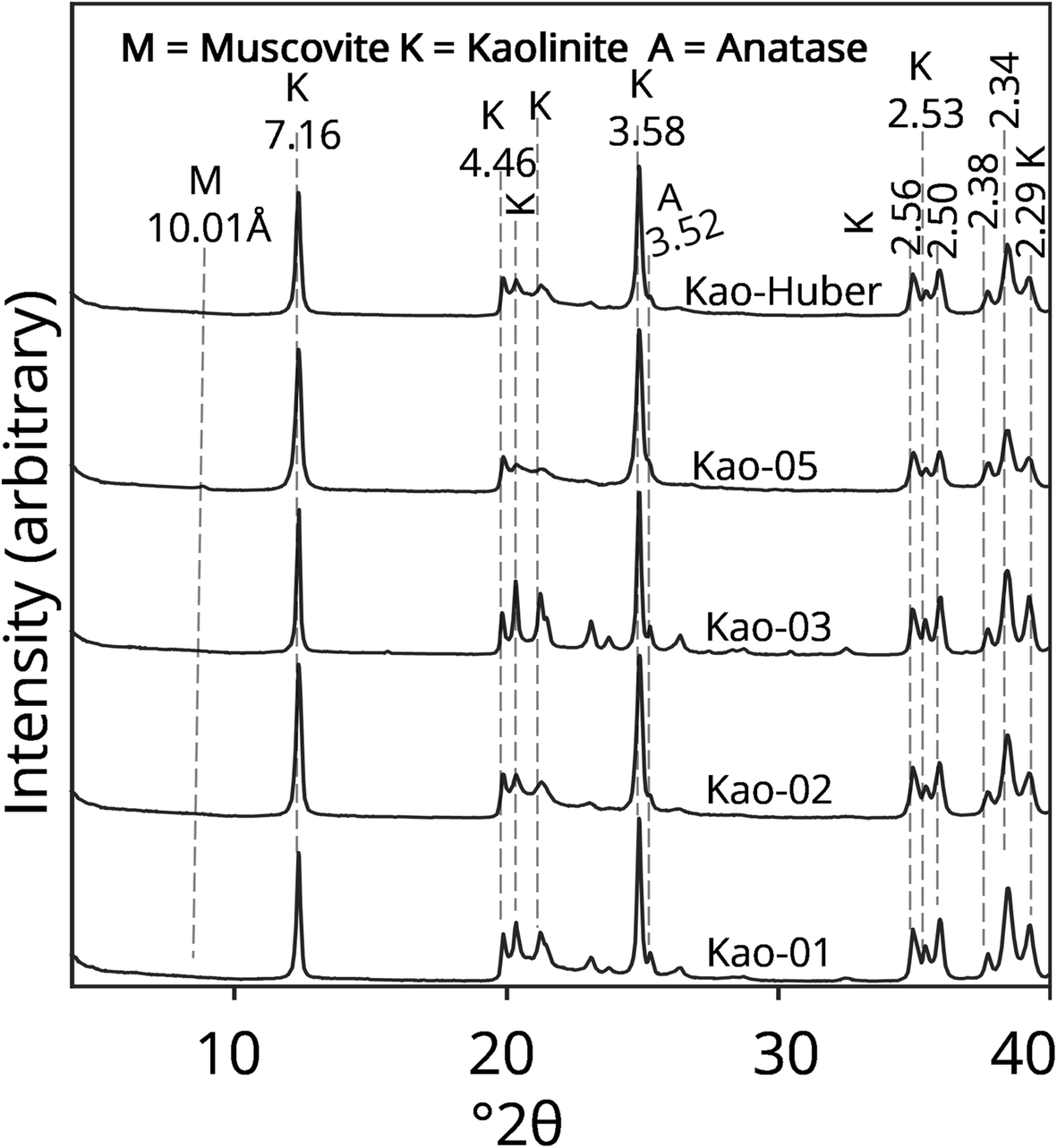
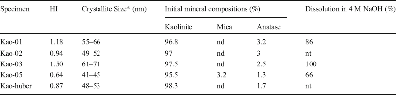
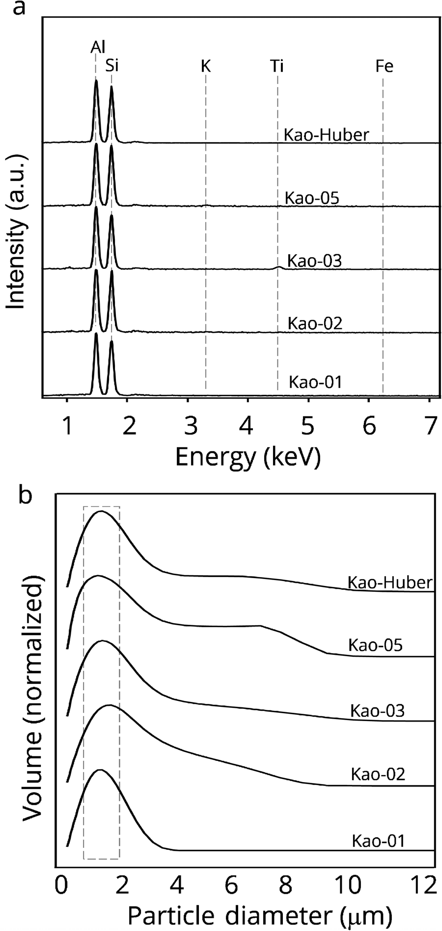
From the XRD patterns of the 1–2 μm fraction of the five kaolinites (Fig. 3), the extent of structural disorder followed the trend: kao-05 > kao-huber > kao-02 > kao-01 > kao-03. The less disordered samples had well resolved peaks and a low background of between 4.46 and 3.58 Å compared to the highly disordered samples. The kaolinite content in all the samples was >95% and the crystallite size (00l) ranged from 41 to 71 nm based on XRD peak fitting (Table 1). All the samples had a similar chemistry (Fig. 4a), similar particle-size distribution (Fig. 4b), and contained a small amount of accessory minerals (anatase and mica) (Table 1, Fig. 3). These analyses indicated that the particle size and chemistry of the samples could be regarded as similar and should have similar effects on the reactivities in the intercalation and dissolution in this study.
XRD patterns of the 1–2 μm fraction of the kaolinites tested

The Hinckley Index (HI), the proportion of minerals, and the dissolution of the 1–2 μm fraction of five kaolinites

*= along 00l; nd not detected, nt not tested
a The EDS spectra and b normalized (volume = 0 to 1) particle-size distributions of the 1–2 μm kaolinite fractions

Effect of Disordering in Kaolinite on the Rate of Intercalation by CH3COOK
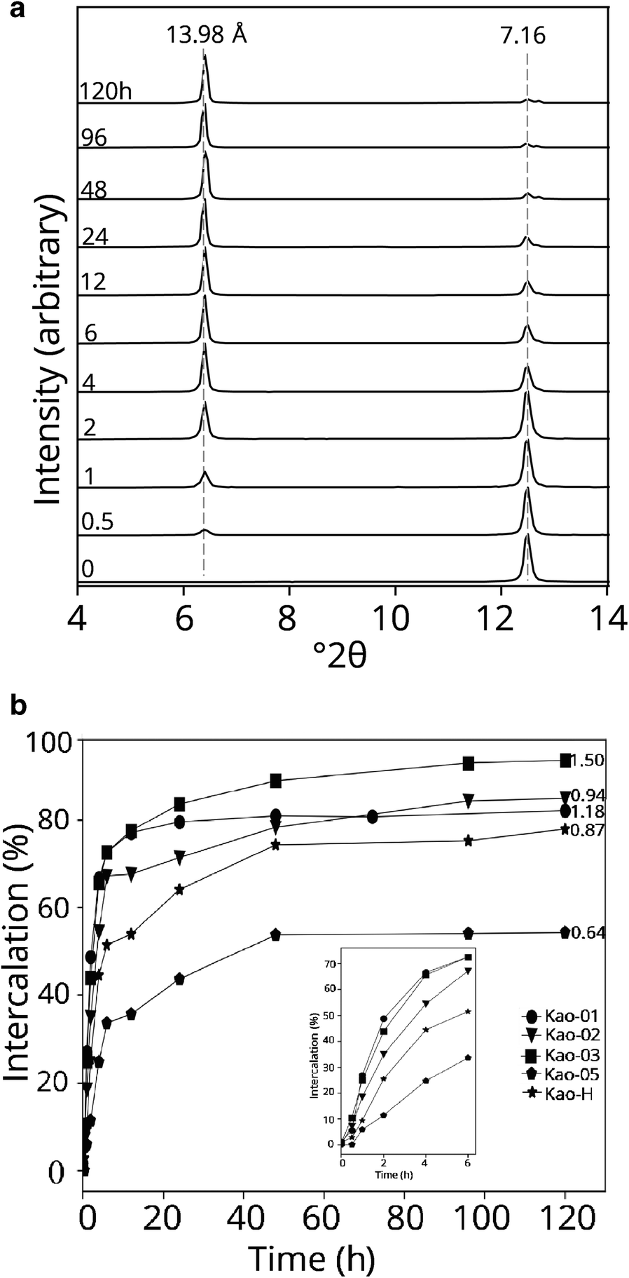
As intercalation progressed, the kaolinite 7.16 Å reflection decreased in intensity while the reflection at ~14 Å of the kaolinite-CH3COOK became more intense (Fig. 5a). The latter peak was due to the increase in the interlayer spacing as the CH3COOK molecules accrued in the interlayer of kaolinite. In the first 6 h of the intercalation experiment, the samples with a low degree of structural disorder intercalated more quickly than samples with a higher degree of disorder; the most ordered sample (kao-03) intercalated twice as much as the most disordered (kao-05) sample (Fig. 5b). By the end of the experiment, samples with the lowest extent of disordering recorded the greatest degree of intercalation (Fig. 5b).
Example of: a in situ XRD patterns of kao-03 during the intercalation experiment; and b percentages of intercalation of the 1–2 μm fractions of tested kaolinites

Effect of Disordering on the Kaolinite Dissolution and Transformation in NaOH Solution — XRD and FTIR Data
The majority of kaolinite in all of the specimens was dissolved in the 4 M NaOH solution under current experimental conditions, but small differences in the percentages of residual kaolinite was difficult to quantify accurately. The dissolution of kaolinite led to the formation of sodalite (Na8Al6Si6O24Cl2):

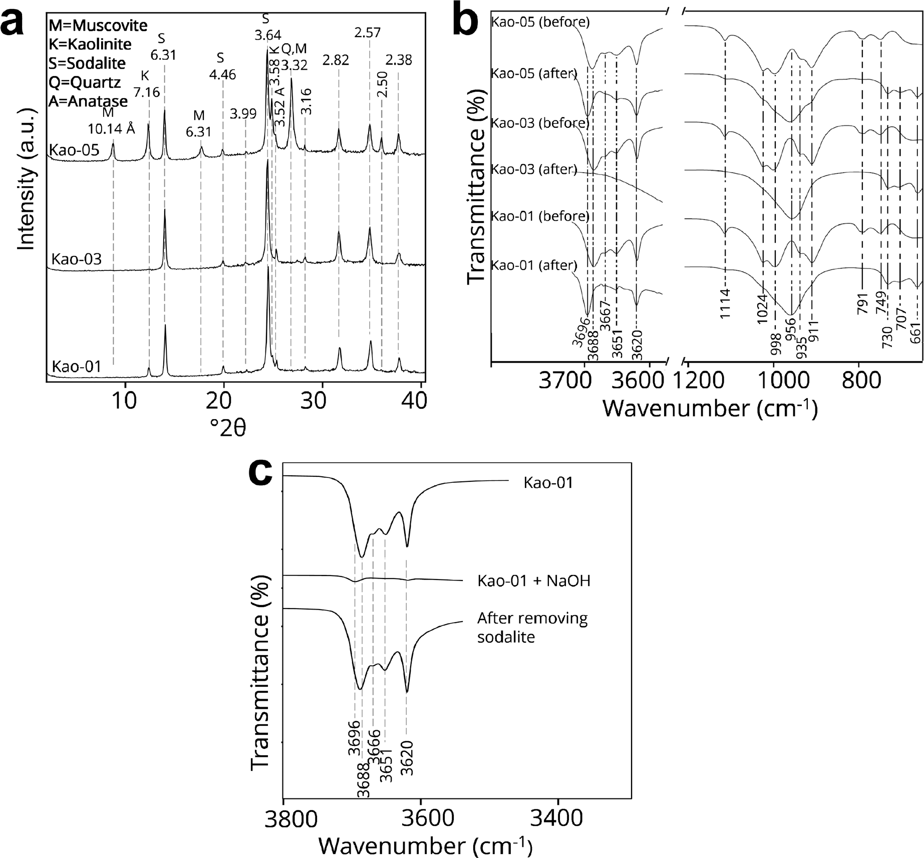
The most disordered kaolinite samples were more difficult to dissolve and transform to sodalite (Table 1). The most ordered sample (kao-03) was completely dissolved by the end of the experiment with no obvious kaolinite peaks on the XRD pattern (Fig. 6a).
a XRD patterns of the 1–2 μm fractions of kao-01, -03, and -05 after dissolution by 4 M NaOH; b FTIR spectra of the 1–2 μm fractions of kao-01, -03, and -05 before and after the dissolution by 4 M NaOH, and c FTIR spectra of kao-01 before (upper) and after (middle) dissolution in NaOH and after (lowest) washing in HCl. The 3700–3600 cm–1 section is magnified for clarity.

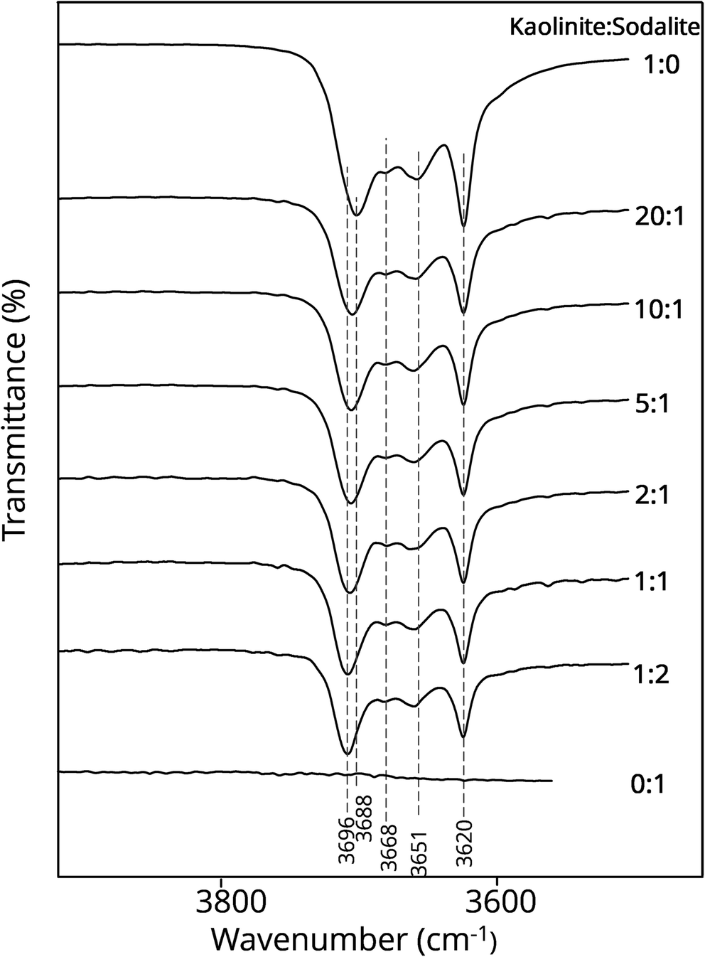
The following assignment of the FTIR bands of the original kaolinite samples (with the suffix ‘before’ in Fig 6b) was based on Farmer (Reference Farmer1974). The band at 3620 cm–1 (v 1) is from the stretching vibrations of inner OH groups, the bands at 3651 (v 2), 3667 (v 3), and 3688 (v 4) cm–1 are from stretching vibrations of inner-surface OH groups. In agreement with the XRD patterns, the kaolinite peaks in the FTIR spectra decreased or disappeared after dissolution (suffix ‘after’ in Fig. 6b). The four OH-stretching bands were missing from the FTIR spectrum of kao-03 after the dissolution, two of the bands remained in kao-01 (v 1 and v 4), while the four bands remained in kao-05 except for a reduction in intensity. This corresponds to the complete dissolution of kaolinite in kao-03 (least disordered sample) and least dissolution in kao-05 (most disordered sample). The kaolinite bands at lower wavenumbers disappeared after dissolution to give rise to bands at 956–960, 730, 707, and 661 cm–1 which have been observed previously after the transformation of kaolin to sodalite (Li et al., Reference Li, Zeng, Yang, Wang and Luo2015; Sari et al., Reference Sari, Suprapto and Prasetyoko2018). Another observation in the OH-stretching region is the shift of v 1 from 3688 cm–1 before the NaOH dissolution to 3696 cm–1 after NaOH dissolution, which was due to the matrix effect by sodalite. This matrix effect, observed in ATR mode, can be reproduced by artificially mixing kaolinite with pure sodalite (Fig. 7). When the kaolinite-sodalite mixture was washed with HCl to dissolve the sodalite, the in-phase OH band returned to 3688 cm–1 (Fig. 6c).
Infrared (ATR) spectra of various ratios of kaolinite-sodalite mixture

Intercalation of Kaolinite Residue after NaOH Dissolution
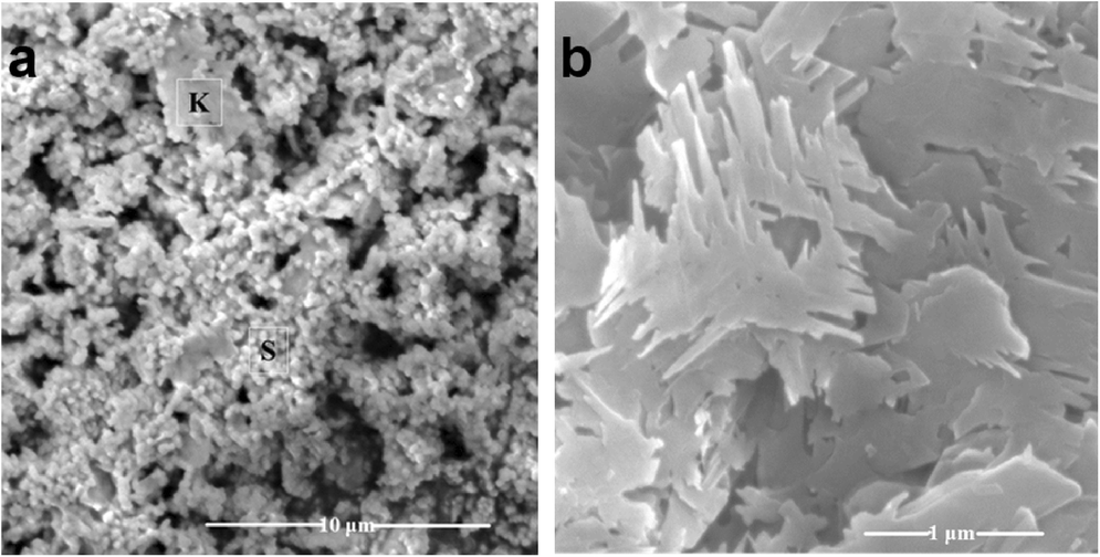
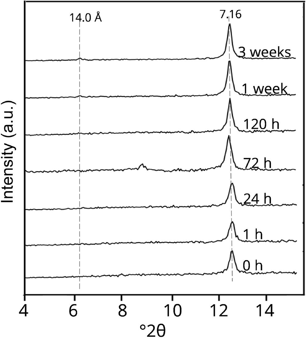
The SEM images of kao-01 after dissolution in NaOH (Fig. 8a) and after washing the kaolinite-sodalite matrix with HCl (Fig. 8b) indicated that the sodalite crystals formed in the former were fully dissolved by the acid wash. The kaolinite residue was characterized by jagged edges. The intercalant molecules could not surmount the interlayer binding strength of the kaolinite layers (despite observing the intercalation experiment for several weeks) (Fig. 9). The FTIR spectra of the kaolinite residue (Fig. 6c) registered similar band positions to those of the starting kaolinite except for the changes in the relative intensities of the OH bands. The intensities of v 4 /v 1 and v 2 were also reduced.
SEM images of a sodalite (s, granular) precipitate and residual kaolinite (k, platy) in kao-01 after dissolution in 4 M NaOH and b the kaolinite residue after removing the sodalite with HCl

In situ XRD patterns of the less reactive kaolinite residue of NaOH-reacted kao-01 during the intercalation experiment

Discussion
The kaolinite samples used in the present study were relatively pure with just traces of mica and anatase. The chemistry of the samples confirmed the ideal composition (Al2Si2O5(OH)4) and suggested little or no substitution of Fe for Al in the octahedral sheet. Such substitutions would have influenced the electrostatic energy and the degree of structural disorder of the mineral (Gaite et al., Reference Gaite, Ermakoff, Allard and Muller1997; Iriarte et al., Reference Iriarte, Petit, Huertas, Fiore, Grauby, Decarreau and Linares2005) and consequently the reactivity. The influence of particles on the HI values and reactivity is also constrained given the similar particle-size distribution of the current kaolinite samples.
For intercalation to proceed, the intercalant molecules must first break the interlayer H-bond binding kaolinite layers. The ease of breaking this bond can be used as an indirect probe of the average interlayer binding strength. Going by the results of the first 4 h of intercalation (inset Fig. 5b), the disordered samples were more resistant to intercalation compared to ordered samples. The rate and degree of intercalation followed a similar trend to the degree of stacking order: kao-05 < kao-huber < kao-02 < kao-01 ≈ kao-03. Once the H-bonding between kaolinite layers is broken, intercalation proceeds. Yet, 100% intercalation is seldom observed, which can only mean that the H-bonding between some layers was not surmounted. This suggests that the interlayer binding strength is not uniform across layers. The results reported here suggest that disordered samples had greater average interlayer binding strength (i.e. stability) than the ordered samples. This conclusion contradicted Frost et al. (Reference Frost, Van Der Gaast, Zbik, Kloprogge and Paroz2002), who reported that the highly ordered Birdwood kaolinite intercalated very slowly and to a lesser degree (20%) after 18 days. A likely reason for this exception is that Birdwood consisted mostly of small (0.5 μm) particles. Such very fine fractions have been well known not to intercalate easily. The pursuit of an explanation for this anomalous intercalation by very fine fractions has yielded several theories (Weiss et al., Reference Weiss, Becker, Orth, Mai, Lechner and Range1969; Wiewióra & Brindley, Reference Wiewióra and Brindley1969; Raussell-Colom & Serratosa, Reference Raussell-Colom, Serratosa and Newman1987; Deng et al., Reference Deng, White and Dixon2002; Frost et al., Reference Frost, Van Der Gaast, Zbik, Kloprogge and Paroz2002 Zhang & Xu, Reference Zhang and Xu2007).
The percent dissolution of kaolinite in 4 M NaOH followed a similar trend as the degree of structural disorder: kao-05 > kao-01 > kao-03. This result agreed with the intercalation experiment and suggested that highly disordered kaolinite had a greater binding strength than ordered kaolinite. The sharp, nearly symmetrically branched needle morphology of the residual kaolinite in the NaOH dissolution experiment suggested that only selected areas of the original kaolinite particle were dissolved by the NaOH (Fig. 8b). Under the experimental conditions used here, some layers within the highly disordered specimen were clearly more resistant to dissolution. The few available reports on structural order-dissolution relationships concluded mostly that disordered kaolinites were easier to dissolve compared to ordered specimens (Kittrick, Reference Kittrick1966; Sutheimer et al., Reference Sutheimer, Maurice and Zhou1999). Hence, the current results contradicted these studies. An explanation is that in those studies, given that highly disordered samples were characterized by smaller particle sizes, the greater dissolution observed for disordered samples was more likely to be due to larger surface areas in the disordered specimens. When the different size fractions of kao-01 were dissolved in 4 M NaOH, the finer fractions showed more complete kaolinite dissolution (data not shown). By studying samples with a similar size distribution, the influence of size on dissolution was constrained.
Source of Differences in Interlayer Binding Strength
Having shown that the degree of structural disorder had a direct relationship with the reactivity, which, in turn, was related to the binding strength of kaolinite layers, a need remains to explain the sources of the observed differences in the binding strength of kaolinite of varying degrees of structural disorder. Though disorder due to the presence of stacking of enantiomorph layers t 1 t 2 is the most common type in kaolinite (Kogure et al., Reference Kogure, Elzea-Kogel, Johnston and Bish2010; Kogure, Reference Kogure2011; Sakharov et al., Reference Sakharov, Drits, McCarty and Walker2016), these two types of displacements had little or no effect on the electrostatic energy of the system or on the hydrogen bonding between the layers because the C and Ce are identical layers that differ only in translation vector and are related by a pseudo-mirror plane passing through the center of the vacant octahedral site (Drits & Tchoubar, Reference Drits, Tchoubar, Drits and Tchoubar1990; Giese, Reference Giese1982). This also explains why the OH-stretching bands of several kaolinites are at almost the same frequencies, with only differences in relative intensity. Hence, the observed differences in reactivity/binding strength of kaolinite layers could not be explained by differences in the proportion of t 1 t 2 stacking disorder. On the other hand, dickite-like sequences (CB or BC) modify the relative position of the atoms located on both sides of the disordered layer (Drits & Tchoubar, Reference Drits, Tchoubar, Drits and Tchoubar1990) and, as such, alter significantly the H-bonding between the layers. This form of stacking disorder is expected to influence the reactivity and stability of kaolinite layers.
Just as in the presence of a B-layer in the C-kaolinite, the t 0 layer displacement leads to the creation of a new layer with atoms in a different position compared to t 1 or t 2 displacements and, thus, influences the binding strength of the layers. The insertion of t 0 amongst t 1 has the same effect on the calculated XRD patterns as inserting B-layers (Sakharov et al., Reference Sakharov, Drits, McCarty and Walker2016) and improves agreement between calculated and observed low-frequency regions (White et al., Reference White, Kearley, Provis and Riley2013).
Though the incorporation of dickite-like fragments or t 0 displacements were expected to change the binding energetics of the layer in kaolinite, they have been shown to exist only in small percentages in sedimentary kaolinites (Plançon et al., Reference Plançon, Giese, Snyder, Drits and Bookin1989; Kogure & Inoue, Reference Kogure and Inoue2005a; Kogure et al., Reference Kogure, Elzea-Kogel, Johnston and Bish2010). The dickite-like character is more common in hydrothermal kaolinites formed at relatively high temperatures (Kogure & Inoue, Reference Kogure and Inoue2005b). By modeling XRD patterns, Sakharov et al. (Reference Sakharov, Drits, McCarty and Walker2016) showed that the proportion of t 0 displacements could not be great. A model containing 15–20% of t 0 displacements resulted in calculated XRD patterns that deviated significantly from the experimental XRD patterns of KGa-1, KGa-1b, and KGa-2. The same authors also showed that the incorporation of 10% of B-layers led to a reduction in intensity and shifting of 131 reflections compared to the experimental pattern. Such shifts are observable in highly disordered kaolinite such as the Charente sedimentary kaolinite, however; this was shown to contain up to 15% of these layers (Drits & Tchoubar, Reference Drits, Tchoubar, Drits and Tchoubar1990).
Thermodynamic and computational approaches have been used to investigate the stability of dickite and kaolinite layers, but the results are often contradictory. Using an electrostatic energy model, Giese (Reference Giese1973) showed computationally that the binding strength of dickite (CB) layers is 1.25 times greater than that of kaolinite (CC) layers while density functional state (DFT) calculations predicted a similar stacking energy for dickite or kaolinite layers (Sato et al., Reference Sato, Ono, Johnston and Yamagishi2004). Thermodynamic approaches have shown that either dickite (Kiseleva et al., Reference Kiseleva, Orogodova, Krupskaya, Melchakova, Vigasina and Luse2011; Zotov et al., Reference Zotov, Mukhamet-Galeev and Schott1998) or kaolinite is thermodynamically more stable (De Ligny & Navrotsky, Reference De Ligny and Navrotsky1999; Fialips et al., Reference Fialips, Navrotsky and Petit2001, Reference Fialips, Majzlan, Beaufort and Navrotsky2003). On the other hand, direct and simple experiments such as intercalation and dissolution appeared to suggest that dickite layers were more stable compared to kaolinite. For example, dickite layers were more difficult to intercalate than kaolinite (Cruz-Cumplido et al., Reference Cruz-Cumplido, Sow and Fripiat1982). Despite the size similarity of the kaolinite and dickite particles in Keokuk kaolinite, the kaolinite particles were shown (Fraser et al., Reference Fraser, Wilson, Roe and Shen2002) to be more susceptible to dissolution by HF. Experimental results from dehydroxylation reactions, using infrared emission spectroscopy, showed that kaolinite lost inner-surface hydroxyls and inner hydroxyls simultaneously whereas dickite lost the inner surface hydroxyls before inner hydroxyls (Frost & Vassallo, Reference Frost and Vassallo1996). This further suggests that the structure of dickite layers is more stable than that of kaolinite layers. Given that dickite-like sequences are also a source of disorder in kaolinites, the observed differences in reactivity as a function of stacking disorder could be explained at least partly. The failure of the kaolinite residue from the NaOH dissolution experiment to intercalate (Fig. 9) supports the view that the layer binding strength of the residual kaolinite was indeed greater than that of the dissolved ones. Compared to the starting material, the kaolinite residue appeared to exhibit more dickite-like character as shown by the decrease in the intensities of v 1 /v 4 (1.185 to 0.969) band as v 2 became slightly weaker, and the v 2 band increased slightly by ~6% (Fig 6c). The distribution of the intensities of the OH-stretching bands in the residue was characteristic of FTIR patterns of dickite stacking containing kaolinite (Cuadros et al., Reference Cuadros, Vega and Toscano2015; Fraser et al., Reference Fraser, Wilson, Roe and Shen2002; Prost et al., Reference Prost, Dameme, Huard, Driard and Leydecker1989). In synthetic mixtures of kaolinite and dickite, as the proportion of the latter increased, the v 1/v 4 ratio decreased and the v 2 band became weaker. A shift in v 1 to higher frequencies and the disappearance of v 2 was also observed as the proportion of dickite became greater than 90% (Cuadros et al., Reference Cuadros, Vega and Toscano2015). Similar observations were made in the present study but no v 1 shift was noted, suggesting that the less reactive residue still contained a significant proportion of kaolinite layers.
Given the more dickite-like character of the residue, as observed in the IR spectrum, the present study concludes that the average layer binding strength of disordered kaolinites was greater than that of ordered kaolinites. One of the sources of this greater binding strength in disordered kaolinites is the presence of dickite-like stackings in disordered kaolinites.
Conclusions
The present study investigated the influence of structural disorder on the reactivity of kaolinites. To limit the contribution of particle size to the reactivity, only the 1–2 μm fraction of five kaolinites with similar particle-size distributions was used. Using saturated CH3COOK solution as an intercalant and 4 M NaOH as a solvent, disordered kaolinites were more difficult to intercalate or dissolve compared to the more ordered kaolinites with very similar particle-size distributions. The kaolinite residue after 4 M NaOH dissolution was more difficult to intercalate than the starting kaolinite. Infrared data also suggested that the non-reactive portion in the NaOH dissolution contained more dickite-like features than the starting kaolinite. Having constrained the effect of the particle size on the reactivity of kaolinite, disordered kaolinites with a high degree of structural disorder had greater interlayer binding strength than the more ordered kaolinites. In addition to the common enantiomorphic stacking disorder, the former contains more dickite-like sequences in its stacking compared to the latter and contributed to the lower reactivity in the intercalation by CH3COOK and dissolution by NaOH.
ACKNOWLEDGMENTS
The authors are grateful to the Norman Borlaug Institute, Texas A&M University, for the fellowship that sponsored the first author’s graduate study, and the NSF grant DBI0116835 which supported the acquisition of the FE-SEM.
Funding
Funding sources are as stated in the Acknowledgments.
Declarations
Conflict of Interest
The authors declare that they have no conflict of interest.