INTRODUCTION
According to Darwin (Reference Darwin1851), the genus Pollicipes Leach, 1817, within the order Scalpelliformes (Buckridge & Newman, Reference Buckridge and Newman2006), includes three species of stalked barnacles: Pollicipes pollicipes (Gmelin, 1789) in the north-eastern Atlantic; Pollicipes polymerus Sowerby, 1833 in the north-eastern Pacific; and Pollicipes elegans Lesson, 1830 in the central-eastern Pacific (Foster, Reference Foster1978). In these hermaphrodite, pedunculate cirripedes, the flexible peduncle, which is covered by scales, contains ovarian tissue and is attached to the substratum by a basal disc. These species possess a capitulum that is covered by calcified plates that protect the body and have filamentary processes on the prosoma (Barnes, Reference Barnes1996). Based on morphological differences, such as the structure of the maxillule, the presence of a complemental male and the absence of filamentary processes on the prosoma, Pollicipes spinosus Quoy & Gaimard, 1834 and its synonym Pollicipes sertus Darwin, Reference Darwin1851 have been assigned to Calantica spinosa (Quoy & Gaimard, 1834), while Pollicipes mitella (Linnaeus, 1767), which has highly sculptured plates and multi-articulated caudal appendages, is included into the separate genus Capitulum Gray, 1825 (Foster, Reference Foster1978).
The only species with an Atlantic distribution is P. pollicipes (= P. cornucopia Leach, 1817) (Barnes, Reference Barnes1996; Foster, Reference Foster1978). The species is present in aggregations on rocky intertidal sites that are exposed to strong swells. The distribution records were compiled by Barnes (Reference Barnes1996) and range from the northern coast of Brittany in France (along the Bay of Biscay coastline) to the Atlantic coasts of Spain and Portugal (up to Cape San Vicente in southern Portugal). Along the Atlantic African coast, it occurs from the Strait of Gibraltar to Dakar (15°N). Although some earlier citations have recorded it in the western Mediterranean, Azores and East Asia, Barnes (Reference Barnes1996) suggested that such records might have occurred as a result of artificial transportation.
The population genetics structure of P. pollicipes along its distribution-range in the Atlantic was recently studied by Quinteiro et al. (Reference Quinteiro, Rodríguez-Castro and Rey-Méndez2007). A northern assemblage of open populations was defined based on an analysis of the mitochondrial genetic variation of populations sampled from the Canary Islands (28°N) to Brittany (47°N). Furthermore, a highly differentiated population was collected at the Cape Verde Islands (16°N). Phylogenetic analysis supported the clustering of the Cape Verde haplotypes into a divergent clade, distinct from the rest of the P. pollicipes haplotypes. A coalescent analysis identified this population as the oldest dating back to 4.37 million years. This phylogeographical pattern is generally unexpected from a co-specific status and Quinteiro et al. (Reference Quinteiro, Rodríguez-Castro and Rey-Méndez2007) consequently hypothesized about the possibility of a cryptic speciation in the Cape Verde population. The retention of a mitochondrial ancestral lineage in the Cape Verde population, its phylogenetic position with respect to other Pollicipes species and the presence of morphological differentiation are important issues that need to be studied further.
Partial cytochrome c oxidase I (COX1) mitochondrial gene sequences have been demonstrated to be effective for discriminating phyla at the species level, even in Crustacea (Costa et al., Reference Costa, deWaard, Boutillier, Ratnasingham, Dooh and Hebert2007). They are therefore the sequences of choice for the DNA barcoding approach in molecular taxonomy. Use of additional and alternative nuclear loci are, nevertheless, needed in order to verify the consistency of gene patterns and the absence of specific processes that affect mitochondrial DNA (Ballard & Whitlock, Reference Ballard and Whitlock2004). To that end, the 5.8S rDNA and the flanking interspaces sequences (ITS), located at the nuclear ribosomal operon were analysed.
In the present study we carry out a characterization of the Cape Verde population of stalked barnacles based on the morphological description and the molecular analysis of the mitochondrial cytochrome oxidase subunit I (COX1) and the nuclear (ITS1, 5.8S rDNA and ITS2) sequences.
MATERIALS AND METHODS
Sampling
Barnacles were collected using the sampling procedure detailed in Quinteiro et al. (Reference Quinteiro, Rodríguez-Castro and Rey-Méndez2007). Complete specimens were preserved in 70% ethanol until morphological and molecular characterization and analysis.
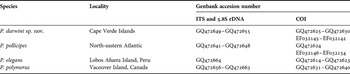
Adult specimens (total length >2 cm) of new species (N = 50) were collected from the Ponta do Sol site on the island of Santo Antão, in the Cape Verde Islands (17°12′02″N/22°05′44″W, 04/03/2005) and included both holotype and paratype individuals. The sampling procedure used for collecting P. pollicipes specimens from the north-eastern Atlantic is detailed elsewhere (Quinteiro et al., Reference Quinteiro, Rodríguez-Castro and Rey-Méndez2007). In addition, samples of P. elegans (N = 10) were also obtained from Lobos Afuera Island, Perú (6°57′04″N/80°41′20″W, 5 August 2008) and of P. polymerus (N = 10) from Vancouver Island, Canada (1 April 2007).
Morphological characterization
External morphological characters were observed and dissections carried out with the aid of Zeiss and Leica MZ16 binocular microscopes. Structures were observed and photographed from semi-permanent preparations using glycerine gel (10g gelatine, 60 ml of distilled water, 54 ml of glycerine and 0.5 g phenol crystal as preservative agent) and drawn with the aid of a camera lucida attached to a Leica MZ16 binocular microscope.
Material collected in the Cape Verde Islands was morphologically compared with specimens of P. pollicipes from the Iberian coast and the Canary Islands, and also with specimens of P. elegans from Peru; furthermore, a specimen held in the collections of the Muséum National d'Histoire Naturelle, Paris (MNHN), was also studied.
Genetic analysis
Total DNA was isolated from 30 mg of peduncle muscle by means of the DNeasy Tissue kit (Qiagen), in order to obtain a set of 10 individual DNAs per species.
Partial mitochondrial COX1 sequences for P. pollicipes and specimens of the new species (N = 13), reported by Quinteiro et al. (Reference Quinteiro, Rodríguez-Castro and Rey-Méndez2007), were used and supplementary COX1 data were obtained from new specimens (N = 7) using the LCO1490 and HCO2198 primers (Folmer et al., Reference Folmer, Black, Hoeh, Lutz and Vrijenhoek1994). Additional homologous sequences from P. polymerus and P. elegans species were also obtained. Finally, multicopy nuclear ribosomal interspacers 1 and 2 (ITS1 & 2), and 5.8S rDNA sequences were amplified, for one specimen of each of the Pollicipes species, using primers POL18S1F (5′–TGAACCTGCGGAA GGATCAT–3′) and POL18S2R (5′–CCACTTCACTCGCCGTTACTG–3′).
Polymerase chain reaction (PCR) amplifications were performed in a 15 µl volume using a GeneAmp 9700 thermocycler (Applied Biosystems). Reactions contained 1 × Green GoTaq Buffer (Promega), 1.5 mM MgCl2, 200 µM each of dNTP, 0.3 units of GoTaq (Promega), 0.1 µM of each primer, and 0.5 µl of the DNA solution. The PCR profile, for COX1 amplification, included a 2 minutes step at 95°C followed by 35 cycles of 95°C for 40 seconds, 50°C for 40 seconds and 72°C for 1 minute. Time was reduced by 10 seconds for each PCR step and annealing was increased to 55°C for the amplification of nuclear sequences.
Amplifications from the nuclear ribosomal operon array were gel purified using Promega Wizard SV Gel and PCR Clean-Up System (Promega). About 30 ng of PCR products were used for cloning with the pGEM-T Easy Vector system (Promega). A number of 8 clones were selected from each one transformation. Plasmids were isolated with the EZNA Plasmid Miniprep kit (Omega Bio-Tek). DNA sequencing was performed with the BigDye 3.1 sequencing kit (Applied Biosystems). Extension products were purified with DyeEx 2.0 Spin columns (Qiagen) and electrophoresis and automated detection were carried out in an ABI PRISM 377XL (Applied Bisystems). The obtained electropherograms were revised and sequences edited and aligned with BioEdit 7.0.1 (Hall, Reference Hall1999), using default parameter values. Sequences were deposited in GenBank (Table 1).
Pollicipes species included in this study and the Genbank accession numbers for the obtained nuclear and mitochondrial DNA sequences.

Phylogenetic relationships were inferred using distance, maximum parsimony, and maximum likelihood methods, based on mitochondrial COX1 and nuclear ITS data sets, using PAUP* version 4.0b10 (Swofford, Reference Swofford2002). The nucleotide substitution model that best fitted the data was evaluated with the aid of Modeltest version 3.7 under the Akaike's information criterion (Posada & Crandall, Reference Posada and Crandall1998), wherein: (a) for COX1 data, a general time reversible (GTR) model (Lavane et al., Reference Lavane, Preparata, Saccone and Serio1984) with proportion of invariable sites (I) = 0.5854 and a gamma distribution of variable sites with shape parameter = 1.0804, was used; and (b) for ITS sequences, the Tamura–Nei model (Tamura & Nei, Reference Tamura and Nei1993) with proportion of invariable sites (I) = 0.6796, was selected. Using these model parameters, we constructed a neighbour-joining (NJ) (Saitou & Nei, Reference Saitou and Nei1987) tree from a distances matrix. A maximum likelihood (ML) tree was furthermore obtained by heuristic search with the tree-bisection-reconnection (TBR) branch-swapping algorithm. A consensus tree was also obtained under parsimony criterion from the sequence data set using a heuristic parsimony search with closest addition sequence, being considering gaps as missing. Starting trees were obtained by stepwise addition. Clustering support was estimated using the bootstrapping method (Felsenstein, Reference Felsenstein1985) with 2000 replicates. Outgroup taxa for COX1 analysis included the closely related C. spinosa (Quoy & Gaimard, 1834) (AY428047), P. (= Capitulum) mitella (Linnaeus, 1767) (NC008742), Catomerus polymerus (Darwin, 1854) (EU423229), Astrobalanus imperator (Darwin, 1854) (EU423232) and Semibalanus balanoides (Linnaeus, 1767) (AF242697).
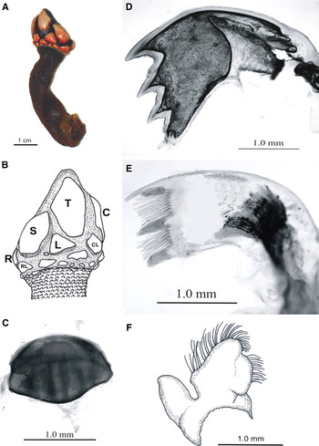
Pollicipes darwini sp. nov. (A) Adult specimen; (B) arrangement of capitular plates (R, rostrum; S, scutum; T, tergum; L, lateral; C, carina; RL, rostrolatus; CL, carinolatus); (C) scale of the peduncule; (D) mandible; (E) first maxilla; (F) second maxilla.

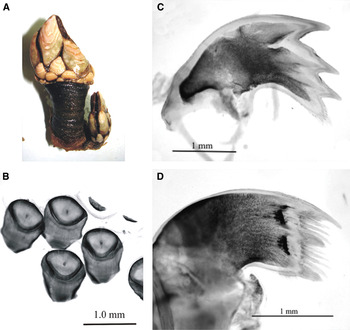
Pollicipes pollicipes. (A) Adult specimen; (B) scale of the peduncule; (C) mandible; (D) first maxilla.

TYPE MATERIAL
Holotype: adult (total length >2.0 cm). Cabo Verde S1, Santo Antão, Cape Verde Islands (17°12′02″N/22°05′44″W, 4 March 2005); exposed rocky shore; preserved in 70% ethanol; Museo Nacional de Ciencias Naturales, Madrid (MNCN-20.04/8354). Collected by M. Rey-Méndez, 4 March 2005.
Paratypes: MNCN 20.04/8355-8365, (total length > 2.0 cm); same data as holotype. MNCN 20.04/8365 is a slide preparation of the buccal appendages.
COMPARATIVE MATERIAL EXAMINED
Pollicipes pollicipes (Gmelin, 1789): 76 specimens; PPOL Sample III (MEI): PPOL40-PPOL49 (Ferrol, A Coruña, Spain, 43°35′46″N/8°12′29″W, 3 December 1998); PPOL130-PPOL139 (Cedeira, A Coruña, Spain, 43°37′45″N/8°08′36″W, 21 September 1998); PPOL sample VII (AF): PPOL900-PPOL955 (Fuerteventura, Canarias Islands, Spain 28°43′35″N/13°59′45″W, 1 September 2005), personal collection at the Molecular Systematics Laboratory, University of Santiago de Compostela, Spain.
Pollicipes elegans Lesson, 1830: 11 specimens; PELE Sample I: PELE1-PELE10 (Lobos Afuera Island, Peru, 6°57′04″N/80°41′20″W, 5 August 2008), personal collection at the Molecular Systematics Laboratory, University of Santiago de Compostela, Spain; MNHN-Ci319 (A. Gruvel det. 1902, patria ignota 1900).
DIAGNOSIS
Capitullum plates are whitish-orange and separated by extensive membranous interspaces. Peduncule is dark brown with scales of semicircular shape and the proximal edge clearly convex. Palpus is without bristles.
ETYMOLOGY
In honour of the eminent naturalist Charles Darwin who's exhaustive and complete work on the genus Pollicipes remains as a reference today, and to commemorate his 200th birth anniversary, as well as the 150th anniversary of the publication of On the Origin of Species.
DESCRIPTION
Peduncle muscular, narrow, dark brown (Figure 1A); covered by membrane interlaced with closely-placed semicircular scales; scales with the proximal edge clearly convex and curved in frontal view, base expanded, distal portion thinned, slightly curved in lateral view (Figure 1C).
Capitulum is 1/3–1/4 total length; capitular plates including one carina, one rostrum, paired terga, scuta, carinolatera, rostrolatera and latera. Capitulum base is surrounded by 15–30 small plates. Plates whitish-orange, orange in eroded portions, not overlapping, widely separated by reddish-black membranous interspaces. Tergum largest plate, carinal margin slightly curved, scutal margin curved, more pronounced in basal portion of margin; occludent margin almost straight. Scutum is approximately 2/3 of tergum, lanceolate; tergal and lateral margins undifferentiated and similar to the curved occludent margin; basal margin almost straight; adductor muscle pit deep in the internal face. Carina almost equal to tergum, almost straight near basal area, slightly curved distally. Rostrum is triangular, very small, basal margin convex. Remaining latera is triangular or sub-triangular, transversally arranged with respect to peduncle (Figure 1B).
Prosoma with 13–14 pairs of long and thin filamentary appendages, the longest ones lie nearest the thorax; first pair of cirri with well developed basal lanceolate appendages; following pair of cirri with small basal protuberances; distant pairs progressively less developed.
Mouth located close to the adductor muscle. Buccal pieces placed next to the bulky labrum. Thin palpus is without bristles, located between the adductor muscle and the filamentary appendages of the first pair of cirri. Mandibles are highly curved, yellow, palm shaped and containing three sharp and strong teeth (Figure 1D). The first tooth is the most developed with the tip backwards, as compared to the other two prominent teeth. Third tooth is a blunt protuberance with very thick bristles; outer margin with groups of thin bristles. Lanceolate palpus shows a high number of bristles on the inner margin and at the apex. First pair of yellow maxillae has a strong thin tooth at its upper angle (Figure 1E); three tufts of bristles originated from small prominences along the edge; first tuft is right next to the tooth; isolated tufts are separated by 6 and 8 pairs of spiny thick bristles. Second pair of maxillae presents a bi-lobulated shape (Figure 1F); anterior lobe adjacent to the first maxilla, well developed, containing numerous bristles; posterior lobe is quite small and lacks bristles.
Six pairs of bi-rami cirri; the number of segments increment progressively until the last pair with 18–20 segments; first pair short, with 7–9 segment; second pair with 13–15 segments. Cirri covered by tufts of bristles, short along the external part, tall on the internal side; bristles decrease in number from the first to the last segment, which show 1–2 bristles.
Purple penis has clear rings. The basal part is lacking bristles. Two rows of bristle tufts emerg from each ring along the upper half; there is 1 single bristle tuft at the apex. Two filiform caudal appendages flanking the penis, consisting of four segments with a sharp apex; bristle tufts between articulations.
Oviductal sacs (seem in one specimen) are pale yellow, on either side of the prosoma, on level with the cirri, disc shaped, 13 mm diameter.
DISTRIBUTION
Found in the Cape Verde Islands.
MOLECULAR ANALYSIS
The alignment of the partial mitochondrial COX1 sequences obtained from P. pollicipes, P. elegans, P. polymerus, P. darwini sp. nov. and the closely related outgroup species (C. spinosa, C. polymerus, C. mitella, A. imperator and S. balanoides) resulted in 584 base pair (bp) length. A total of 357 characters were constant, 29 were parsimony-uninformative and 198 were parsimony-informative.
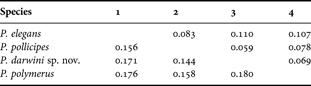
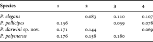
The mean intraspecific distance within the north-eastern Atlantic in P. pollicipes (0.006) species was lower than in the P. darwini sp. nov. from the Cape Verde sample (0.011). The net mean distance between these groups is one order of magnitude higher, at 0.144 (SE = 0.015). Similar values were obtained for the interspecific comparisons, thus distances between P. elegans and P. polymerus with respect to the North Atlantic P. pollicipes were 0.156 (SE = 0.016) and 0.158 (SE =0.015), respectively. These distance values are slightly higher when compared to P. darwini sp. nov. (Table 2).
Matrix of mean net genetic distances among pairs of species belonging to the genus Pollicipes, based on partial COX1 sequences (below diagonal) and ITS1, ITS2 and 5.8S rDNA sequences (above diagonal).
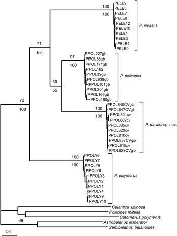
Four species-specific, divergent and well-supported clades (>97% bootstrapping values) were obtained from the COX1 sequences using all described methodologies of phylogenetic inference. Each of the recovered clades included all individuals belonging to their respective, presently recognized Pollicipes species. In addition, a divergent clade included all the Cape Verde specimens from P. darwini sp. nov., being equidistant with respect to the other Pollicipes species. Although, the P. pollicipes and P. darwini sp. nov. clades are clustered as sister groups, the low bootstrapping support values (<55%) suggest a polytomy involving both species plus P. elegans. Similarly, the basal position obtained for P. polymerus is supported by relatively low bootstrapping values (<71%). The monophyly of the genus Pollicipes obtains a 72% bootstrapping support under distance criterion, even though it is recovered in all parsimony trees (Figure 3).
Phylogenetic relationships among the species included in the Pollicipes genus (P. elegans, P. pollicipes and P. polymerus), considering the new species P. darwini (sp. nov.). The depicted tree is a consensus of the trees obtained from the analysis of mitochondrial COX1 using the distances, parsimony and maximum likelihood criteria. The number above and below the branches shows the bootstrapping values, after 2000 replicates, obtained for neighbour-joining distance and parsimony tree, respectively, with PAUP. The outgroup COX1 sequences were collected from Genbank.

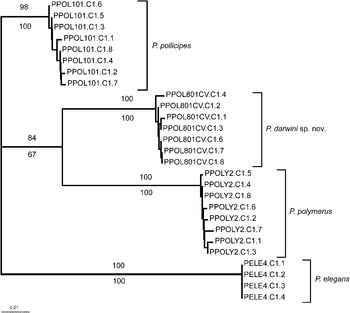
As expected from nuclear sequences, the divergences estimated from ITS and 5.8S rDNA sequences, 816 bp length including gaps, are lower than those obtained from mitochondrial data. A number of 620 characters were constant, 10 variable characters were parsimony-uninformative, whereas 114 were parsimony-informative. The intraspecific distance among detected alleles in Pollicipes species ranged from 0 to 0.003. The interspecific distance between the Cape Verde population and the north-eastern Atlantic Pollicipes was 0.059 (SE = 0.009), whereas that for P. polymerus and P. elegans were 0.069 (SE = 0.010) and 0.110 (SE = 0.012), respectively (Table 2). From the unrooted tree, based on nuclear sequences, the sister species of P. darwini sp. nov. is P. polymerus, although the bootstrapping support (<84%) of this cluster is the lower value observed in the tree (Figure 4).
Phylogenetic unrooted tree suggesting the relationships between the species included in the Pollicipes genus: P. elegans, P. pollicipes, P. polymerus and the new species P. darwini (sp. nov.). This is a consensus of the trees obtained from the analysis of nuclear ITS1, 5.8S rDNA and ITS2 sequences using the distances, parsimony and maximum likelihood criteria and PAUP software. Bootstrapping support from 2000 replicates was indicated above branches for neighbour-joining distance, and below branches for parsimony tree.

The specific status of P. darwini sp. nov. is thus supported by the presence of taxonomically diagnostic morphological characters, monophyletic and divergent clustering from molecular mitochondrial and nuclear data and by the congruence between these two approaches.
DISCUSSION
The morphological description of the Pollicipes darwini sp. nov. specimens from Cape Verde fully coincides with the taxonomic characters described by Darwin (Reference Darwin1851), Foster (Reference Foster1978), Barnes (Reference Barnes1996) and Southward (Reference Southward2008) for the genus Pollicipes. However, there are morphological characters that certainly differentiate P. darwini sp. nov. from the rest of the species belonging to the genus Pollicipes. Morphological comparison is focused on differentiation from the most similar taxa in which P. darwini sp. nov. were initially included, P. pollicipes, being considered as a sample from the southernmost limit of their distribution area (Quinteiro et al., Reference Quinteiro, Rodríguez-Castro and Rey-Méndez2007), and from the poorly characterized P. elegans.
The number and the distribution of capitulum plates are the same as that in the biogeographically closely related P. pollicipes; however, they show a differentiated shape (Figures 1A, B & 2A). Moreover, the close and overlapped plates of P. pollicipes plates are strongly calcified and have conspicuous growth marks. In contrast, the plates in P. darwini sp. nov. are well separated and show an extensive membranous area between them. Its fragile plates are furthermore orange, especially in eroded areas, while the plates of P. pollicipes are colourless. Morphological differences in the peduncle are even more noticeable: the peduncle plates in P. pollicipes are thin and stylized in the basal portion and wider in the distal portion, whereas in P. darwini sp. nov. they are wider than long and with smooth, blunt edges (Figures 1C & 2B). Finally, the dark, almost black, peduncle colour in P. pollicipes contrasts with the dark reddish colour observed in P. darwini sp. nov.
Internally, differences are less marked but are significant. The palpus of P. pollicipes is covered with bristles, whereas they are absent in P. darwini sp. nov. Moreover, the first tooth of P. pollicipes is located behind and arises much further back than in the mandible of P. darwini sp. nov. (Figures 1D, E, F & 2C, D).
Filamentary appendages show noticeable differences among evaluated species. In the basal portion of the first pair of cirri in P. darwini sp. nov. there are three well developed and lanceolate filaments; in P. pollicipes two well-developed and lanceolate filaments and a blunt and poorly developed one. Pollicipes elegans on the other hand has three lanceolate filaments plus one thin and very large one, which is as long as the palpus.
The capitulum of P. elegans is differentiated from that of P. darwini sp. nov. by the presence of a membranous and large bulging lip located at the distal portion of the tergum in P. elegans. In addition, the plates observed in P. elegans, are almost sub-hexagonal, longer and thinner than those of P. darwini sp. nov.
The observed monophyly of the genus Pollicipes in the COX1 tree, agrees with Darwin's (1851) concept of ‘natural genus’ with respect to these species and the current taxonomy (Barnes, Reference Barnes1996; Foster, Reference Foster1978; Newman, Reference Newman and Southward1987). The exclusion of Pollicipes mitella in the separate genus Capitulum (Foster, Reference Foster1978) is also supported by molecular data. Both, the monophyletic clustering and the divergence estimated from COX1 sequences confirm the profound genetic isolation of the Cape Verde population from the north-eastern Atlantic populations of P. pollicipes observed through the AT-rich sequences of the mitochondrial putative control region (Quinteiro et al., Reference Quinteiro, Rodríguez-Castro and Rey-Méndez2007). However, there are difficulties for inferring the phylogenetic relationships between the species of the genus Pollicipes. Thus, P. polymerus appears as the sister species of the rest of the genus in the basal portion of the phylogenetic tree, but with a relatively low support. Similarly, there is no consensus about the sister relationship between P. pollicipes and P. darwini sp. nov.
The specimens belonging to the P. darwini sp. nov. maintain a range of interspecific genetic distances similar to those observed among the previously described and recognized species: P. pollicipes, P. elegans and P. polymerus. The mean genetic distance among species of the Pollicipes (16.41 ± 0.13%) is similar to the mean distance among congeneric species genus of Decapoda (17.16 ± 0.18%) (Costa et al., Reference Costa, deWaard, Boutillier, Ratnasingham, Dooh and Hebert2007), among species of the acorn barnacle, Tetraclita, (15–19%) (Chan et al., Reference Chan, Tsang and Chu2007a, Reference Chan, Tsang and Chub) and among probable cryptic species of Chthalamus (7.5%–14%) (Tsang et al., Reference Tsang, Chan, Wu, Ng, Chatterjee, Williams and Chu2008). The level of mean intraspecific variation (0.87% ± 0.25%) is similar to values observed in Tetraclita species (<1.5%) (Chan et al., Reference Chan, Tsang and Chu2007a) and slightly higher than estimations for other crustacean taxa (0.46%), but the interspecific to intraspecific ratio of sequences divergence is high (20.01), within the range estimated for crustaceans (Costa et al., Reference Costa, deWaard, Boutillier, Ratnasingham, Dooh and Hebert2007). These divergence values suggest the high resolution of this sequence for species delineation within the genus Pollicipes.
Nuclear sequences show a similar divergence and clustering pattern that suggests an absence of processes that affect mitochondrial phylogenetic pattern such as introgression, incomplete mitochondrial lineage sorting and retention of ancestral haplotypes (Ballard & Whitlock, Reference Ballard and Whitlock2004). Thus, the divergence between P. darwini sp. nov. and north-eastern Atlantic P. pollicipes falls within the range of the estimated interspecific distances and is similar to that estimated for the Pacific P. polymerus. With respect to the branching pattern, an alternative and weakly supported hypothesis was observed, of a sister relationship between P. darwini sp. nov. and P. polymerus.
The alternative relationships recovered in the basal area of the topologies suggested a lack of resolution in the analysed sequences for testing Newman's hypothesis (1987), suggesting that P. elegans is the most primitive species of the genus, and the hypothesis of a sister relationship between P. pollicipes and the P. darwini sp. nov. Besides the potential effect of the absence of adequate outgroups in the nuclear analysis, the deep speciation patterns that take place within the genus Pollicipes (Newman, Reference Newman and Southward1987) probably remain obscured under the phylogenetic signal contained in the analysed data (Fischer & Steel, Reference Fischer and Steel2009). Moreover, the occurrence of simultaneous and temporally very close speciation events usually results in the observed star-like topologies.
Morphological distinctiveness and diagnostic characters, congruent divergence level and monophyletic clustering, at both nuclear and mitochondrial loci, support the validity of P. darwini sp. nov. Consequently, an intriguing taxonomic symmetry is observed between the Pacific and Atlantic oceans, where a single and different species is distributed in the north-eastern and central-eastern coast of each basin. Just like in other Pollicipes species, this new species is currently being exploited, since it is considered to be an appreciated seafood resource. A management plan for fisheries sustainability must therefore be considered. Such a plan should be aimed at preserving this evolutionarily significant unit of the Macaronesian and global marine biodiversity.
ACKNOWLEDGEMENTS
We are grateful to the following for the essential sampling work: BANCOMAC project, Programme Interreg IIIB Macaronesian space, Chris Neufeld, Rich Palmer and Cesar Martín Quevedo Vera (Instituto Tecnológico Pesquero de Perú). We are indebted to Javier Souto and Dr Eugenio Fernández Pulpeiro for their effort in the morphological description, including photo and drawing work. Dr Diana S. Jones and O. Reverter-Gil are acknowledged for helpful critical comments.