Introduction
There is only one true case of primary mastoid cyst reported in the English language literature.Reference Nomura, Takemoto and Komatsuzaki1 The aetiology of mastoid cyst can be primary (i.e. congenital or developmental) or secondary to chronic infection, inflammation or trauma.
Here, the authors report a case of primary mastoid cyst in a middle-aged, Caucasian woman who presented with sinusitis symptoms in the absence of any otological clinical features.
Case report
A 34-year-old, Caucasian woman was referred by her general practitioner to an otolaryngologist for management of chronic sinusitis. She reported an 18-month history of chronic frontal headaches with no history of any ear infection or trauma. Apart from rhinitis, the rest of the ENT examination was normal for both ears.
Audiology was also normal for both ears.
A computed tomography (CT) sinus scan, organised by the referring general practitioner, showed pansinusitis. However, upon re-examining the CT scan, the otolaryngologist identified a soft tissue opacity with abnormal calcification along the inferior tip of the left mastoid process; this abnormal finding had not been reported by the radiologist.
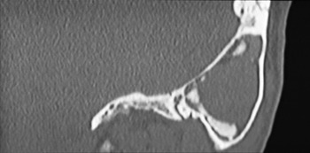
The otolaryngologist arranged further imaging, including a CT scan of the petrous temporal bone (Figure 1), magnetic resonance imaging of the temporal bone and single photon emission CT. Radiological imaging showed a soft tissue, proteinaceous mass within the mastoid tip, without any evidence of bony destruction or osteomyelitic changes.
Coronal computed tomography scan of the left temporal bone showing opacification of the mastoid cavity and calcification at the mastoid tip.

Complete blood examination and inflammatory marker testing were all within normal limits.
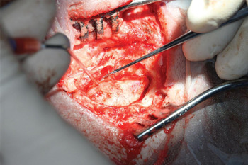
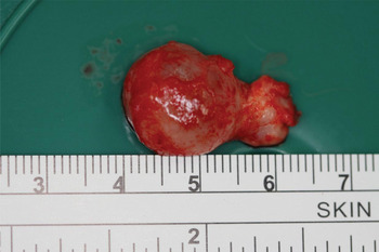
As the radiological imaging raised the possibility of neoplastic disease, the patient underwent mastoid exploration undertaken by the senior authors. Intra-operative findings revealed a lobulated, cystic lesion completely filling the entire mastoid process (Figure 2). The cyst did not extend into the epitympanum. The actual aditus ad antrum and mastoid antrum were not obstructed. There were abnormal bony ridges at the inferior mastoid cavity which correlated with the abnormal calcification seen on CT imaging. The cyst was removed in its entirety, and measured up to 2.3 cm along its maximal diameter (Figure 3).
Intra-operative photograph showing the mastoid cyst in situ.

Intra-operative photograph showing the completely excised mastoid cyst, measuring 2.3 cm along its maximal diameter.

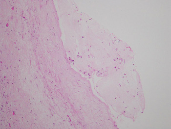
Histological examination of the cyst was reported to show a simple, benign cyst with a fibrous wall, which was low in cellularity and devoid of epithelium along its internal aspect. The cystic wall was lined with occasional lymphocytes and foam cells (Figure 4). Cut section revealed an intact cyst which contained clear, watery fluid and fibrinous exudate with no identifiable cells. Biopsy of the bony ridges showed normal cancellous bone with no evidence of inflammation or atypia.
Photomicrograph of the mastoid cyst wall showing a fibrous lining with foam cells. (H&E; ×10)

At six-month post-operative follow up, there were no signs of recurrence of mastoid disease either clinically or radiologically. The patient's previous sinus disease had also been completely cleared with medical therapy.
Discussion
Mastoid cysts are rare and usually secondary to infection or trauma.Reference Nomura, Takemoto and Komatsuzaki1, Reference Zimmerman and Proud2 Primary mastoid cyst is even rarer, with only one true case previously reported.
Zimmerman and Proud reported two cases of paediatric mastoid cyst in the presence of chronic serous otitis media.Reference Zimmerman and Proud2 They described these two cases as primary mastoid cysts. However, we feel that Zimmerman and Proud's case series should be classified as secondary mastoid cyst due to the presence of chronic otitis media. The presence of chronic inflammatory changes can result in poor ventilation and drainage of the mastoid, resulting in cyst formation similar to the mucocele formation found in obstructed paranasal sinuses.Reference Stankiewicz, Newell and Park3
Nomura et al. reported one case of congenital mastoid cyst.Reference Nomura, Takemoto and Komatsuzaki1 This patient presented only with painless, retroauricular swelling, with no history or clinical evidence of any infective or inflammatory component. Therefore, this was a case of true primary mastoid cyst.
• Mastoid cysts are rare
• They can be primary (developmental or congenital) or secondary to trauma or infection
• Patients may have no otological symptoms
• Management includes radiological imaging and mastoid exploration to establish diagnosis
• Complete cyst removal can have minimal morbidity and recurrence
The present patient represents an interesting case of a mastoid lesion which was evident on the CT sinus scan but missed by the radiologists. This case highlights the importance of otolaryngologists reviewing radiological films themselves instead of relying on the radiologist's report. In the absence of any previous trauma or infection, the case reported in this paper represents a true primary mastoid cyst which was most likely congenital or developmental in nature. The cyst probably expanded slowly over time with minimal pressure effect and therefore minimal ear symptoms. Primary mastoid cyst is a highly unusual new pathological entity.
In the present case, the observed soft tissue opacification together with calcification of the mastoid cavity raised the possibility of a neoplastic process. At that time, the differential diagnosis for the mass also included a mastoid cyst, histiocytosis, cholesteatoma or cholesterol granuloma. In such circumstances, it is therefore prudent for the clinician to exclude a sinister cause.
The management of mastoid lesions should include a complete otological history and physical examination, imaging, and mastoid exploration to establish the diagnosis. In order to confirm the diagnosis of a mastoid cyst, the authors recommend a postauricular approach, cortical mastoidectomy and mastoid exploration. Complete extirpation of the cyst can then be safely performed with minimal morbidity and risk of recurrence.