Introduction
Cystic neck masses can be challenging to manage, due in part to their wide differential diagnosis. All clinicians should be mindful of the pitfall of head and neck squamous cell carcinoma (SCC) presenting as a cystic neck mass, particularly with an occult primary, and especially in the era of human papillomavirus (HPV) -driven oropharyngeal cancer. However, a cystic neck mass may also be the presenting feature of nodal metastases from papillary thyroid cancer (PTC). These metastases can have a similar appearance on ultrasound to simple cysts or lymphatic malformations, and the primary thyroid tumour may be sonographically occult. Often there is insufficient solid component in the cystic neck node for meaningful fine-needle aspiration (FNA) or core biopsy. If biopsy is performed, the result may be non-diagnostic, inconclusive or falsely negative. If no squamous content is detected, then there is less concern for SCC, and there is potential that the cystic node is misclassified as benign.
We propose cyst fluid thyroglobulin (Tg) testing as a solution to this problem. This is a different test to the established technique of FNA-Tg washout.Reference Pacini, Fugazzola, Lippi, Ceccarelli, Centoni and Miccoli1 To perform this assay, the contents of FNA aspirate are washed with a known volume of normal saline, and the Tg concentration of this fluid is measured. FNA-Tg washout is useful in diagnosis of papillary thyroid cancer but requires successful solid tissue sampling.Reference Wang, Duan, Li, Yue, Liu and Lai2 The aim of this case series is to demonstrate the value of Tg measurement of ultrasound-guided, directly aspirated cyst fluid in diagnosis or exclusion of papillary thyroid cancer.
Methods
Prospective case series (n = 10) of patients presenting with a lateral cystic neck mass with or without a thyroid abnormality, where thyroglobulin measurement of aspirated cyst fluid was part of the diagnostic workup. Cyst fluid Tg measurement was made by biochemical analysis of ultrasound-guided, directly aspirated and unaltered cyst fluid.
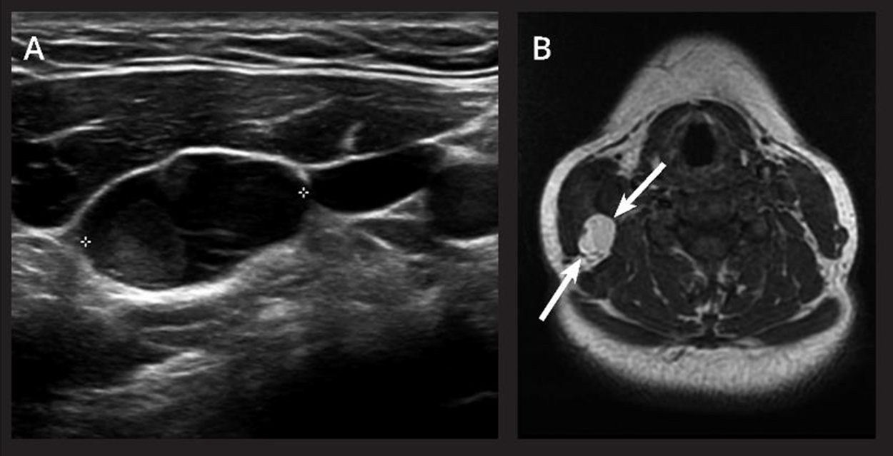
Case 1. (A) Greyscale image showing a cystic lesion lateral neck (calipers) with mural nodule. The thyroid (not shown) was normal. (B) Axial T1W magnetic resonance imaging (MRI) image showing T1 hyperintense internal content in the lesion in keeping with proteinaceous content (colloid) typical of a metastatic papillary thyroid carcinoma cystic nodal metastasis (white arrows).

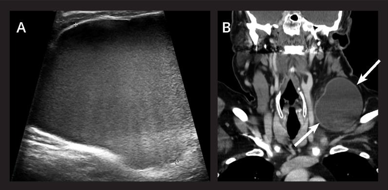
Case 2. (A) Greyscale ultrasound (US) image showing partial coverage of a large, thin-walled cystic lesion with homogeneous internal echoes giving a pseudosolid appearance but no true solid components. Doppler imaging (not shown) confirmed no internal vascularity. (B) Corresponding coronal image from contrast enhanced CT shows a relatively thin-walled cystic lesion spanning level III–IV (white arrows) with dependent debris.

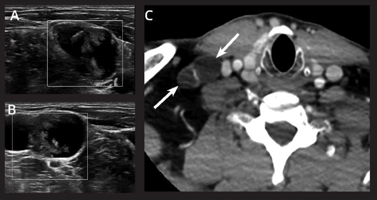
Case 3. (A,B) Greyscale with colour doppler showing a cystic lesion lateral neck with vascularised papillary projections. Normal thyroid appearances (not shown). (C) Axial contrast enhanced computed tomography (CT) scan showing the corresponding cystic lesion right neck level IV (white arrows).

Results
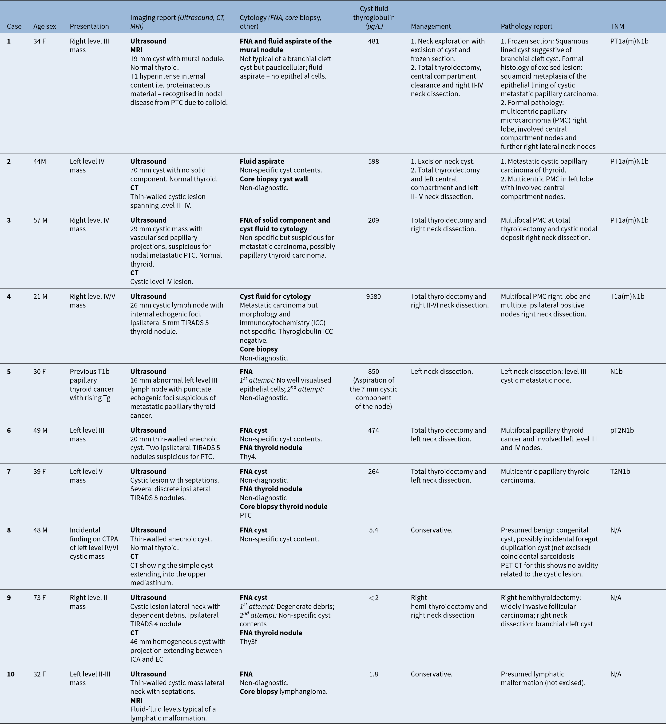
N1b PTC diagnosis was predicted by markedly elevated Tg in cyst fluid aspirate in seven cases, all greater than 200 µg/L, confirmed at total thyroidectomy and neck dissection (cases 1–7). PTC nodal metastasis was refuted by a low Tg result in three benign, congenital cystic lesions, all less than 6 µg/L (cases 8, 9, 10). For full summary of results, see Table 1.
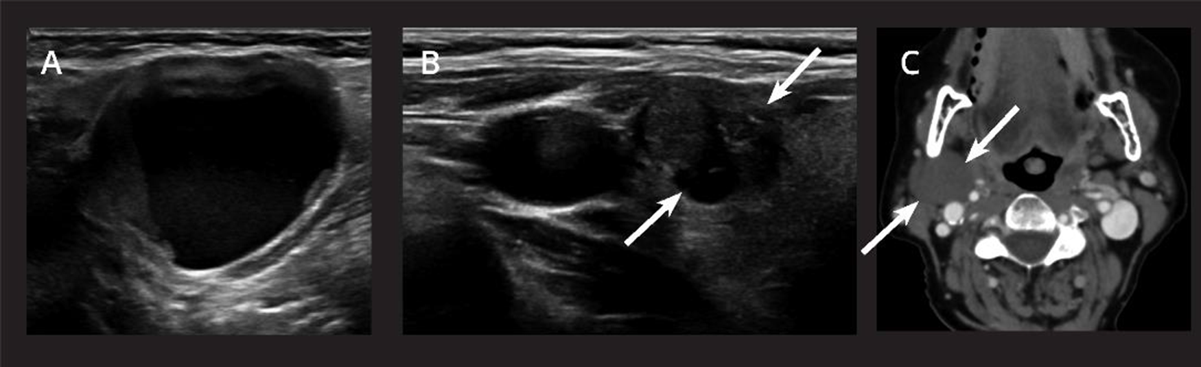
Case 9. (A) Greyscale image showing a cystic lesion lateral neck with dependent debris. (B) Greyscale image showing a TIRADS 4 nodule ipsilateral thyroid lobe (white arrows). (C) Axial slice from contrast enhanced computed tomography (CT) scan showing a simple looking cyst right level II (white arrows) with a small projection extending between internal carotid artery (ICA) and external carotid artery (ECA) typical of a branchial cleft cyst.


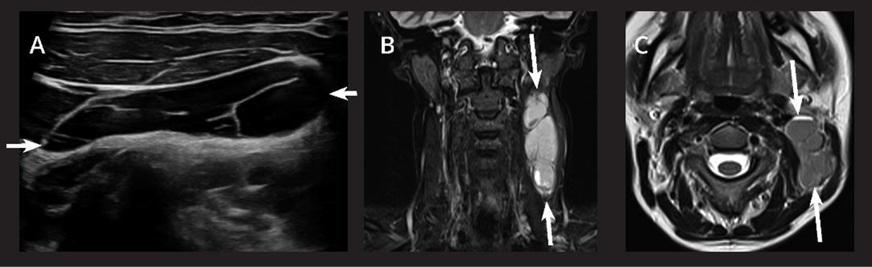
Case 10. (A) Greyscale image showing a thin-walled cystic mass lateral neck (white arrows) with septations. (B) Coronal short tau inversion recovery (STIR) image depicting a loculated hyperintense lesion (white arrows). (C) Axial T2W image demonstrating fluid-fluid levels in the lesion typical of a lymphatic malformation.

Discussion
This case series is the first prospectively collected data to suggest directly aspirated cervical cyst fluid Tg testing as a positive or negative diagnostic indicator of PTC. Elevated Tg in cyst fluid accurately predicted PTC diagnosis in seven cases. Very low Tg result was found in three benign congenital cystic lesions.
Directly aspirated cyst fluid thyroglobulin testing is distinct from the established technique of FNA-Tg washout, which requires successful solid tissue biopsy. The diagnostic value of FNA-Tg washout for metastatic cervical lymph nodes in PTC is higher than that of ultrasound and FNA cytology (FNAC) alone. When there is an adequate biopsy target, in combination, FNAC and FNA-Tg washout can provide nearly 100 per cent diagnostic sensitivity.Reference Wang, Duan, Zhou, Liu, Lai and Ye3 Wang et al. (2022) propose a diagnostic cutoff value of FNA-Tg washout of 3.96 µg/L (sensitivity 92.48 per cent, specificity 75.00 per cent).Reference Wang, Duan, Li, Yue, Liu and Lai2 Lack of successful solid tissue biopsy is the pitfall of FNA-Tg washout, and there is no current diagnostic alternative in the absence of this.
Cyst fluid thyroglobulin has been previously reported to be a positive diagnostic predictor of PTC in non-cervical cysts. Ntotsikas et al. (2023) report association between high cyst fluid Tg and diagnosis of metastatic PTC in a patient with a solitary cystic lesion in the brain.Reference Ntotsikas, Lazarioti, Daraki, Drakos, Tsakalomatis and Syntzanaki4 Aspiration of the brain cyst demonstrated Tg level of greater than 300 µg/L and pathology confirmed PTC after resection.
Retrospective evidence exists supporting the use of directly aspirated cyst fluid Tg as a marker for PTC. Li et al. (2022) analysed single centre clinical biochemistry data which included Tg levels in centrifuged cyst aspirate.Reference Li, Liu, Zhang, Long, Jiang and Su5 They reported that Tg levels in centrifuged cyst aspirate of cases of PTC-related cystic metastases were significantly higher (all >500 µg/L) than in 32 other cervical cystic lesions (range, 2.9–40.1 µg/L; p < 0.01). From this it was suggested that cyst fluid Tg may be a useful adjust to diagnosis with a cutoff value of 100 µg/L.Reference Li, Liu, Zhang, Long, Jiang and Su5 It is unclear, however, exactly how many confirmed PTC cases were included, how these were diagnosed (pathological vs biochemical) and the diagnoses of the non-PTC cystic lesions.
Limitations exist. Tg fluid analysis are assay dependent, so cutoff levels cannot necessarily be compared across different methods in use. Further work is required to establish a validated diagnostic threshold. Complicating assessment of this threshold is the influence of plasma Tg on cyst fluid Tg.
These data support cyst fluid thyroglobulin testing as a valuable adjunct for evaluation of cystic neck masses and pre-operative diagnosis of PTC cystic nodal metastases, particularly in the absence of an adequate biopsy target.
• Diagnosing N1b papillary thyroid cancer can be difficult
• Lateral cystic neck masses may be misdiagnosed as benign, especially if sonographically non-specific or insufficient solid component for biopsy
• Fine-needle aspiration thyroglobulin washout is an established diagnostic technique but requires successful solid tissue biopsy
• Directly aspirated cyst fluid thyroglobulin testing is a valuable adjunct in pre-operative papillary thyroid cancer diagnosis, particularly in the absence of an adequate biopsy target
Acknowledgements
None.
Funding
This research did not receive any specific grant from funding agencies in the public, commercial or not-for-profit sectors.
Competing interests
The authors declare that they have no known competing financial interests or personal relationships that could have appeared to influence the work reported in this paper.