




No CrossRef data available.
Article contents
The interaction between the gelatin-binding domain of fibronectin and the attachment of Pasteuria penetrans endospores to nematode cuticle
Published online by Cambridge University Press: 28 November 2001
Abstract
Pasteuria penetrans is a Gram-positive endospore-producing bacterium that is a parasite of root-knot nematodes. Attachment of endospores to the cuticle of the nematode is the first stage in the infection process. Western blot analysis with monoclonal and polyclonal antibodies that recognize the 30 kDa heparin-binding domain (HBD) and the 45 kDa gelatin-binding domain (GBD) fragments of human fibronectin (Fn) revealed a series of polypeptides of approximately 40, 45 and 55 kDa present in crude cuticle extracts of Meloidogyne javanica 2nd-stage juveniles. The results suggest that the structure of the nematode fibronectin is different to the fibronectins so far characterized. Pre-treatment of endospores of Pasteuria with either the HBD or the GBD was found to inhibit binding to the nematode cuticle. The larger GBD fragment was the most effective at blocking adhesion. Pre-treatment of the GBD fragment with gelatin prevented the GBD fragment from inhibiting endospore attachment to the nematode cuticle.
Information
- Type
- Research Article
- Information
- Copyright
- © 2002 Cambridge University Press
INTRODUCTION
Nematodes extracted from soil have surprisingly few organisms adhering to their cuticles. Pasteuria penetrans is a Gram-positive obligate bacterial parasite of nematodes that produces endospores which, in the initial phase of infecting the nematode, must adhere to its cuticle (Sayre & Starr, 1988). This bacterium has the potential to be developed into a biological control agent of plant-parasitic nematodes (Stirling, 1991). In standard attachment assays, the endospores of an isolate of the bacterium have been shown to adhere to one population of the nematode better than another population (Stirling, 1991; Davies, Kerry & Flynn, 1988) and this is not linked to the phylogeny of the nematode (Davies et al. 2000). Studies with a panel of monoclonal antibodies raised to the surface of a population of endospores have shown a high degree of heterogeneity to both the surface of the endospore and also the nematode cuticle (Davies, Redden & Pearson, 1994). It was shown that 1 monoclonal antibody, from the panel, reduced the binding of endospores to the nematode cuticle and that fibronectin competes with this monoclonal antibody for the same binding site on the surface of the endospore (Davies & Redden, 1997). Hydrophobic interactions have been shown to be responsible for the binding of fibronectin to the surface of endospores and fibronectin-treated endospores showed reduced binding to the cuticle of 2nd-stage juveniles (Davies, Afolabi & O'Shea, 1996).
Fibronectin is an extracellular glycoprotein (Mr∼440 kDa) which is present in a soluble form in plasma and an insoluble form in extracellular matrices. It is involved in many important physiological processes including the organization and maintenance of tissue architecture in mammals (Hynes 1990; Akiyama & Yamada, 1995). Fibronectin (Fn) is composed almost entirely of 3 types of protein domains (or modules) Fn1, Fn2 and Fn3 which combine to form larger domains containing binding sites for a variety of molecules including denatured collagen (Gelatin; GBD) and heparin (HBD) (Potts & Campbell, 1994, 1996). Fibronectin has an important role in the pathogenicity of many bacteria (Hasty et al. 1989; Courtney et al. 1990) by binding to microbial surface components which specifically recognize adhesive matrix molecules (Patti et al. 1994). The Caenorhabditis elegans databases do not show the presence of a heterodimeric fibronectin glycoprotein similar to that reported in mammals with Mr∼440 kDa. There are, however, a number of predicted proteins which contain Fn3 type domains. Western blot analysis of extracts of cuticle from C. elegans and the plant-parasitic nematode Meloidogyne incognita, probed with a polyclonal antibody raised to human Fn revealed a Fn like heterodimer with Mr∼440 kDa and a smaller polypeptide with Mr∼45 kDa (Davies et al. 1996). Two monoclonal antibodies, raised to 2nd-stage juveniles of root-knot nematodes (Meloidogyne spp.) were shown to recognize the 440 kDa heterodimer of fibronectin and also a series of smaller polypeptides in Western blot analysis of 2nd-stage juvenile cuticle extracts of M. incognita (Davies et al. 1996). This paper reports experiments that test the hypothesis that one of the domains of fibronectin, either the GBD or the HBD, is present in the extracellular matrix of the cuticle of 2nd-stage juveniles of Meloidogyne and is responsible for the adhesion of endospores of Pasteuria.
MATERIALS AND METHODS
Nematodes and Pasteuria endospore populations
Second-stage juveniles were collected from the egg masses obtained from a population of M. javanica which were tested for purity by native gel electrophoresis and stained for non-specific esterases (Davies & Beadle, 1995). The nematodes were routinely cultured in the glass-house at 25 °C on tomato plants (cv. Pixie) in peat/sand (1:1, v/v) compost at Rothamsted. Egg masses were placed in a hatching tray and allowed to hatch in tap water at room temperature (Hooper, 1986). Endospores of P. penetrans PP Theis (Accession number PP-ORS-21414 isolated from Theis in Senegal) were collected by homogenizing infected root-knot females dissected from root galls and counted on a haemocytometer slide. A stock suspension of 7×10⁶ spores/ml was stored at 4 °C.
Cuticle characterization of Fn by Western blot analysis
Freshly hatched 2nd-stage juveniles of M. javanica (50000) were pelleted by centrifugation and 500 μl of cuticle extraction buffer (0.125 M Tris-HCl, pH 6.8, 0.1% SDS, 5% 2-mercaptoethanol (2ME), 10% glycerol) were added and proteins extracted following standard procedures (Hemmer et al. 1991; Davies et al. 1996). The protein concentration in the cuticle extract was quantified using a Bio-RadTM protein assay (Bio-Rad Laboratories Ltd, Hemel Hempstead, UK). Subsequently the sample was centrifuged (1000 g for 5 min) and the supernatant containing the protein was collected and stored at −45 °C. Polyacrylamide gel electrophoresis in the presence of sodium dodecyl-sulphate (SDS–PAGE) was performed using 10% (w/v) separating gel (pH 8.8) and 4% (w/v) stacking gel (pH 6.8). Samples of 100 μl of cuticle extract, Fn (Sigma Cat. No. F-1141), the GBD fragment of Fn (45 kDa; Sigma. Cat. No. F-0162) or the HBD fragment of Fn (30 kDa; Sigma Cat. No. F-9911) from stocks of 1 mg/ml, were diluted with 1:1 (v/v) sample buffer (50 mM Tris–HCl, pH 6.8, 2% (w/v) SDS, 2% (v/v) 2-ME, 10% (v/v) glycerol and 0.002% (w/v) bromophenol blue and heated in Eppendorf tubes for 5 min at 100 °C. Samples (approximately 5 μg) were loaded on to the stacking gel. Pre-stained SDS–PAGE molecular weight marker mix (Bio-RadTM; Cat. No. 161-0372) was run with each gel. Following electrophoresis the gels were either transferred to the fixative (40% methanol and 10% acetic acid) for silver staining using a silver staining kit (Bio-RadTM), or electroblotted onto nitrocellulose membranes in continuous buffer (Davies et al. 1996) using a Semi-phorTM blotting system (Hoeffer Scientific Instruments) for 1 h at 100 mA. After electrotransfer the membranes were blocked for 1 h at room temperature by incubating the membrane in PBS containing 0.05% (v/v) Tween (PBST) and 5% (w/v) milk powder (PBSTM), and then probed with either a rabbit anti-fibronectin polyclonal antibody (Sigma; Cat No F-3648) or tissue culture supernatant containing monoclonal antibodies Mab6.2 or Mab32.3 (Davies et al. 1996). Following overnight incubation the membranes were washed (x2) in PBST before probing with either goat anti-rabbit (Sigma; Cat No A-6387) or rabbit anti-mouse (Sigma; Cat No A-2418) IgG whole molecule conjugated to alkaline phosphatase (1:500 in PBSTM for 2 h) as appropriate. Following incubation the membranes were washed (×2) in PBST for 30 min and visualization of the proteins recognized by the antibodies was done by applying Sigma FastTM BCIP/NBT alkaline phosphatase substrate (Sigma B-5655). The reaction was stopped by 3 washes in distilled water.
Inhibition of endospore attachment by Fn, the GBD fragment and the HBD fragments of Fn
Stock solutions of 20 μg/ml of Fn, GBD and HBD fragments were prepared in either 100 mM KCl or 100 mM KSCN in siliconized Eppendorf tubes (Hewlett & Dickson 1993). Thiocyanate (SCN) at low concentrations (100 mM) is an inhibitor of hydrophobic interactions (Hatefi & Hanstein, 1969). Each treatment was further serially diluted to achieve a dilution series of 20.0, 2.0, 1.0, 0.4, 0.2, 0.0 μg/ml in the respective buffers. The stock endospore (Pp Thies) suspension of 106 endospores/ml was made in either 100 mM KCl or 100 mM KSCN and 250 μl of each suspension were added to each of the dilution series. Endospores were incubated at room temperature for 1 h, centrifuged (1000 g for 2 min) and the supernatants removed. Two suspensions of 2000/ml, 1- to 2-day-old, 2nd-stage juveniles of M. javanica were prepared in 100 mM KCl and 100 mM KSCN. The nematodes were then added to the respective dilution series containing the Fn, GBD or HBD pre-treated endospores. The contents of the Eppendorf tubes were then centrifuged for 2 min at 9500 g (Hewlett & Dickson, 1993), and stored at 4 °C until examined. Spore attachment was recorded for 20 individual 2nd-stage juveniles using a high power microscope (×400).
Competitive inhibition of the GBD fragment by gelatin
Experiment 1 was performed to examine whether gelatin affected endospore binding. Gelatin was titrated (0, 0.015, 0.03, 0.06, 0.12, 0.25, 0.5, 1.0, 2.0%) against endospores in 100 mM KCl, following the technique described. Second-stage juveniles of M. javanica were then exposed to the pre-treated spore suspension. Inhibition studies were repeated using 2% gelatin, firstly pre-treating the endospores with gelatin before adding fresh nematodes, and secondly pre-treating the 2nd-stage juveniles with gelatin before adding endospores.
Experiment 2 was undertaken to test the hypothesis that the gelatin-binding receptor of the GBD fragment was responsible for the binding of the GBD fragment to endospores. A dilution series of the GBD fragment (20, 40, 80, 160, 320 and 640 μl/ml was made in 250 μl of 100 mM KCl and the same volume of 2% gelatin was added to it. The stock spore suspension (250 μl of 106 endospores/ml in 100 mM KCl) was further added to the above treatments and incubated for 1 h at room temperature. The pre-treated spores were centrifuged (9500 g for 2 min) and 500 μl of the supernatant was discarded from each Eppendorf tube. Second-stage juveniles (ca 400 in 250 μl of 100 mM KCl) of M. javanica were added to the treatments. Attachment tests were performed as described above.
RESULTS
Cuticle characterization of Fn by Western blot analysis
Western blots of human fibronectin and the HBD and GBD fragments onto nitrocellulose were probed with the polyclonal antibody raised to human fibronectin. The polyclonal antibody not only recognized the heterodimer of fibronectin with Mr∼440 kDa, but also the GBD fragment with Mr∼45 kDa and the HBD fragment with Mr∼30 kDa (Fig. 1). There was also some contamination from the undigested 70 kDa heparin and gelatin-binding fragment from which the HBD and the GBD fragments were produced. Both monoclonal antibodies 6.2 and 32.2 clearly recognized the HBD with Mr∼30 kDa (Fig. 1, lane 4; only Mab 32.2 shown) and the GBD with Mr∼45 kDa (Fig. 1, lane 5) less clearly. Although both monoclonal antibodies also recognized the fibronectin heterodimer each with Mr∼220 kDa, this was extremely faint and very difficult to visualize (Fig. 1, lane 6). Western blots of cuticle extract showed the polyclonal antibody raised to human Fn recognized 3 polypeptides of Mr∼55, 45 and 40 kDa (Fig. 2). The monoclonal antibodies clearly recognized the polypeptide with Mr∼55 kDa in the cuticle extract (lanes 1–3) and also the polypeptides of Mr∼40 and 45 kDa but much less clearly (Fig. 2).

Fig. 1. Western blot of gelatin-binding fragment of fibronectin (GBD; lanes 1 and 4) and heparin-binding fragment of fibronectin (HBD; lanes 2 and 5), fibronectin (Fn; lanes 3 and 6) probed with polyclonal antibody raised to human fibronectin (lanes 1–3), monoclonal antibodies to Meloidogyne Fn Mab 32.2 (lanes 4–6).

Fig. 2. Cuticle extracts from 2nd-stage juveniles of Meloidogyne electro-blotted onto nitrocellulose membrane and probed with a polyclonal antibody (lanes 4 and 5) which recognize human fibronectin and monoclonal antibodies 6.2 (lane 1) and 32.2 (lanes 2 and 3).
Inhibition of endospore attachment by Fn, the GBD and HBD fragments of Fn
Twice the number of endospores adhered to 2nd-stage juveniles in 100 mM KCl (13.7±4.2) than those in 100 mM KSCN (6.8±4.6). Endospores pre-treated with either Fn or HBD fragment were inhibited from adhering to the nematode cuticle. However, the GBD fragment was most effective at reducing endospore attachment (Fig. 3).

Fig. 3. Inhibition of Pasteuria endospore attachment to the cuticle of Meloidogyne javanica by the gelatin-binding domain (black), the heparin-binding domain (grey) and whole molecule of fibronectin at different concentrations (ANOVA P<0.001; S.E.D. = 1.17).
Competitive inhibition of GBD fragment by gelatin
The pre-treatment of endospores with gelatin had a considerable effect on endospore attachment to 2nd-stage juveniles. Gelatin (1%) reduced endospore attachment by over 90%, whereas pre-treatment of 2nd-stage juveniles reduced attachment by less than 20% (Fig. 4). The titration of the GBD fragment of Fn against endospores in the presence of 1% gelatin in 100 mM KCl showed a rapid increase in endospore attachment to a maximum attachment level of nearly 14 endospores per 2nd-stage juvenile at 40 μg/ml of the GBD fragment and thereafter a rapid decrease in endospore attachment. The level of endospore attachment in the presence of 40 μg/ml of the GBD fragment in the presence of 1% gelatin was within 10% of endospore attachment in the absence of gelatin.

Fig. 4. The number of endospores of Pasteuria penetrans 1% adhering to 2nd-stage juveniles of Meloidogyne javanica in the presence of gelatin when titrated against the gelatin-binding domain of fibronectin (GBD) together with the control treatment which contained neither gelatin nor GBD (ANOVA P<0.01; S.E.D. = 1.16).
DISCUSSION
The biochemical mechanism by which endospores of Pasteuria penetrans adhere to the nematode cuticle is poorly understood. Fibronectin is a molecule involved in the adhesion of bacteria to the extracellular matrix, which is important in the process of bacterial infection (Hasty et al. 1989; Courtney et al. 1990; Patti et al. 1994; Deitsch, Moxon & Wellems, 1997). Fibronectin adheres to endospores of P. penetrans by hydrophobic interactions and it has been hypothesized that it is involved in their attachment to the cuticle of root-knot nematodes (Davies et al. 1996). Immunological studies have shown that antibodies to fibronectin recognize a series of antigens present in the cuticle extract of C. elegans and M. incognita with Mr∼45 and 58 kDa. This is of similar molecular size to the gelatin and heparin-binding fragments of mammalian fibronectin. The object of this study was, therefore, to determine whether one of the domains of fibronectin, either the GBD or the HSD is present in the extracellular matrix of the cuticle of 2nd-stage juveniles of root-knot nematodes and, therefore, a receptor for endospores present on the nematode cuticle. The results suggest that there are a series of polypeptides of Mr between 30 and 55 kDa in the extracellular matrix of the nematode cuticle which were recognized by a polyclonal antibody raised to human fibronectin. One of these polypeptides with Mr∼45 kDa, recognized by the polyclonal antibody raised to human fibronectin, was also recognized by both of the monoclonal antibodies which had been raised to root-knot nematodes and in the same Western blots these monoclonal antibodies recognized the human HBD fragment of Mr∼30 kDa but not the GBD fragment of Mr∼45 kDa. A schematic representation (Fig. 5) of mammalian fibronectin (Petersen et al. 1983; Skorstengaard et al. 1994; Yu et al. 1997) showed that the monoclonal antibodies raised to root-knot nematodes could be reacting to the 29 kDa heparin I fragment, made up of 5 Fn-1 repeated units. The antibodies did not recognize the 31 kDa heparin II fragment, made up of 3 Fn-III repeated units as the HBD fragment had been produced from the 70 kDa heparin and gelatin-binding fragment of the N-terminal sequence. The 45 kDa gelatin-binding domain fragment of mammalian fibronectin, consisting of 2 Fn-II repeated units with a single Fn-I unit on the N-terminal side and 3 Fn-I repeated units on the C-terminal side, was not recognized by either monoclonal antibody. It is interesting that the anti-fibronectin antibodies used in this study appeared to recognize, albeit poorly, a fibronectin heterodimer-like molecule with Mr∼440 kDa, which appears also to be present in C. elegans (Davies et al. 1996) together with a series of other peptides. This work, therefore, suggests that the overall structure of invertebrate fibronectin present in root-knot nematodes does not follow the same pattern of repeated subunits as seen in the mammalian fibronectin molecule.

Fig. 5. Schematic representation of mammalian fibronectin. Location of the gelatin and heparin-binding fragments which were used in the attachment assays, are in bold. The fragment to which the monoclonal antibodies Mabs 6.2 and 32.3, raised to root-knot nematodes, is arrowed. Regions of homology are indicated by rectangles, squares and circles; Fn-III domain labelled RGDs represent repeated sequences of arginine, glycine and aspartic acid (adapted from Petersen et al. 1983; Skorstengaard et al. 1994; Yu et al. 1997).
The inhibition assays showed that the GBD fragment was far more effective in inhibiting endospore attachment than the HBD fragment. It can be seen, however, from the Western blot that the HBD fragment was contaminated with some undigested 70 kDa substrate, together with some of the GBD fragment. It is, therefore, likely that a highly purified form of the HBD fragment would be even less capable of inhibiting endospore attachment than the results of our test imply. The GBD fragment could be inhibited from reducing spore attachment by gelatin suggesting that the binding site on the GBD fragment was either the same site to which the endospore adhesins were binding or was so close as to hinder endospore recognition sterically. Gelatin itself inhibited spore attachment, which suggests that gelatin only has one type of binding site involved in endospore binding.
Several antigens in the cuticle extracts of root-knot nematodes which recognized antibodies to fibronectin (Davies et al. 1996) had similar molecular weights to those that, in earlier studies, had been shown to be sensitive to digestion by collagenase (Reddigari et al. 1986). The role of collagen as a receptor for Pasteuria adhesion is ambiguous; Persidis et al. (1992) suggested that collagen may be responsible, but the incubation of 2nd-stage juveniles in collagenases did not inhibit endospore attachment (Davies & Danks, 1993). As gelatin (denatured collagen) has now been shown to bind to endospores and to prevent them from binding to nematode cuticle it is unclear whether fibronectin (or one of its domains) or collagen is responsible for the attachment of endospores. Gelatin has been shown to bind to fibronectin through hydrophobic interactions (Isaacs, Brew & Ingham, 1989) and similar hydrophobic interactions have been shown to be responsible for the binding of fibronectin to endospores (Davies et al. 1996). It is certainly intriguing that these two molecules, both present in some form in the nematode cuticle, interact in such a way as to mutually cancel out each other's ability to inhibit endospores from binding.
We would like to thank Thierry Mateille (IRD) and Saliou N'Diaye (ENSA, Thies) whose collaboration led to the isolation of PP-ORS-21414. S.M. was in receipt of a Rothamsted International Fellowship. IACR-Rothamsted receives grant-aided support from the Biotechnology and Biological Sciences Research Council of the UK which in turn is supported by the British Tax Payer.
References
AKIYAMA, S. K. & YAMADA, K. M. (1995). Fibronectin and fibronectin fragments. In Extracellular Matrix: a Practical Approach (ed. HARALSON, M. A. & HASSELL, J. R.), pp. 175–185. IRL Press at Oxford University Press, Oxford.
COURTNEY, H. S., HASTY, D. L. & OFEK, I. (1990). Hydrophobicity of group A streptococci and its relationship to adhesion of streptococci to host cells. In Microbial Cell Hydrophobicity (ed. DOYLE, R. I. & ROSENBERG, M.), pp. 361–386. American Society for Microbiology, Washington.
DAVIES, K. G., AFOLABI, P. & O'SHEA, P. S. (1996). Adhesion of Pasteuria penetrans to the cuticle of root-knot nematodes (Meloidogyne spp.) inhibited by fibronectin: a study of electrostatic and hydrophobic interactions. Parasitology 112, 553–559.Google Scholar
DAVIES, K. G. & BEADLE, J. (1995). Use of a cellulose acetate electrophoresis system for the simultaneous characterisation of individual root-knot (Meloidogyne spp.) females using three isozymes. Fundamental and Applied Nematology 18, 549–551.Google Scholar
DAVIES, K. G. & DANKS, C. (1993). Carbohydrate/protein interactions between the cuticle of infective juveniles of Meloidogyne incognita and spores of the obligate hyperparasite Pasteuria penetrans. Nematologica 39, 53–64.CrossRefGoogle Scholar
DAVIES, K. G., FARGETTE, M., BALLA, G., DAUDI, A., DUPONNOIS, R., GOWEN, S. R., MATEILLE, T., PHILLIPS, M. S., SAWADOGO, A., TRIVINO, C., VOUYOUKALOU, E. & TRUDGILL, D. L. (2000). Cuticle heterogeneity as exhibited by Pasteuria spore attachment is not linked to the phylogeny of parthenogenetic root-knot nematodes (Meloidogyne spp.) Parasitology 122, 111–120.Google Scholar
DAVIES, K. G., KERRY, B. R. & FLYNN, C. A. (1988). Observations on the pathogenicity of Pasteuria penetrans, a parasite of root-knot nematodes. Annals of Applied Biology 112, 1491–1501.CrossRefGoogle Scholar
DAVIES, K. G. & REDDEN, M. (1997). Diversity and partial characterisation of putative virulence determinants in Pasteuria penetrans, the hyperparasite of root-knot nematodes. Journal of Applied Microbiology 83, 227–235.CrossRefGoogle Scholar
DAVIES, K. G., REDDEN, M. & PEARSON, T. K. (1994). Endospore heterogeneity in Pasteuria penetrans related to attachment to plant-parasitic nematodes. Letters in Applied Microbiology 19, 370–373.CrossRefGoogle Scholar
DEITSCH, K. W., MOXON, E. R. & WELLEMS, T. E. (1997). Shared themes of antigenic variation and virulence in bacterial, protozoal, and fungal infections Microbiology and Molecular Biology Reviews 61, 281–282.Google Scholar
HASTY, D. L., BEACHEY, E. H., COURTNEY, H. S. & SIMPSON, W. A. (1989). Interactions between fibronectin and bacteria. In Fibronectin in Health and Disease (ed. CARSONS, S. E.), pp. 89–112. CRC Press. Boca Raton, Florida.
HATEFI, Y. & HANSTEIN, W. G. (1969). Solubilization of particulate proteins and non-electrolytes by chaotropic agents. Proceedings of the National Academy of Sciences, USA 62, 1129–1136.CrossRefGoogle Scholar
HEMMER, R. M., DONKIN, S. G., CHIN, K. J., GRENACHE, D. G., BHATT, H. & POLITZ, S. M. (1991). Altered expression of an L1-specific, O-linked cuticle surface glycoprotein in mutants of the nematode Caenorhabditis elegans. Journal of Cell Biology 115, 1237–1247.CrossRefGoogle Scholar
HEWLETT, T. E & DICKSON, D. W. (1993). A centrifuge method for attaching endospores of Pasteuria spp. to nematodes. Journal of Nematology 25(Suppl.), 785–788.Google Scholar
HOOPER, D. J. (1986). Extraction of nematodes from plant material. In Laboratory Methods for Working with Plant and Soil Nematodes (ed. SOUTHEY, J. F.), pp. 51–58. Ministry of Agriculture Fisheries and Food, London, H.M.S.O.
ISAACS, B. S., BREW, S. A. & INGHAM, K. C. (1989). Reversible unfolding of the gelatin-binding domain of fibronectin: structural stability in relation to function. Biochemistry 28, 842–850.CrossRefGoogle Scholar
PATTI, J. M., ALLEN, B. L., MCGAVIN, M. J. & HÖÖK, M. (1994). MSCRAMM-mediated adherence of microorganisms to host tissues. Annual Review of Microbiology 48, 585–617.CrossRefGoogle Scholar
PERSIDIS, A., LAY, J. G., MANOUSIS, T., BISHOP, A. H. & ELLAR, D. J. (1992). Characterisation of potential adhesins of the bacterium Pasteuria penetrans, and of putative receptors on the cuticle of Meloidogyne incognita, a nematode host. Journal of Cell Science 100, 613–622.Google Scholar
PETERSEN, T. E., THOGERSEN, H. C., SKORSTENGAARD, K., VIBEPEDERSEN, K., SAHL, P., SOTTRUPJENSEN, L. & MAGNUSSON, S. (1983). Partial primary structure of bovine plasma fibronectin – 3 types of internal homology. Proceedings of the National Academy of Sciences, USA 80, 137–141.CrossRefGoogle Scholar
POTTS, J. R. & CAMPBELL, I. D. (1994). Fibronectin structure and assembly. Current Opinion in Cell Biology 6, 648–655.CrossRefGoogle Scholar
POTTS, J. R. & CAMPBELL, I. D. (1996). Structure and function of fibronectin modules. Matrix Biology 15, 313–320.CrossRefGoogle Scholar
REDDIGARI, S. R., JANSMA, P. L., PREMACHANDRAN, D. & HUSSEY, R. S. (1986). Cuticular collagenous proteins of second-stage juveniles and adult females of Meloidogyne incognita: isolation and partial characterisation. Journal of Nematology 18, 294–302.Google Scholar
SAYRE, R. M. & STARR, M. P. (1988). Bacterial diseases and antagonisms of nematodes. In Diseases of Nematodes, Vol. 1, (ed. POINAR, G. O. & JANSSON, H. B. JR.), pp. 69–101. CRC Press, Boca Raton, Florida.
SKORSTENGAARD, K, HOLTET, T. L., ETZERODT, M. & THOGERSEN, H. C. (1994). Collagen-binding recombinant fibronectin fragments containing type-II domains. FEBS Letters 343, 47–50.CrossRefGoogle Scholar
STIRLING, G. R. (1991). Biological Control of Plant-Parasitic Nematodes: Progress, Problems and Prospects. CAB International, Wallingford.
YU, J. L., JOHANSSON, S. & LJUNGH, A. (1997). Fibronectin exposes different domains after adsorption to a heparinized and unheparinized poly(vinyl chloride) surface. Biomaterials 18, 421–427.CrossRefGoogle Scholar

Fig. 1. Western blot of gelatin-binding fragment of fibronectin (GBD; lanes 1 and 4) and heparin-binding fragment of fibronectin (HBD; lanes 2 and 5), fibronectin (Fn; lanes 3 and 6) probed with polyclonal antibody raised to human fibronectin (lanes 1–3), monoclonal antibodies to Meloidogyne Fn Mab 32.2 (lanes 4–6).

Fig. 2. Cuticle extracts from 2nd-stage juveniles of Meloidogyne electro-blotted onto nitrocellulose membrane and probed with a polyclonal antibody (lanes 4 and 5) which recognize human fibronectin and monoclonal antibodies 6.2 (lane 1) and 32.2 (lanes 2 and 3).

Fig. 3. Inhibition of Pasteuria endospore attachment to the cuticle of Meloidogyne javanica by the gelatin-binding domain (black), the heparin-binding domain (grey) and whole molecule of fibronectin at different concentrations (ANOVA P<0.001; S.E.D. = 1.17).

Fig. 4. The number of endospores of Pasteuria penetrans 1% adhering to 2nd-stage juveniles of Meloidogyne javanica in the presence of gelatin when titrated against the gelatin-binding domain of fibronectin (GBD) together with the control treatment which contained neither gelatin nor GBD (ANOVA P<0.01; S.E.D. = 1.16).
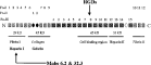
Fig. 5. Schematic representation of mammalian fibronectin. Location of the gelatin and heparin-binding fragments which were used in the attachment assays, are in bold. The fragment to which the monoclonal antibodies Mabs 6.2 and 32.3, raised to root-knot nematodes, is arrowed. Regions of homology are indicated by rectangles, squares and circles; Fn-III domain labelled RGDs represent repeated sequences of arginine, glycine and aspartic acid (adapted from Petersen et al. 1983; Skorstengaard et al. 1994; Yu et al. 1997).
 You have
Access
- 19
- Cited by