1. Introduction
Viruses are simple lifeless entities that cannot reproduce on their own and therefore depend on host cells to provide them with the necessary life support mechanisms. Viruses are categorized as either prokaryotic [also called bacteriophage, or ‘phage’ for short, originating from the Greek word phagein (to eat)] or eukaryotic (i.e. viruses that infect plant and animal cells). Simplified, all viruses consist of a protein shell (capsid) that protects the viral genome (DNA or RNA). To infect, the viral genome must enter the cell, either alone or as part of a viral nucleoprotein complex. This entry is assisted by receptors that are present on the host cell surface (Hendrix et al. Reference Hendrix, Roberts, Stahl and Weisberg1983; Flint et al. Reference Flint, Enquist, Racaniello, Skalka and Flint2004b; Ptashne, Reference Ptashne2004). Many phages are equipped with a tubular ‘tail’ structure through which the viral genome passes into the cell; the capsid is not internalized. In contrast, most animal viruses are internalized into the host cell essentially intact through endocytosis or membrane fusion mechanisms. In the latter case, the capsid is covered with a lipid envelope that fuses with the host cell plasma membrane. Plant viruses utilize insects as vectors that aid in mechanical penetration of the plant cell wall (Hendrix et al. Reference Hendrix, Roberts, Stahl and Weisberg1983; Flint et al. Reference Flint, Enquist, Racaniello, Skalka and Flint2004b; Ptashne, Reference Ptashne2004). Once the viral genome has entered the cell, the virus hijacks the host cell's machinery and synthesizes multiple copies of viral genome. Transcription and translation of viral genes yields proteins that assemble into new viral particles that exit the host cell. This may occur by a budding process that is not necessarily detrimental to the cell or by cell lysis, which is a lethal event (Hendrix et al. Reference Hendrix, Roberts, Stahl and Weisberg1983; Flint et al. Reference Flint, Enquist, Racaniello, Skalka and Flint2004a; Ptashne, Reference Ptashne2004).
Viruses present a major threat to human health and welfare. The extent of this threat becomes obvious if one considers the morbidity and mortality caused by the human immunodeficiency virus (HIV), hepatitis virus, influenza as well as threats from SARS (severe acute respiratory syndrome) and H5N1 (avian influenza virus) (WHO, 2007). The potential for global spread of lethal viral infections is accentuated by the modern aviation network and traveling habits, thus putting millions of people at risk (Brockmann et al. Reference Brockmann, Hufnagel and Geisel2006). Moreover, viral infections are associated with serious chronic conditions such as cancer, obesity and cardiovascular diseases that also seriously compromise human health (WHO, 2007). Aside from vaccination, current antiviral strategies either target the receptors through which viruses enter host cells, or target specific enzymes that are critical to viral replication. For instance, abacavir and retrovir inhibit HIV reverse transcriptase, acyclovir inhibits DNA polymerase of herpes simplex virus, and oseltamivir and zanamivir inhibit neuraminidase of the influenza virus (Flint et al. Reference Flint, Enquist, Racaniello, Skalka and Flint2004c). The main limitation of these kinds of antiviral agents is that they are highly ‘specialized’, as they target specific events in replication cycle of individual viruses. Thus, their use is limited to a specific virus or a small group of viruses. Furthermore, viruses frequently undergo mutations that alter the target sites. Therefore, there is a growing need to develop less specialized viral intervention strategies that will enable targeting a broad range of viruses with little susceptibility to mutation. For instance, drugs that directly interfere with the assembly of the viral capsid and/or packaging of the viral DNA into the capsid could provide a ‘broad spectrum’ antiviral agent.
Biophysical virology is a rather new field that seeks to define the physical mechanisms controlling virus development. This knowledge can provide information essential to the rational design of new antiviral strategies with less specificity for a limited number of viruses. Biological and physical simplicity relative to other viral systems have made the bacteriophages an attractive model system in biophysical virology. Notwithstanding, the fundamental mechanistic and structural features of phages can be directly applied to the eukaryotic viruses. For many years phages have served as reliable experimental models that have significantly contributed to the advent of modern molecular biology. To mention a few examples, initial studies on gene regulation and protein synthesis were conducted using phages (Hendrix et al. Reference Hendrix, Roberts, Stahl and Weisberg1983; Echols, Reference Echols2001; Ptashne, Reference Ptashne2004). Further, Hershey and Chase for the first time demonstrated that DNA is the hereditary material using phages (Hershey & Chase, Reference Hershey and Chase1952). Today, phages provide ideal systems to study biophysical virology and this review focuses on recent advances in this field.
A common feature of many phages is that the microns-long genome is confined within a nanometer-size capsid by means of an ATP-driven motor (Riemer & Bloomfield, Reference Riemer and Bloomfield1978). In the case of phage λ and many other double-stranded DNA (dsDNA) phages, this confinement creates internal pressures of tens of atmospheres (atm), which is exerted on the inner capsid walls (Kindt et al. Reference Kindt, Tzlil, Ben-Shaul and Gelbart2001; Smith et al. Reference Smith, Tans, Smith, Grimes, Anderson and Bustamante2001; Purohit et al. Reference Purohit, Kondev and Phillips2003b; Tzlil et al. Reference Tzlil, Kindt, Gelbart and Ben-Shaul2003; Evilevitch et al. Reference Evilevitch, Castelnovo, Knobler and Gelbart2004). DNA is packed to nearly crystalline densities inside the capsid; given the limited volume of the rigid viral capsid, such levels of compression lead to strong bending of the DNA and DNA–DNA repulsion between its negatively charged strands (Kindt et al. Reference Kindt, Tzlil, Ben-Shaul and Gelbart2001; Tzlil et al. Reference Tzlil, Kindt, Gelbart and Ben-Shaul2003). The high force generated by the pressurized genome is in turn necessary to initiate the infection process through DNA ejection into the cell, which is energetically a downhill process that does not require ATP (Evilevitch et al. Reference Evilevitch, Lavelle, Knobler, Raspaud and Gelbart2003, Reference Evilevitch, Gober, Phillips, Knobler and Gelbart2005). This common physical property of many dsDNA phages is likely applicable to eukaryotic dsDNA viruses that use motor proteins to package their genome, e.g. the herpesviruses and possibly adenovirus (Catalano, Reference Catalano2005b). The physical properties of the DNA-filled capsid suggests that high internal pressure induced by tightly packed DNA must be physically matched by the mechanical strength of the capsid, which must withstand this pressure (Ivanovska et al. Reference Ivanovska, De Pablo, Ibarra, Sgalari, Mackintosh, Carrascosa, Schmidt and Wuite2004, Reference Ivanovska, Wuite, Jonsson and Evilevitch2007). This raises several fundamental questions that will be discussed in this review:
• What is the physical basis for the internal pressure generated by packaged DNA? What internal and external factors control this pressure?
• How can phage capsids, which are held together by non-covalent hydrophilic/hydrophobic interactions between capsid-protein subunits, withstand this high pressure and how does it affect survival of viruses outside of the host cell?
• How is DNA packaged and ejected from phage? Is the internal pressure required for infection in vivo?
This review is an interdisciplinary account of theoretical and experimental aspects of biophysical virology, where most of the experiments have been performed on phages λ, φ29, T4, T5, T7 and SPP1. We discuss recent work on the mechanism of DNA packaging and ejection in Section 2 and mechanical properties of viral capsids are reviewed in Section 3. We also discuss some of the eukaryotic viruses for which relevant measurements have been made. Viral infectivity and virion stability are discussed from the perspective of internal genome stress and its consequences for the virus. We believe that a deeper understanding of this common physical parameter for many dsDNA motor-packaged viruses will lead to new possibilities for development of antiviral strategies as well as novel tools for gene therapy and nanotechnology.
2. DNA packaging and ejection
2.1 Theoretical considerations
In this section, we review the physical concepts underlying the current understanding of DNA packaging properties. Indeed it has long been a challenging theme for physicists, because it implies basic concepts of polymer physics that can be explicitly addressed in the case of bacteriophages. Early systematic studies traced back to works of Earnshaw and Harrison (Earnshaw et al. Reference Earnshaw, Casjens and Harrison1976, Reference Earnshaw, King, Harrison and Eiserling1978; Earnshaw & Harrison, Reference Earnshaw and Harrison1977; Earnshaw & Casjens, Reference Earnshaw and Casjens1980) highlighted a concentric layered structure of the DNA within the phage λ capsid, and a strong correlation between genome length and spacing between consecutive DNA layers. This study led to the proposal of several packaging models: the DNA genome has been envisaged as being wound into a spool (Earnshaw & Harrison, Reference Earnshaw and Harrison1977; Hendrix, Reference Hendrix1978; Riemer & Bloomfield, Reference Riemer and Bloomfield1978; Earnshaw & Casjens, Reference Earnshaw and Casjens1980; Harrison, Reference Harrison1983; Gabashvili & Grosberg, Reference Gabashvili and Grosberg1991, Reference Gabashvili and Grosberg1992), a ball (Richards et al. Reference Richards, Williams and Calendar1973; Earnshaw & Harrison, Reference Earnshaw and Harrison1977), and a liquid crystal with hairpins or folds (Earnshaw & Harrison, Reference Earnshaw and Harrison1977; Black et al. Reference Black, Newcomb, Boring and Brown1985; Serwer, Reference Serwer1986; Sun et al. Reference Sun, Louie and Serwer1999). The first quantitative estimate of the energetics of DNA packaging was proposed by Riemer & Bloomfield (Reference Riemer and Bloomfield1978) on theoretical grounds. However, the lack of precise information regarding the true conformation of packaged DNA has hampered direct comparison with experimental data. It was, nevertheless, realized that the Gibbs free energy of the system is dominated by the balance of two main contributions associated to both the small size of viral capsid and high DNA density: (i) bending energy that tends to crowd DNA on the capsid wall in order to minimize DNA curvatures, and (ii) DNA–DNA self-interactions that tend to oppose this self-crowding.
This is nicely illustrated by bacteriophage λ: in this case, the internal capsid diameter is ~55 nm, i.e. of the order of the DNA persistence length (50 nm), which represents the typical length scale of spontaneous bending due to thermal fluctuations. Packaging of DNA lengths longer than the persistence length inside such a small volume will therefore take a significant amount of mechanical work. Similarly, the volume fraction of DNA inside the capsid for wild-type (wt) λ is roughly 0·59, implying small distances between consecutive DNA layers, in the order of 2·7 nm in the case of an inverse spool conformation to be described below. At these very short separations, dense hexagonal phases of DNA exhibit strong electrostatic and hydration self-interactions, as was shown using the osmotic stress method by Rau and Parsegian (Rau & Parsegian, Reference Rau and Parsegian1992; Strey et al. Reference Strey, Podgornik, Rau and Parsegian1998). As discussed below, the buffer conditions, particularly the multivalent ion content, strongly influence these self-interactions, that can be either repulsive or attractive (Bloomfield, Reference Bloomfield1997; Evilevitch et al. Reference Evilevitch, Fang, Yoffe, Castelnovo, Rau, Parsegian, Gelbart and Knobler2008). The balance between these two characteristic energies leads to an internal ‘pressure’ on the order of several tens of atmospheres, equivalent to an internal force of roughly tens of pN (recall that 1 atm=0·1 pN/nm2). We note that in addition to these two major energetic contributions to DNA packaging, the entropy cost associated with DNA packaging inside the capsid has not been thoroughly investigated analytically and this might also play some non-negligible role in the total Gibbs free energy, as suggested by recent simulation works (Forrey & Muthukumar, Reference Forrey and Muthukumar2006).
Using an inverse spool conformation, Odijk was able to cast analytically these two dominant energies into a single predictive model for bacteriophage T7 (Odijk, Reference Odijk1998). In this proposed conformation, DNA is wound in the interior of the capsid in an ordered way, filling first layers close to the walls followed by layers of decreasing radius, finally leaving a cylindrical hole inside the capsid devoid of any DNA. It has been well established by Cerritelli et al. (Reference Cerritelli, Cheng, Rosenberg, McPherson, Booy and Steven1997) that this conformation is the most likely and is the result of a central protein connector that imposes internal cylindrical symmetry to the DNA condensate. In Odijk's model, total bending energy is calculated as the sum of elementary bending energies of circles filling the inverse spool region associated with the DNA condensate. Similarly, DNA self-interactions are assumed to be of purely electrostatic origin, and are evaluated explicitly using a classical Poisson–Boltzmann approach. The balance between these two energies specifies the optimal spacing between neighboring turns of DNA in the condensate and the main predictions of this model are (i) the spacing between DNAs in the hexagonal packing within the inverse spool and (ii) the inner radius of the condensate. The resulting theoretical values are in quantitative agreement with Cerritelli's experimental data. While this model was the first to address the structural properties of packaged DNA, a quantitative discussion of the energetics in connection with particular structure was still missing at that time.
The next step towards this direction came from single-molecule experiments. Indeed, original experiments performed by Smith et al. (Reference Smith, Tans, Smith, Grimes, Anderson and Bustamante2001) renewed the interest of the physics community in the problem of viral DNA packaging, by providing direct single-molecule measurements of the in vitro packaging rates in phage φ29. From these studies, an internal force-resisting packaging on the order of tens of pN can be inferred, thus providing quantitative estimates of the energy associated with packaging (as will be discussed in the next section). Based on these experimental results, several theoretical models of DNA packaging have been proposed in recent years, addressing a variety of issues by both analytical models and numerical simulations.
Tzlil et al. (Reference Tzlil, Kindt, Gelbart and Ben-Shaul2003) extended Odijk's original model to quantitatively include the energetics. First, they allowed for shape variations of the inverse spool condensate ranging from ideal spool to toroid, and second, they used more realistic DNA–DNA interactions based on osmotic stress experiments (Rau & Parsegian, Reference Rau and Parsegian1992; Strey et al. Reference Strey, Podgornik, Rau and Parsegian1998). The outcome of this extended model is to describe the relative contribution of each of the energies (bending, self-interaction, and surface tension in the case of attractive self-interactions induced by multivalent ions, etc.). A similar model was proposed by Purohit and colleagues (Purohit et al. Reference Purohit, Kondev and Phillips2003a, Reference Purohit, Kondev and Phillipsb, Reference Purohit, Inamdar, Grayson, Squires, Kondev and Phillips2005), further extending the theoretical discussion on experimental quantities affecting the amplitude of internal force: capsid size and shape, genome length, discreteness of DNA condensate and buffer conditions. This work showed a strong reduction of internal force-resisting packaging if DNA–DNA interactions are switched from repulsive to attractive conditions. Indeed in the former case, the repulsive self-interactions result in a high price for self-crowding as soon as a few persistence lengths of DNA are packaged. In the latter case, attractive interactions induce the condensation of DNA into a toroid in a bulk solution, whose dimensions are slightly larger than the capsid size in the case of bacteriophage λ. This means that early stages of DNA packaging have much less crowding energy, because the capsid influence on the toroid is negligible. Since more DNA is internalized in the capsid, the DNA toroidal condensate becomes compressed, its shape changes from a toroid to an inverse spool, and consequently free energy increases sharply.
One of the first simulations was performed by Kindt et al. (Reference Kindt, Tzlil, Ben-Shaul and Gelbart2001). In this work, DNA packaging is mimicked by Brownian molecular dynamics by inserting a semi-flexible chain made of beads into a sphere in a stepwise fashion. By an appropriate choice of physical parameters (persistence length, capsid size, interactions between beads), these simulations are able to reproduce quantitatively variations of the internal force observed in single-molecule experiments as nucleic acid is translocated into the capsid. They showed, for example, that if attractive DNA–DNA self-interactions are assumed, due to the presence of multivalent ions in the buffer, the packaging proceeds mainly in two steps: during the first two thirds of genome packaging, the force increases smoothly, the attractive interactions helping to reduce the cost of packaging, as discussed above. Then the force increases sharply during the last third of packaging, in qualitative agreement with the experiments by Smith et al. (Reference Smith, Tans, Smith, Grimes, Anderson and Bustamante2001).
Many more numerical simulations of semi-flexible packaging inside a container were subsequently proposed (Arsuaga et al. Reference Arsuaga, Tan, Vazquez, Sumners and Harvey2002; Klug & Ortiz, Reference Klug and Ortiz2003; LaMarque et al. Reference Lamarque, Le and Harvey2004; Spakowitz & Wang, Reference Spakowitz and Wang2005; Ali et al. Reference Ali, Marenduzzo and Yeomans2006; Forrey & Muthukumar, Reference Forrey and Muthukumar2006; Locker & Harvey, Reference Locker and Harvey2006; Petrov et al. Reference Petrov, Boz and Harvey2007a, Reference Petrov, Lim-Hing and Harveyb). Unlike experimental studies to date, the use of computer simulations allows: changing at will specific parameters of the system, such as capsid shape, twisting DNA during packaging, constant rate or constant force packaging, strength of electrostatic interactions, etc., which allows theoretical testing of the relevance of these parameters. Consequently these works have provided important structural insights on DNA conformation inside the capsid. However, precise experimental results on DNA structure within viral capsids are not yet available for direct comparison. Nevertheless, most of the simulations showed that semi-flexible chain organization inside cavities might not be as well ordered as assumed in the previous analytical models, unless attractive self-interactions are included (Forrey & Muthukumar, Reference Forrey and Muthukumar2006) or elongated capsid shape (Petrov et al. Reference Petrov, Lim-Hing and Harvey2007b).
Another complementary way of providing quantitative data on DNA packing energetics is to investigate DNA ejection from the capsid. Since DNA packing properties are expected to produce mechanical pressure in the order of several tens of atmospheres within the capsid, it was proposed by Kindt and colleagues (Kindt et al. Reference Kindt, Tzlil, Ben-Shaul and Gelbart2001; Tzlil et al. Reference Tzlil, Kindt, Gelbart and Ben-Shaul2003) that this pressure could be balanced during in vitro-triggered DNA ejection by osmotic pressure in the solution bathing the capsids. This was confirmed by Evilevitch et al. (Reference Evilevitch, Lavelle, Knobler, Raspaud and Gelbart2003, Reference Evilevitch, Gober, Phillips, Knobler and Gelbart2005), where incomplete DNA ejection from the bacteriophage λ capsid was observed (see Section 2.3 below). Using these data together with an appropriate modeling of the osmotic force resisting ejection from the bacteriophage (Castelnovo et al. Reference Castelnovo, Bowles, Reiss and Gelbart2003; Evilevitch et al. Reference Evilevitch, Castelnovo, Knobler and Gelbart2004), it was possible to compare the analytical models mentioned above with experimental data. This osmotic force is associated with the thermodynamic PV work of creating a cavity large enough to accommodate DNA, where P is the osmotic pressure outside the capsid and V is DNA volume. An excellent quantitative agreement was observed with λ deletion mutants (Grayson et al. Reference Grayson, Evilevitch, Inamdar, Purohit, Gelbart, Knobler and Phillips2006), thus validating the current theoretical description.
As mentioned above, buffer conditions strongly influence the amplitude of internal force-resisting packaging. This was shown recently using the osmotic suppression technique with bacteriophage λ (Evilevitch et al. Reference Evilevitch, Fang, Yoffe, Castelnovo, Rau, Parsegian, Gelbart and Knobler2008) by increasing the concentration of various multivalent ions (magnesium, spermine) in solution. The experimental data were successfully interpreted within the framework of previous theoretical models by changing the parameters of DNA self-interactions, providing further credibility to the inverse spool model; however, recent studies indicate that our understanding of DNA packaging properties is still incomplete. First, a striking feature of all the simulations mentioned so far is that the final conformation of packaged DNA might deviate significantly from the inverse spool, which is the basis of the successful model described above (Forrey & Muthukumar, Reference Forrey and Muthukumar2006). Further, it appears that the order of magnitude of internal force-resisting packaging is not very sensitive to the final conformation. These facts suggest that some rather general formulation of this force beyond the inverse spool models is needed. Indeed, Castelnovo & Evilevitch (Reference Castelnovo and Evilevitch2007) showed that osmotic suppression data on bacteriophages λ and T5 are equally well described using a structureless model (i.e. no hexagonal packing is assumed) of DNA condensate. In this simple model, DNA self-interactions are assumed to be of excluded volume or hard-core type, and the only length-dependent contribution to the internal force comes from bending penalty. Therefore this work questions the reason for the agreement of structured and structureless models, which remains an open question.
2.2 DNA packaging
As described above, the physical situation in many viruses and especially the majority of bacteriophages implies that a negatively charged, microns-long DNA molecule is tightly packed within a nanometer-size capsid. This unusual situation raises the question: ‘How is a long, rigid DNA molecule packaged into a very small capsid and what are the forces associated with it?’
The assembly of an infectious virus is a complex process that requires the temporal and coordinated activities of numerous proteins of both viral and host origin (Calendar, Reference Calendar2006; Knipe & Howley, Reference Knipe and Howley2007). Assembly pathways vary among the virus types, but common features are observed within certain groups, such as the complex dsDNA viruses including adenovirus, the herpesviruses and many bacteriophages (Calendar, Reference Calendar2006; Knipe & Howley, Reference Knipe and Howley2007). In these cases, viral DNA is actively translocated into the interior of a pre-assembled procapsid structure by terminase enzymes, a process known as DNA packaging (Catalano, Reference Catalano and Catalano2005a). The packaging substrate is most often a linear concatemer of viral genomes, linked in a head-to-tail fashion, synthesized by a rolling-circle DNA replication mechanism (Calendar, Reference Calendar2006; Knipe & Howley, Reference Knipe and Howley2007); adenovirus and phage φ29 are notable exceptions in which monomeric genomes are synthesized and used as the packaging substrate (Anderson & Grimes, Reference Anderson, Grimes and Catalano2005; Calendar, Reference Calendar2006; Berk, Reference Berk, Knipe and Howley2007). In all cases, the terminase enzymes are responsible for specific recognition of viral DNA and translocation of a single genome into the capsid interior, fuelled by ATP hydrolysis (Black, Reference Black1989; Fujisawa & Morita, Reference Fujisawa and Morita1997; Catalano, Reference Catalano and Catalano2005a).
Virus assembly has been best characterized in the bacteriophages where in vitro virus assembly assays have been developed for λ (Becker et al. Reference Becker, Murialdo and Gold1977), T4 (Rao et al. Reference Rao, Thaker and Black1992), T7 (Shibata et al. Reference Shibata, Fujisawa and Minagawa1987), SPP1 (Droge & Tavares, Reference Droge and Tavares2000) and φ29 (Guo et al. Reference Guo, Grimes and Anderson1986), among others. These assays use crude extracts of virus-infected cells as a source of assembly components and purified viral DNA that can be packaged to afford an infectious virus. Extracts of cells infected with mutant, assembly-incompetent virus can be ‘complemented’ with purified protein or with cell extracts that replace the defective protein and assemble an infectious virion. These systems have allowed interrogation of the virus assembly pathways and have provided significant insight into the nucleoprotein complexes involved in DNA packaging. Recently, a defined biochemical system has been developed in which seven purified proteins (viral and host), purified procapsids and tails and commercially available viral DNA can be assembled into an infectious λ virus under strictly defined biochemical reaction conditions (Gaussier et al. Reference Gaussier, Yang and Catalano2006). In addition, defined biochemical systems that allow rigorous examination of the DNA packaging step have been developed in bacteriophages λ (Hwang & Feiss, Reference Hwang and Feiss1995; Yang & Catalano, Reference Yang and Catalano2003), φ29 (Guo et al. Reference Guo, Grimes and Anderson1986), SPP1 (Oliveira et al. Reference Oliveira, Alonso and Tavares2005) and T7 (Hamada et al. Reference Hamada, Fujisawa and Minagawa1986). This sets the stage to systematically probe the enzymatic, biochemical, biophysical and structural features of the virus assembly pathway in the complex dsDNA viruses. Unfortunately, in vitro assembly systems are not available for any of the eukaryotic viruses, which has limited mechanistic description of these pathways; however, existing evidence strongly suggests that the general features of virus assembly are conserved between the prokaryotic and eukaryotic virus groups.
Genome packaging is an essential step in the assembly of the large dsDNA viruses. As with most complex biological processes, DNA packaging can be described in three phases – initiation, active packaging and termination. Initiation starts with terminase assembly at a packaging initiation site in concatemeric viral DNA (pac). This site represents the concatemeric joint – the end of one genome and the beginning of the next in the concatemer. In all characterized cases, the terminase enzymes are composed of large (49–72 kDa) and small (18–21 kDa) subunits (Fujisawa & Morita, Reference Fujisawa and Morita1997; Catalano, Reference Catalano and Catalano2005a; Calendar, Reference Calendar2006). The small subunits are responsible for recognition of a pac sequence and site-specific assembly of the functional terminase complex (terminase holoenzyme) on viral DNA. The holoenzymes are heteroligomers of the two subunits whose stoichiometry has not been conclusively defined in any virus, although it is likely that they form ring-like structures that encircle the duplex DNA (Chai et al. Reference Chai, Lurz and Alonso1995; Lin et al. Reference Lin, Simon and Black1997; Morais et al. Reference Morais, Tao, Olson, Grimes, Jardine, Anderson, Baker and Rossmann2001; Maluf et al. Reference Maluf, Gaussier, Bogner, Feiss and Catalano2006). The large subunits possess a nuclease activity that cleaves the duplex at or near pac to ‘mature’ the first genome end in preparation for packaging. In some viruses such as phage λ, duplex cleavage is site-specific and defined single-stranded overhangs are formed (the ‘sticky’ end of λ) (Daniels et al. Reference Daniels, Schroeder, Szybalski, Sanger, Coulson, Hong, Hill, Petersen, Blattner, Hendrix, Roberts, Stahl and Weisberg1983; Feiss & Catalano, Reference Feiss, Catalano and Catalano2005). In others, such as phages P22 and T4 the cleavage appears to be more random (Rentas & Rao, Reference Rentas and Rao2003; Casjens & Weigele, Reference Casjens, Weigele and Catalano2005).
Bacteriophage φ29 presents an interesting variation to the above paradigm in that the packaging substrate is a monomeric genome that possesses terminal proteins covalently attached to the 5′ ends of the duplex, a consequence of a protein-primed DNA replication strategy. The so-called packaging ATPase protein shares functional similarity to all of the large terminase subunits – it interacts with the terminal protein (30 kDa) and the portal ring, it possesses ATPase activity and it is required to translocate viral DNA into the capsid. In this case, the terminal protein may be viewed as a DNA recognition protein (i.e. the small terminase subunit) in the extreme sense as it is covalently attached to the genome ends.
In all cases, the matured duplex end remains associated with terminase, which next binds to a doughnut-shaped portal structure situated in a unique vertex of the procapsid. The dodecameric portal ring serves as a conduit for DNA entry into (and out of) the capsid interior and while the details are unclear, current models presume that the bona fide packaging motor is composed of both terminase and portal subunits. [The portal from phage φ29 presents an interesting variation in that a small RNA (pRNA) is also required to assemble a functional packaging motor.]
Terminase assembly and duplex cleavage triggers the active packaging phase in which the packaging motor releases the matured genome end and threads the duplex through the portal and into the capsid interior. The large terminase subunits uniformly possess an ATPase activity and it is presumed that the holoenzymes transduce the chemical energy of ATP hydrolysis to physical translocation of DNA. Primary sequence analysis reveals significant homology among the large terminase subunits, both prokaryotic and eukaryotic, particularly in the N-terminal ‘ATPase domains’ (Mitchell et al. Reference Mitchell, Matsuzaki, Imai and Rao2002). These domains display features common to the ATPase domains of the DEAD box helicases including Walker A, Walker B, adenine base-binding, catalytic carboxylate signatures and importantly the ‘motif III’ sequence that couples ATP hydrolysis to DNA translocation (Story et al. Reference Story, Li and Abelson2001; Mitchell et al. Reference Mitchell, Matsuzaki, Imai and Rao2002). Consistently, the crystal structure of the N-terminal domain of the large terminase subunit of bacteriophage T4 reveals structural similarities with monomeric helicases and an ‘inchworm’ DNA translocation mechanism has been proposed based on these similarities (Sun et al. Reference Sun, Kondabagil, Gentz, Rossmann and Rao2007). However, the terminase enzymes probably function as oligomers, and ring structures composed of four, five and six large subunits have been proposed to represent the functional packaging motors in phages λ (Maluf et al. Reference Maluf, Gaussier, Bogner, Feiss and Catalano2006), φ29 (Morais et al. Reference Morais, Tao, Olson, Grimes, Jardine, Anderson, Baker and Rossmann2001) and T4 (Sun et al. Reference Sun, Kondabagil, Gentz, Rossmann and Rao2007), respectively, which suggests functional homology to the hexameric ring helicases (Patel & Donmez, Reference Patel and Donmez2006). This arrangement would allow the terminase subunits to act cooperatively in a multi-site, sequential translocation mechanism where ATP hydrolysis mediates passage of the duplex from one subunit to the next, ‘escorting’ 1–2 bases of DNA through the portal channel with each cycle (Enemark & Joshua-Tor, Reference Enemark and Joshua-Tor2006; Patel & Donmez, Reference Patel and Donmez2006). A sequential hydrolysis/escort mechanism has similarly been proposed for the hexameric RNA packaging motor of bacteriophage φ4 (Kainov et al. Reference Kainov, Mancini, Telenius, Lisal, Grimes, Bamford, Stuart and Tuma2008).
While it is clear that terminase enzymes are required for DNA translocation, the extent to which the portal ring is involved remains controversial. Around 30 years ago, Hendrix noted a symmetry mismatch between the dodecameric portal ring, the fivefold vertex in which it resides and the tenfold screw symmetry of the DNA that passes through it (Hendrix, Reference Hendrix1978). Based on this observation, he proposed that DNA packaging is mediated by rotation of the portal ring within the capsid vertex. More recent models based on the crystal structure of the phage φ29 portal further suggest that portal rotation is powered by ATP hydrolysis (ostensibly by the large terminase subunit) and that the portal itself is responsible for DNA translocation (Simpson et al. Reference Simpson, Tao, Leiman, Badasso, He, Jardine, Olson, Morais, Grimes, Anderson, Baker and Rossmann2000); however, Black and co-workers have recently demonstrated that physically constraining the portal ring within the capsid has little effect on DNA packaging, a result not favoring models where portal rotation is required for DNA translocation (Baumann et al. Reference Baumann, Mullaney and Black2006). Whatever the case, it is clear that the portal serves as the sole conduit for DNA into and out of the capsid interior and it is likely that the portal proteins activate the DNA translocase activity of terminase holoenzyme (see Feiss & Catalano, Reference Feiss, Catalano and Catalano2005). Minimally, therefore, the portal ring may function as a ‘sliding clamp’ structure reminiscent of those involved in processive DNA replication (Jeruzalmi et al. Reference Jeruzalmi, O'Donnell and Kuriyan2002), with the obvious difference that the portal directs the duplex into a confined space. The portal may further serve as a ‘head-full’ sensor that terminates motor translocation and activates the nuclease activity of terminase to finalize the genome packaging reaction. This is discussed further below.
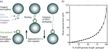
In most cases, DNA packaging triggers an expansion process where the roughly spherical procapsid takes on the angularized icosahedral shape of the mature virion and roughly doubles its volume. Capsid expansion is typically accompanied by the addition of ‘decoration’ proteins to the capsid surface that ostensibly serve to stabilize the expanded capsid structure against the tremendous internal pressures generated by the packaging motor (Evilevitch et al. Reference Evilevitch, Lavelle, Knobler, Raspaud and Gelbart2003). Indeed, single-molecule optical tweezer (Fuller et al. Reference Fuller, Raymer, Rickgauer, Robertson, Catalano, Anderson, Grimes and Smith2007b) (see Fig. 1a) and bulk biochemical experiments (Yang & Catalano, unpublished observations) strongly suggest that capsid integrity is compromised when the penultimate ~10–15% of a λ genome is packaged into the capsid in the absence of the decoration protein. The surface decoration proteins are a common feature in the dsDNA viruses and are conserved in both prokaryotic and eukaryotic dsDNA viruses. An interesting exception is phage HK97 for which there are no decoration proteins. Instead, the major capsid proteins are covalently cross-linked in an autocatalytic reaction (Duda, Reference Duda1998; Wikoff et al. Reference Wikoff, Liljas, Duda, Tsuruta, Hendrix and Johnson2000). This wholesale cross-linking of the capsid proteins likely serves an identical function in this unique virus, i.e. stabilization of the highly pressurized, DNA-filled capsid.
(a) Illustration of the optical-tweezers set-up for packaging force measurement. Microspheres carrying prohead–ATPase complex and DNA molecule are captured in two optical traps. DNA packaging is initiated by bringing together the microspheres and then separating them to probe for DNA binding followed by packaging. The force acting on the DNA is recorded. (Adopted from Fuller et al. Reference Fuller, Raymer, Rickgauer, Robertson, Catalano, Anderson, Grimes and Smith2007b). (b) Mean internal force vs. packaged DNA fraction in phage φ29. (Adopted from Rickgauer et al. Reference Rickgauer, Fuller, Grimes, Jardine, Anderson and Smith2008.)

Upon packaging the entirety of the viral genome, the motor complex enters the packaging termination phase, which includes terminase-mediated duplex cleavage to release the DNA-filled capsid. There are two basic mechanisms by which the packaging machinery stops and enters the termination phase. The first is a unit-length packaging mechanism, which is exemplified by bacteriophage λ (Becker & Murialdo, Reference Becker and Murialdo1990; Feiss & Catalano, Reference Feiss, Catalano and Catalano2005). In these cases, genome packaging both initiates and terminates at a specific pac sequence in the concatemer, which in the case of λ is known as the cohesive end site, or cos. Since the cos site represents the junction between individual genomes in the concatemer, initiation and termination specifically at this site results in the packaging of a single, unit-length (100%) viral genome with ‘sticky’ (cohesive) ends. The herpesviruses likely utilize a unit-length packaging mechanism (Baines & Weller, Reference Baines, Weller and Catalano2005). The second mechanism is referred to as head-full packaging, which is exemplified by bacteriophage p22 (Casjens & Weigele, Reference Casjens, Weigele and Catalano2005). In these cases, the terminase enzyme similarly initiates packaging at a specific pac sequence; however, the translocating motor bypasses the downstream pac site and does not stop until the capsid (i.e. head) is filled to capacity (head full). Thus, the DNA isolated from head-full packaging viruses is 103–105% of the length of a mature viral genome and possesses 1–3 kb direct terminal repeats (terminally redundancy) (Casjens & Weigele, Reference Casjens, Weigele and Catalano2005; Catalano, Reference Catalano and Catalano2005a; Droge & Tavares, Reference Droge, Tavares and Catalano2005). We note that there is an element of head-full packaging even in bacteriophage λ as shortened genomes are inefficiently packaged (Cue & Feiss, Reference Cue and Feiss1997).
In both unit-length and head-full packaging mechanisms there must be a ‘signal’ that triggers entry into the termination phase. In the former case, one can envision that the translocating terminase enzyme, which is responsible for specific recognition and cleavage of the pac sequence, plays a dominant role. In the latter case, several investigators have suggested that the portal is responsible for sensing the filled capsid, stopping translocation and triggering duplex cutting by the terminase enzyme (Simpson et al. Reference Simpson, Tao, Leiman, Badasso, He, Jardine, Olson, Morais, Grimes, Anderson, Baker and Rossmann2000; Lander et al. Reference Lander, Tang, Casjens, Gilcrease, Prevelige, Poliakov, Potter, Carragher and Johnson2006). However, is it not clear how this process occurs in either case, and the two models are not mutually exclusive.
There are limits to the amount of DNA that can be packaged into a capsid, although it is unclear exactly what physically constrains this process. One suggestion is that the decoration proteins not only provide physical stability to the DNA-filled capsid (see above), but also serve to prevent ‘over-filling’ by the terminase motors, which are extremely powerful (Sternberg & Weisberg, Reference Sternberg and Weisberg1977). Indeed, single-molecule optical-tweezer studies on the viral packaging motors from bacteriphages φ29 (Rickgauer et al. Reference Rickgauer, Fuller, Grimes, Jardine, Anderson and Smith2008), λ (Fuller et al. Reference Fuller, Raymer, Rickgauer, Robertson, Catalano, Anderson, Grimes and Smith2007b) and T4 (Fuller et al. Reference Fuller, Raymer, Kottadiel, Rao and Smith2007a) clearly indicate that the motors are capable of generating forces of between 50 pN and 100 pN (see Fig. 1b). This is consistent with the observation that the DNA-filled capsid is tightly packed and highly pressurized, experimentally measured as tens of atmospheres in phage λ (Evilevitch et al. Reference Evilevitch, Lavelle, Knobler, Raspaud and Gelbart2003). Thus, the capsid shell, in the presence of decoration proteins or physically cross-linked, may provide a structure that is sufficiently stable to withstand the internal pressure and which serves to provide a negative force sufficient to stall the packaging motor precisely at the point at which a complete genome has been packaged. This is discussed in further detail below. Presumably, the force required to further translocate DNA into the filled capsid is greater than that obtainable from the hydrolysis of ATP.
The terminase enzymes are not part of the mature virus and dissociate from the DNA-filled capsids after packaging is complete. Finishing proteins add, ostensibly to the portal vertex, to prevent release of the tightly packaged, highly pressurized DNA. It is unclear how this process occurs, but it is likely that the finishing proteins displace terminase in a step-wise manner such that at least one of the proteins maintains contact with the DNA and the portal during the exchange process. Additionally, recent cryo-electron microscopy studies suggest that conformational reorganization of the portal structure occurs at this point, changing from a patent conduit that allows DNA passage into the capsid to a DNA clamp that precludes duplex escape (Zheng et al. Reference Zheng, Olia, Gonen, Andrews, Cingolani and Gonen2008).
While much is known about the process of DNA packaging, several questions remain unanswered. For over 50 years, the study of bacteriophage biology has yielded significant insight into virus development and has been at the forefront of genetics and molecular biology. As outlined above, bacteriophages now provide well-developed systems that allow an ensemble of genetic, biochemical, biophysical and structural techniques to be brought to bear on virus assembly mechanisms and allow interrogation of the process at the molecular level. Given the remarkable similarities between these dsDNA viruses, both prokaryotic and eukaryotic, a detailed understanding of the principles governing virus assembly in one virus will undoubtedly have far-reaching implications for all viruses.
2.3 DNA ejection
In order to infect a cell, viruses must release the nucleic content of their capsids into the host cell. Tailed bacteriophages attach to specific receptors on the surface of the host, which triggers DNA ejection into the cell. Extracting and purifying such receptors provides an important tool to study phage ejection mechanism in vitro; however, only a few phage receptors have been successfully purified. For example, the LamB receptor of λ (Roa & Scandella, Reference Roa and Scandella1976), the FhuA receptor of T5 (Boulanger et al. Reference Boulanger, Le Maire, Bonhivers, Dubois, Desmadril and Letellier1996) and the YueB780 receptor of SPP1 (Sao-Jose et al. Reference Sao-Jose, Lhuillier, Lurz, Melki, Lepault, Santos and Tavares2006) can be purified in functional form. Although the mechanism of complete DNA ejection from phages in vivo is not entirely clarified, there is experimental and theoretical evidence that DNA internal force is responsible for initiating the ejection process and for translocation of the first DNA fraction into the cell. Among the main questions related to the DNA ejection process are:
• Is internal force stored in the capsid responsible for DNA release from phages in vivo?
• How does osmotic pressure, salt and DNA length affect the extent and rate of DNA ejection?
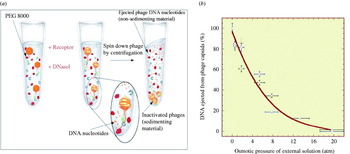
Capsids are permeable to water and smaller ions but are impermeable to larger molecules like polyethylene glycol (PEG) (Evilevitch et al. Reference Evilevitch, Lavelle, Knobler, Raspaud and Gelbart2003). DNA packaging experiments, described above, demonstrate that forces inside a phage capsid can reach values greater than 50 pN (Smith et al. Reference Smith, Tans, Smith, Grimes, Anderson and Bustamante2001; Fuller et al. Reference Fuller, Raymer, Rickgauer, Robertson, Catalano, Anderson, Grimes and Smith2007b) (corresponding to ~50 atm internal pressure) when a genome is fully packaged. Binding of the cellular receptor to the phage tail opens the tube and the internal force of the pressurized DNA, F eject, promotes full ejection of the genome. When the ejection is triggered in the presence of added osmolytes, which generate an external osmotic pressure, this ejection force will be balanced by the osmotic force resisting ejection of DNA from the capsid. The osmotic force resisting ejection has been attributed to a decrease of hydration properties of DNA inside the capsid resulting from a change in the chemical activity of water hydrating DNA induced by osmolytes (Evilevitch et al. Reference Evilevitch, Fang, Yoffe, Castelnovo, Rau, Parsegian, Gelbart and Knobler2008). DNA ejection will thus proceed until the forces are equal, F eject=F resist. PEG is an inert osmotic agent that is widely used to set osmotic pressure in biological systems and to mimic bacterial cytoplasm (Zimmerman, Reference Zimmerman1993). In order to address the question as to whether phage DNA ejection can be suppressed by osmotic pressure, we have used PEG 8000 (MW=8000 Da) which is too large to enter the capsid and for which the osmotic pressure has been well calibrated (Parsegian et al. Reference Parsegian, Rand, Fuller and Rau1986; Rau & Parsegian, Reference Rau and Parsegian1992; Evilevitch et al. Reference Evilevitch, Lavelle, Knobler, Raspaud and Gelbart2003). In our experimental set-up, purified phage λ particles were incubated with increasing concentrations of PEG 8000 in the presence of DNaseI (Evilevitch et al. Reference Evilevitch, Lavelle, Knobler, Raspaud and Gelbart2003). Once the tail tube is opened with the LamB receptor, DNA is ejected into solution and digested into nucleotides by DNaseI, leaving the unejected DNA fraction intact (undigested) inside the capsid. The fraction of DNA ejected is determined by measuring the concentration of nucleotides (digested DNA) by UV absorbance after removing DNA-containing capsids by centrifugation (see Fig. 2a). We found that the fraction of DNA ejected from phage λ (capsid outer diameter 63 nm containing 41·5 kb DNA, 86% the length of the wt 48·5 kb genome) decreases monotonically with increasing osmolyte concentration and is completely inhibited at osmotic pressure corresponding to 20 atm (see Fig. 2 b) (Evilevitch et al. Reference Evilevitch, Lavelle, Knobler, Raspaud and Gelbart2003). Analysis of DNA length remaining in the capsid, using pulsed-field gel electrophoresis, nicely confirmed this data (Evilevitch et al. Reference Evilevitch, Gober, Phillips, Knobler and Gelbart2005). Osmotic suppression measurements were also performed with bacteriophage T5 (capsid outer diameter 90 nm containing 113 kb DNA), where the fraction of ejected DNA was determined as a function of osmotic pressure set by PEG 8000 (Castelnovo & Evilevitch, Reference Castelnovo and Evilevitch2007). Similar to phage λ, the fraction of DNA ejected progressively decreased with increase in osmotic pressure exerted by the osmolyte; however, the pressure required to completely inhibit DNA ejection was only 6 atm (compared to 20 atm for λ) (Castelnovo & Evilevitch, Reference Castelnovo and Evilevitch2007). Modeling internal ejection force for phages λ and T5 as a function of the DNA remaining inside the capsid and rescaling these data with regard to the capsid size and DNA filling density, produces a ‘master plot’ showing that despite differences in internal pressure the osmotic suppression data for these two different phages is superimposable (Castelnovo & Evilevitch, Reference Castelnovo and Evilevitch2007). This nicely illustrates that same physical parameters control DNA ejection from two distinctly different bacteriophages and suggests that there is a general mechanism of DNA ejection that applies to many dsDNA viruses that use ATP-driven motors to package their genomes. These osmotic suppression experiments also partially recapitulate ejection behavior in vivo by mimicking the colloidal milieu of the host cell cytoplasm, which also exerts osmotic force on phage DNA during ejection and packaging processes. For instance, osmotic pressure inside E. coli cells under normal conditions is at least 2–4 atm (Neidhardt, Reference Neidhardt1996). We have demonstrated that wt phage λ can passively eject only 60% of its genome against this pressure (Grayson et al. Reference Grayson, Evilevitch, Inamdar, Purohit, Gelbart, Knobler and Phillips2006). Hence, the pressure induced by the tightly packaged DNA inside the phage capsid is insufficient to complete DNA ejection in vivo.
(a) Illustration of osmotic suppression experiment. (b) Fraction of DNA ejected as a function of the osmotic pressure (calculated from the concentration of PEG 8000). Data are presented for samples taken from three different phage stocks. Horizontal and vertical error bars represent 1 standard deviation and were determined by propagation of errors in the PEG concentration and the absorbance.

Various two-step in vivo DNA internalization scenarios have been suggested for phage (Molineux, Reference Molineux2001; Gonzalez-Huici et al. Reference Gonzalez-Huici, Salas and Hermoso2004; Kemp et al. Reference Kemp, Gupta and Molineux2004; de Frutos et al. Reference De Frutos, Letellier and Raspaud2005b; Evilevitch, Reference Evilevitch2006; Inamdar et al. Reference Inamdar, Gelbart and Phillips2006; Alcorlo et al. Reference Alcorlo, Gonzalez-Huici, Hermoso, Meijer and Salas2007; Lof et al. Reference Lof, Schillen, Jonsson and Evilevitch2007b). For example, genome transfer from φ29 is described as a push-pull mechanism such that the first ~65% of the genome is ‘pushed’ into the cell by pressure, while the remaining 35% is ‘pulled’ into the cell by enzyme-assisted processes (Gonzalez-Huici et al. Reference Gonzalez-Huici, Salas and Hermoso2004; Alcorlo et al. Reference Alcorlo, Gonzalez-Huici, Hermoso, Meijer and Salas2007). Similarly, it was shown that only 8% of T5 genome is injected into cell in the first step, allowing production of phage-encoded proteins that are necessary for the internalization of the remaining DNA (Lanni, Reference Lanni1968). Interestingly, the in vitro osmotic suppression measurements on phage T5 described above indicate only ~9% of the wt T5 genome (121·75 kbp) is ejected at an osmotic pressure of 3–4 atm (corresponding to cytoplasmic pressure), consistent with the in vivo observations (Castelnovo & Evilevitch, Reference Castelnovo and Evilevitch2007). For bacteriophage T7, experimental evidence suggests that its entire genome is actively pulled into the cell by RNA polymerases, similar to the situation observed for T5 (Kemp et al. Reference Kemp, Gupta and Molineux2004; Molineux, Reference Molineux2001). In addition, there is now evidence that the portion of DNA that is not passively ejected into the cell by pressure can be pulled out of the phage capsid by adsorbing and ratcheting effects of non-specific DNA-binding proteins, such as the HU protein in E. coli (Lof et al. Reference Lof, Schillen, Jonsson and Evilevitch2007b), as well as by the condensing effects of polyamines (i.e. spermidine) also present in cells (de Frutos et al. Reference De Frutos, Brasiles, Tavares and Raspaud2005a; Evilevitch, Reference Evilevitch2006). For instance, the addition of a non-specific DNA-binding protein, HU, to a solution of phage λ has been shown to significantly increase the rate of ejection (Lof et al. Reference Lof, Schillen, Jonsson and Evilevitch2007b).
As has been discussed above, the length of the packaged DNA has a pronounced effect on the internal pressure. The ability to accurately predict the force required to stop DNA ejection or to stop packaging of the DNA into a capsid is clearly important to our fundamental understanding of the viral life-cycle and has many practical applications. For instance, for gene therapy purposes non-viral DNA vectors are packaged into adenovirus capsids that are transfected into mammalian cells (Amalfitano & Parks, Reference Amalfitano and Parks2002). Therefore, it is important to have models that can predict whether vector packaging is feasible. As described above, such direct comparisons and rescaling based on the DNA-filling density and capsid size have been made with phages λ and T5 (Castelnovo & Evilevitch, Reference Castelnovo and Evilevitch2007). Furthermore, one can directly verify the effect of genome length on internal force by osmotic suppression studies on the same phage but with different genome lengths packaged into it. The effect of genome length was verified for phage λ mutants containing 48·5 (wt phage), 41·5 and 37·7 kb DNA (Grayson et al. Reference Grayson, Evilevitch, Inamdar, Purohit, Gelbart, Knobler and Phillips2006). Ejection of DNA from all three λ mutants rapidly decreases up to ~50% ejected fraction and then slowly progresses to completion. It was evident that the length of ejected DNA of the shortest DNA λ mutant (37·7 kb) was always smaller than that of wt λ (48·5 kb) at every PEG concentration. The external osmotic pressure required to completely inhibit DNA ejection was found to be strongly dependent on the genome length. For λ (37·7 kb) it was found to be 10–15 atm while that for λ (48·5 kb) was 20–25 atm. Thus, a 20% longer genome (37·7 kb vs. 48·5 kb) doubles the pressure that suppresses ejection. A nice agreement between these data and the theoretical model description (Kindt et al. Reference Kindt, Tzlil, Ben-Shaul and Gelbart2001; Purohit et al. Reference Purohit, Kondev and Phillips2003b; Tzlil et al. Reference Tzlil, Kindt, Gelbart and Ben-Shaul2003) was found (Grayson et al. Reference Grayson, Evilevitch, Inamdar, Purohit, Gelbart, Knobler and Phillips2006).
Considering that viral capsids are permeable to ions and realizing that electrostatic interactions between DNA strands is the major determining parameter of internal force, salt conditions become equally important, on a par with packaged genome length. The effect of mono-, di- and multivalent DNA counter ions on internal pressure has been investigated. The extent of ejection from phage λ at a fixed osmotic pressure of 4 atm (mimicking cytoplasmic pressure) has been measured as a function of concentration of salts with 50 mm Tris–HCl and 10 mm MgSO4 as standard buffer present in all solutions (Evilevitch et al. Reference Evilevitch, Fang, Yoffe, Castelnovo, Rau, Parsegian, Gelbart and Knobler2008). While Na+ ions had no effect on the fraction of DNA ejected, adding 30 mm Mg2+ ions halved the ejected DNA fraction (which initially was around 60%) and with 1 mm tetravalent spermine the ejection was nearly completely suppressed with only 5% DNA ejected (Evilevitch et al. Reference Evilevitch, Fang, Yoffe, Castelnovo, Rau, Parsegian, Gelbart and Knobler2008). This is explained by the fact that Mg2+ ions screen DNA–DNA repulsive interactions while spermine also induces attraction between DNA strands (Suwalsky et al. Reference Suwalsky, Traub, Shmueli and Subirana1969; Schellman & Parthasarathy, Reference Schellman and Parthasarathy1984; Arscott et al. Reference Arscott, Li and Bloomfield1990; Bloomfield, Reference Bloomfield1991; Sikorav et al. Reference Sikorav, Pelta and Livolant1994; Raspaud et al. Reference Raspaud, Olvera De La Cruz, Sikorav and Livolant1998, Reference Raspaud, Durand and Livolant2005), leading to a significant decrease in the internal ejecting force. In fact, cryo-electron micrographs have shown that a stable DNA toroid remains inside the capsid when λ is incubated with LamB receptor in vitro in the presence of 1 mm spermine (4+) and DNaseI (Evilevitch, Reference Evilevitch2006) (see Fig. 3). This cryo-EM micrograph shows a relaxed DNA toroid inside the phage capsid when 60% of its genome has been ejected allowing d-spacing between neighboring DNA strands in the capsid to be equal to the d-spacing found in the a DNA toroid condensed by spermine in the bulk (Suwalsky et al. Reference Suwalsky, Traub, Shmueli and Subirana1969; Schellman & Parthasarathy, Reference Schellman and Parthasarathy1984; Arscott et al. Reference Arscott, Li and Bloomfield1990; Bloomfield, Reference Bloomfield1991; Sikorav et al. Reference Sikorav, Pelta and Livolant1994; Raspaud et al. Reference Raspaud, Olvera De La Cruz, Sikorav and Livolant1998, Reference Raspaud, Durand and Livolant2005). Interestingly, if DNase is not present in the sample, all of the DNA will be pulled out of the capsid by the larger DNA condensate (condensed by spermine) formed outside phage particles. This is similar to an Ostwald ripening process, with the larger toroid growing at the expense of the smaller one (Evilevitch, Reference Evilevitch2006). Coincidently, early studies found that infection of phage λ in vivo is inhibited in the presence of several polyamines (Harrison & Bode, Reference Harrison and Bode1975). The fact that spermine significantly decreases the DNA ejection force within the capsid and that this force is required to eject DNA into an osmotic pressure gradient of few atmospheres in the cell can explain this data. It should be noted, however, that the presence of polyamines can also affect the transmembrane potential which has been shown to be important for phage T7 infection (Molineux, Reference Molineux2001; Kemp et al. Reference Kemp, Gupta and Molineux2004).
Cryoelectron micrograph of toroidally condensed DNA remaining unstressed in the capsid (wild-type phage, 48·5 kbp), following ejection at zero ‘external’ osmotic pressure in the presence of spermine.

Internal and external forces acting on viral DNA can also be probed by measuring the rate of DNA ejection. The rate of ejection in vitro from phage λ was determined using time-resolved dynamic light scattering as a function of DNA length and temperature (Lof et al. Reference Lof, Schillen, Jonsson and Evilevitch2007a, Reference Lof, Schillen, Jonsson and Evilevitchb). Comparing two genome-length mutants (37·7 and 45·7 kbp) with wt λ(48·5 kbp), it was found that the initial ejection rate increases in almost direct relationship with the initially packaged genome length, while the total time to completely eject DNA was nearly constant for all the genome lengths. Assuming that the net force of ejection is F=F eject−fv, where F eject is the internal DNA ejection force, f is the friction coefficient and v is the rate of ejection, F eject−fv=0 at steady state and the rate of ejection is therefore v=F eject/f. Assuming that the friction coefficient is the same for all three phages (since capsid and tail dimensions, where friction occurs with the DNA, are constant for all three viruses), then the rate of ejection will depend directly on F eject, which has indeed been observed (Lof et al. Reference Lof, Schillen, Jonsson and Evilevitch2007b). This again confirms that initial DNA ejection from phage is entirely dependent on the internal DNA force. In accord with earlier observations of salt effect on internal genome force (Evilevitch et al. Reference Evilevitch, Castelnovo, Knobler and Gelbart2004, Reference Evilevitch, Fang, Yoffe, Castelnovo, Rau, Parsegian, Gelbart and Knobler2008), single-molecule fluoresce measurements (Grayson et al. Reference Grayson, Han, Winther and Phillips2007) of DNA ejection kinetics from λ also show strong ejection rate dependence on salt conditions. It was found that replacing 10 mm Na+ with 10 mm Mg2+ ions increased ejection time nearly tenfold, suggesting that internal force inside phage decreases by a factor of 2. Maximum measured DNA translocation rates for DNA ejection from λ were up to 60 kb/s (with 10 mm Na+ ions in 10 mm Tris–HCl buffer), comparable to translocation rates found for phage T5 (lower bound of 75 kb/s) in an analogous single-molecule fluorescence study (Mangenot et al. Reference Mangenot, Hochrein, Radler and Letellier2005). Similar speeds were found for the opposite process of DNA packaging in φ29 (10 kb/s) (Rickgauer et al. Reference Rickgauer, Fuller, Grimes, Jardine, Anderson and Smith2008). The kinetics of T5 DNA ejection was also investigated using static light scattering (de Frutos et al. Reference De Frutos, Letellier and Raspaud2005b); however, direct comparison with monotonic ejection from λ cannot be made since step-wise ejection behavior was observed in T5 due to the presence of nicks in the DNA. While most of the genome is ejected by internal pressure in vitro in the absence of external osmotic pressure, the last unconstrained DNA part will remain in the tail for a long time before it will diffuse out, since it is not affected by DNA–DNA repulsion and bending forces (Evilevitch, Reference Evilevitch2006). This makes it difficult to determine the total time required for complete ejection in both single-molecule and bulk measurements.
Current internal force models accurately account for changes in ejection rate associated with changes in genome length and salt conditions. However, they fail, to explain the temperature effect on ejection kinetics. While internal force predicts weak temperature dependence, we found nearly exponential increase in ejection rate with increasing temperature (Lof et al. Reference Lof, Schillen, Jonsson and Evilevitch2007b). Comparing the ejection times for λ, we see that there is an almost tenfold rate difference, with most DNA released in 11 min at 15°C compared to 1 min at 37°C. A suggested mechanism that explains such ejection behavior is due to the temperature-sensitive conformational changes induced in the tail-receptor complex forming the pore (including the portal) through which DNA passes. It is likely that the tail-receptor portal can adopt at least two conformations – an ‘open’ conformation through which DNA can freely pass or ‘closed’ conformation blocking the DNA exit (see above). We propose that the probability of occurrence of the ‘open’ conformation increases with increasing temperature, keeping the portal-tail pore open longer and thus speeding up the ejection of DNA from phage. Such a mechanism will be coupled to energy of activation. This also explains why no ejection occurs in vitro from λ at 4°C with receptor present (Lof et al. Reference Lof, Schillen, Jonsson and Evilevitch2007b). Similarly, DNA ejection from phage T5 is completed within minutes at 30–41°C, while at lower temperatures it takes hours to days for completion (de Frutos et al. Reference De Frutos, Letellier and Raspaud2005b); the energy of activation barrier for T5 was estimated as 2·9×10−19 J (de Frutos et al. Reference De Frutos, Letellier and Raspaud2005b).
In contrast to the phages, the entire viral particle enters the host cell in eukaryotic viruses. In many cases, however, the viral genome still needs to be ejected into the cell nucleolus (Ojala et al. Reference Ojala, Sodeik, Ebersold, Kutay and Helenius2000). For some dsDNA eukaryotic viruses, like herpes simplex virus type 1 (HSV-1), it was shown that it injects its DNA into nucleus in a manner analogous to bacteriophage DNA ejection into the host cytoplasm (Cardone et al. Reference Cardone, Winkler, Trus, Cheng, Heuser, Newcomb, Brown and Steven2007; Newcomb et al. Reference Newcomb, Booy and Brown2007). After the HSV-1 nucleocapsid has entered the host cell by a membrane fusion event, it is transported towards the nucleus where it docks to the nuclear pore and ejects its genome into the nucleus, leaving the empty capsid in the cytosol. Thus, HSV-1 strongly resembles many aspects of phage biology in that it also packages its DNA into a pre-assembled capsid using an ATP-driven motor and ejects the genome through a portal located in one of its vertexes (Cardone et al. Reference Cardone, Winkler, Trus, Cheng, Heuser, Newcomb, Brown and Steven2007; Newcomb et al. Reference Newcomb, Booy and Brown2007).
The experiments described above suggest that internal pressure resulting from DNA confinement within the capsid may provide a common mechanism for genome ejection in many dsDNA viruses, specifically those that package their genomes with an ATP-driven motor [the small dsDNA viruses that spontaneously self-assemble a nucleocapsid, e.g. polyoma virus, do not utilize a packaging motor and thus are not under pressure (Oppenheim & Peleg, Reference Oppenheim and Peleg1989)]. Thus the ability of these viruses to infect by injecting their genome into the cell or cell nucleus is determined by DNA internal force set by the packaged DNA density and capsid size. This force also strongly depends on ambient salt conditions. However, this pressure alone appears to be insufficient to deliver the entire genome into the cell. Once the passive pressure-driven ejection step has ceased, cellular DNA-binding proteins and polyamines can complete internalization of the viral genome by ratcheting, adsorption and/or condensation mechanisms.
3. Capsid strength
Theoretical considerations
In this section, we introduce briefly the physical concepts underlying capsid stability. Viral capsids are formed by the self-assembly of one or several proteins (two in the case of phage λ, excluding portal protein) to form a closed shell structure with high symmetry. As outlined by the pioneering work of Caspar & Klug (Reference Caspar and Klug1962), most viral shells exhibit icosahedral symmetry composed of pentameric (i.e. five proteins) and hexameric (i.e. six proteins) structures at the shell's faces. Accordingly, viral capsids are classified using the so-called T numbers: an icosahedral structure can be constructed from a hexagonal lattice by inserting pentagons at sites of the hexagonal lattice that are separated by h steps along one lattice direction and k steps along the other, with h and k non-negative integers. The integer T=h 2+k 2+hk is then the number of unequivalent sites in the resulting shell, which is comprised of 12 pentagons and 10 (T – 1) hexagons. Therefore, e.g., phage λ head is classified as an icosahedral T=7 capsid (h=2 and k=1) (Dokland & Murialdo, Reference Dokland and Murialdo1993).
From a physical point of view, the capsid must withstand several tens of atmospheres internal pressure due to packaged DNA, as discussed above. The interactions between proteins at the capsid surface are mediated by non-covalent hydrophobic, hydrophilic and electrostatic bonding interactions that provide the robustness and stability of the capsid. A variety of approaches have been taken to gain theoretical insight into the question of capsid stability (reviewed in Gibbons & Klug, Reference Gibbons and Klug2007a).
First, capsids were investigated using normal mode analysis starting with atomic information on capsid structure available from crystallographic data in the Protein Data Bank (Tama & Brooks, Reference Tama and Brooks2005). This method allows the main directions of motion of the whole capsid to be determined and has proven a useful method to analyze large-scale conformational changes in some plant viruses (e.g. CCMV, which is an RNA virus). However, this method does not allow precise quantification of capsid robustness. A second approach to investigate capsid stability is to start from a macroscopic continuous description of closed shells. We restrict ourselves in this section to the particular case of thin shells, which is effectively the most relevant one for interpretation of nano-indentation studies on capsids (see Gibbons & Klug, Reference Gibbons and Klug2007b). Within this approach, the capsid is described by two elastic constants characterizing the in plane stretching of the shell, and its bending out of plane. These elastic constants are conveniently combined into the Foppl–von Karman (FvK) dimensionless parameter, which for a spherical shell of radius R:

where E is the 2D Young modulus and κ is the out-of-plane bending modulus. This number represents the ratio between stretching and bending stiffness. Several works were devoted to the analysis of these continuous models with different geometries by varying the FvK number (Lidmar et al. Reference Lidmar, Mirny and Nelson2003; Nguyen et al. Reference Nguyen, Bruinsma and Gelbart2005; Widom et al. Reference Widom, Lidmar and Nelson2007). In particular, it was shown that the FvK number controls the general morphology of the thin shell: at low FvK numbers, the shell is nearly spherical while for larger FvK numbers a significant faceting of the shell is observed. Furthermore, the models constitute a theoretical framework for the interpretation of capsid nano-indentation experiments, since it predicts an almost linear relationship between force and indentation (Ivanovska et al. Reference Ivanovska, De Pablo, Ibarra, Sgalari, Mackintosh, Carrascosa, Schmidt and Wuite2004, Reference Ivanovska, Wuite, Jonsson and Evilevitch2007; Klug et al. Reference Klug, Bruinsma, Michel, Knobler, Ivanovska, Schmidt and Wuite2006; Michel et al. Reference Michel, Ivanovska, Gibbons, Klug, Knobler, Wuite and Schmidt2006). More precisely, the effective spring constant associated to this linear behavior is given by:

where h is the capsid wall thickness and α is a factor depending on indentation geometry.
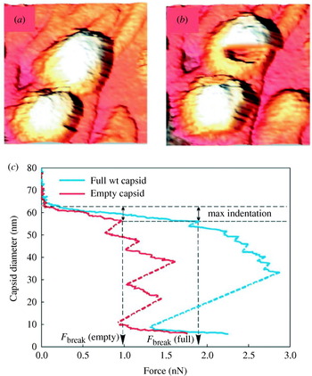
Complementary insights on capsid stability are coming from the use of triangulated surfaces, as described by Buenemann & Lenz (Reference Buenemann and Lenz2007). These authors reproduced a number of experimental observations using triangulated surfaces with elastic (stretching) and bending energy, where each capsomer is represented by several vertices. Moreover, different behavior is observed when the indentation is performed on pentamers or hexamers, the former being softer than the latter. This weakness of pentamers in the icosahedral capsid is predicted to result in a buckling instability, where the main curvature of the shell has been inverted at the pentamers. This type of model allows prediction of the critical breaking force of empty and full capsids. It was theoretically found that pressurized capsids, like in DNA-filled capsids, increase the critical breaking force upon indentation, the pressurized genome helping to resist the imposed deformation. Finally, critical rupture by internal stress (no external force applied), as provoked by osmotic shock experiments, was also investigated. In such an experiment, the internal pressure of a capsid is sharply increased due to a sudden change in the environment, and forces greater than critical breaking force are generated. Interestingly, Buenemann & Lenz (Reference Buenemann and Lenz2007) found that capsid robustness, as determined by critical rupture force, correlates well with the presence of a pressurized or a non-pressurized genome.
3.2 Measuring elasticity and strength of viral capsids
Survival success of viruses hinges in their ability to take control over the host's cellular machineries, and to withstand physical conditions to which they are exposed. The latter is critically dependent on factors such as genome density and capsid strength (De Paepe & Taddei, Reference De Paepe and Taddei2006). More explicitly, safekeeping of viral genetic material, successful delivery of the genome into the host cell, and survival between infection events, during which viruses are susceptible to a variety of mechanical damage, are critical. As discussed above, phages (and quite likely many eukaryotic dsDNA viruses) are exposed to internal genome pressures of tens of atmospheres. While, high internal pressure is required for successful genome injection into a host cell, it was also shown that viability of 12 different phages, defined by capsid rupture, was significantly increasing with increasing density of packaged DNA (De Paepe & Taddei, Reference De Paepe and Taddei2006). Notwithstanding, phages in nature are well suited to host-to-host replication cycle, by virtue of their capacity to withstand environmental stresses, including pH, temperature, osmotic pressure, salt concentration and mechanical shear forces. For example, E. coli, which is host to many tailed phages, is subjected to high shear forces in nature (Thomas et al. Reference Thomas, Trintchina, Forero, Vogel and Sokurenko2002). Therefore, phages that in turn adsorb to the surface of E. coli cells during infection are also exposed to high external shear forces (Ivanovska et al. Reference Ivanovska, Wuite, Jonsson and Evilevitch2007). As discussed above, viral capsids are typically held together by non-covalent bonding interactions (hydrophobic, electrostatic, etc.) and the ensemble of interactions must withstand the internal capsid pressures and in addition a variety of external forces. All of these factors raise some conceptual questions:
• What are the mechanical properties of capsids and how do they depend on capsid dimensions?
• How is packaged genome length correlated with the capsid strength?
Capsids of many viruses can be self-assembled in vitro from their purified proteins (the morphologically defined units of which are also called ‘capsomers’), given the proper buffer conditions (Caspar & Klug, Reference Caspar and Klug1962; Bancroft et al. Reference Bancroft, Hiebert, Rees and Markham1968a, Reference Bancroft, Wagner and Brackerb; Finch & Bancroft, Reference Finch and Bancroft1968; Hiebert et al. Reference Hiebert, Bancroft and Bracker1968; Wagner & Bancroft, Reference Wagner and Bancroft1968; Kushner, Reference Kushner1969). So for instance, cowpea chlorotic mottle virus (CCMV, a plant virus), can self-assemble around its RNA genome without involvement of auxiliary factors (Bancroft et al. Reference Bancroft, Hiebert, Rees and Markham1968a, Reference Bancroft, Wagner and Brackerb; Finch & Bancroft, Reference Finch and Bancroft1968; Hiebert et al. Reference Hiebert, Bancroft and Bracker1968; Wagner & Bancroft, Reference Wagner and Bancroft1968). In contrast, many dsDNA viruses actively package their genomes into a pre-assembled capsid. In these cases, capsid assembly requires the transient association of chaperones, scaffolding proteins and maturation proteases (Hendrix et al. Reference Hendrix, Roberts, Stahl and Weisberg1983; Dokland, Reference Dokland1999; Ptashne, Reference Ptashne2004; Gaussier et al. Reference Gaussier, Yang and Catalano2006). Scaffolding proteins bind to capsid shell proteins inducing co-polymerization into an icosahedral protein shell. Regardless of the mode of assembly, the resultant capsids are comparable in structure (typically icosahedral symmetry) and in non-covalent capsomer–capsomer interactions (Wikoff et al. Reference Wikoff, Duda, Hendrix and Johnson1998, Reference Wikoff, Duda, Hendrix and Johnson1999, Reference Wikoff, Liljas, Duda, Tsuruta, Hendrix and Johnson2000, Reference Wikoff, Che, Duda, Hendrix and Johnson2003, Reference Wikoff, Conway, Tang, Lee, Gan, Cheng, Duda, Hendrix, Steven and Johnson2006; Lata et al. Reference Lata, Conway, Cheng, Duda, Hendrix, Wikoff, Johnson, Tsuruta and Steven2000; Gan et al. Reference Gan, Conway, Firek, Cheng, Hendrix, Steven, Johnson and Duda2004, Reference Gan, Speir, Conway, Lander, Cheng, Firek, Hendrix, Duda, Liljas and Johnson2006; Lee et al. Reference Lee, Gan, Tsuruta, Hendrix, Duda and Johnson2004). The assembly process is often followed by a maturation step (typically induced by DNA packaging), in which the capsid proteins undergo rearrangement (sometimes capsid proteins leave while new ones are added) resulting in capsid expansion and shell wall thinning.
The mechanical properties of capsids have been studied with several techniques including calorimetry (Ross et al. Reference Ross, Cheng, Conway, Firek, Hendrix, Duda and Steven2005, Reference Ross, Conway, Cheng, Dierkes, Firek, Hendrix, Steven and Duda2006; Conway et al. Reference Conway, Cheng, Ross, Hendrix, Duda and Steven2007), osmotic shock (Anderson, Reference Anderson1950; Anderson et al. Reference Anderson, Rappaport and Muscatine1953; Cordova et al. Reference Cordova, Deserno, Gelbart and Ben-Shaul2003) and atomic force microscopy (AFM) (Ivanovska et al. Reference Ivanovska, De Pablo, Ibarra, Sgalari, Mackintosh, Carrascosa, Schmidt and Wuite2004, Reference Ivanovska, Wuite, Jonsson and Evilevitch2007; Carrasco et al. Reference Carrasco, Carreira, Schaap, Serena, Gomez-Herrero, Mateu and De Pablo2006; Klug et al. Reference Klug, Bruinsma, Michel, Knobler, Ivanovska, Schmidt and Wuite2006; Michel et al. Reference Michel, Ivanovska, Gibbons, Klug, Knobler, Wuite and Schmidt2006; Kol et al. Reference Kol, Gladnikoff, Barlam, Shneck, Rein and Rousso2006, Reference Kol, Shi, Tsvitov, Barlam, Shneck, Kay and Rousso2007). For instance, different stages of phage HK97 capsid assembly were analyzed with differential scanning calorimetry (DSC) by determining the transition temperature for capsid denaturation and enthalpy change associated with it (Ross et al. Reference Ross, Cheng, Conway, Firek, Hendrix, Duda and Steven2005, Reference Ross, Conway, Cheng, Dierkes, Firek, Hendrix, Steven and Duda2006; Conway et al. Reference Conway, Cheng, Ross, Hendrix, Duda and Steven2007). It was found that covalent cross-linking following capsid expansion (doubling capsid volume) during maturation significantly enhances capsid stability. In osmotic shock measurements several bacteriophages were first incubated in high salt solution followed by rapid dilution (Anderson, Reference Anderson1950; Anderson et al. Reference Anderson, Rappaport and Muscatine1953; Cordova et al. Reference Cordova, Deserno, Gelbart and Ben-Shaul2003). In cases where the capsid is more permeable to water than to salt ions, rapid uptake of water molecules (diluting the higher salt concentration inside) creates increasing internal stress on the capsid walls resulting in capsid burst. It was found that by virtue of higher capsid permeability, the so-called odd-numbered T phages T1, T3, T5 and T7 (note, that this historical definition refers to the serological and phylogenetic relationship and is not related to earlier defined triangulation number) are able to withstand osmotic shock, while phages T2, T4, and T6 are not. Based on these data, Cordova et al. (Reference Cordova, Deserno, Gelbart and Ben-Shaul2003) provided theoretical estimates of capsid strength using several approximations. As mentioned above, in the context of salt effect on internal genome pressure required for infection, this example also illustrates how capsid permeability to water and ions provides nature with external control over both successful genome release and capsid stability.
While DSC and osmotic shock measurements revealed valuable insights into the mechanical properties of capsids, the data are highly qualitative. On the other hand, by means of AFM it is possible to quantitatively study mechanical properties of viral particles by manipulating individual capsids at the nanometer scale (Ivanovska et al. Reference Ivanovska, De Pablo, Ibarra, Sgalari, Mackintosh, Carrascosa, Schmidt and Wuite2004). In addition to providing high-resolution imaging of viral particles in liquid (see Fig. 4 a, b), the AFM tip can apply force on a single viral particle in order to obtain real-time force–distance (F–Z) curves as the nanometer-size tip of the cantilever scans the specimen surface (Ivanovska et al. Reference Ivanovska, De Pablo, Ibarra, Sgalari, Mackintosh, Carrascosa, Schmidt and Wuite2004) (see Fig. 4 c). Probing phage capsids with an AFM tip generally results in two regimes: reversible deformations are usually observed when force below a certain value is applied, while irreversible rupturing occurs at higher applied force values (Ivanovska et al. Reference Ivanovska, De Pablo, Ibarra, Sgalari, Mackintosh, Carrascosa, Schmidt and Wuite2004, Reference Ivanovska, Wuite, Jonsson and Evilevitch2007). These studies provide three parameters describing capsid mechanical properties. (i) Information about capsid's mechanical limits and strength of capsomer–capsomer bonds is provided by the threshold force beyond which the interactions between capsid proteins collapse, referred to as ‘breaking force’, F break. (ii) Due to essentially linear response to deformation, the spring constant, k, can be obtained by linear fit to the force–distance curves. (iii) Using homogeneous thin-shell theory approximation (Ivanovska et al. Reference Ivanovska, De Pablo, Ibarra, Sgalari, Mackintosh, Carrascosa, Schmidt and Wuite2004) or finite element simulations (Klug et al. Reference Klug, Bruinsma, Michel, Knobler, Ivanovska, Schmidt and Wuite2006), Young's modulus, E, can also be obtained. The spring constant and Young's modulus are measures of the capsid's elasticity, and while k is dependent on materials' geometry, E is an intrinsic geometry-independent material property.
High-resolution AFM imaging of λ phage capsids (3D with shadow): (a) intact phage λ, (b) same phage as in panel (a) but after breakage induced by an AFM tip, (c) F–Z curves of full wild-type vs. empty-phage λ. The data is displayed as observed capsid height vs. force. The deformation of the capsid is linear until a critical deformation is reached, after which a sudden decrease in capsid height can be observed. The dashed line is inserted to guide the eye, because there is no data recorded between these gaps. (Adopted from Ivanovska et al. Reference Ivanovska, Wuite, Jonsson and Evilevitch2007.)

AFM nano-indentation studies of empty viral capsids reveal unique mechanical properties for different classes of viruses, which can in turn be directly related to a virion's replication cycle. For example, we have stressed throughout this review that the presence of internal genome pressure in the complex dsDNA viruses, which is required for DNA ejection into the host cell, is distinct from many eukaryotic single-stranded RNA (ssRNA) viruses, which do not appear to require internal pressure for genome release. Indeed, it was found that Young's modulus of the empty φ29 procapsid is ~1·8 GPa (Ivanovska et al. Reference Ivanovska, De Pablo, Ibarra, Sgalari, Mackintosh, Carrascosa, Schmidt and Wuite2004) (comparable to hard plastics) which is close to the value obtained for empty phage λ capsid ~1 GPa (Ivanovska et al. Reference Ivanovska, Wuite, Jonsson and Evilevitch2007). For comparison, Young's modulus for ssRNA plant virus CCMV was measured as 0·14 GPa (Michel et al. Reference Michel, Ivanovska, Gibbons, Klug, Knobler, Wuite and Schmidt2006) and in animal virus murine leukemia virus (MLV) it was 0·23 GPa (Kol et al. Reference Kol, Gladnikoff, Barlam, Shneck, Rein and Rousso2006). The fact that Young's modulus for these two ssRNA viruses is one order of magnitude smaller than for dsDNA bacteriophages above reflects that the phage capsid is significantly more robust and can tolerate much higher internal force exerted by its genome. At the same time, maximum indentation is similar for all four viruses above, about 20–30% of capsid height. Above this indentation, the AFM cantilever tip causes irreversible capsid rupture with catastrophic drop of force. However, the force required to break an empty λ capsid is 0·8 nN (Ivanovska et al. Reference Ivanovska, Wuite, Jonsson and Evilevitch2007), similar to the force found for CCMV (0·6 nN) (Michel et al. Reference Michel, Ivanovska, Gibbons, Klug, Knobler, Wuite and Schmidt2006) but twofold lower than breaking force found for empty φ29 (~1·5 nN) (Ivanovska et al. Reference Ivanovska, De Pablo, Ibarra, Sgalari, Mackintosh, Carrascosa, Schmidt and Wuite2004). Taking also into account capsid thickness, this provides us with information about capsid rigidity and capsomer–capsomer interactions. The φ29 procapsid, which has a thickness similar to that of λ (~1·8 nm) is able to withstand higher force by being more elastic (also confirmed by measured spring constants: 0·31 N/m for φ29 and 0·13 N/m for λ). This can be related to the fact that λ has slightly higher DNA packaging density and thus higher internal pressure compared to φ29 (Purohit et al. Reference Purohit, Kondev and Phillips2003b) and therefore needs to be more rigid in order to maintain its pressure constant without expansion.
In addition to probing capsid pressure, elasticity and strength, one can also use AFM to study particle maturation steps and infectivity mechanisms in eukaryotic viruses. After budding from the infected cell, the HIV capsid matures undergoing morphological reorganization changing from a thick spherical protein capsid to a much thinner conically shaped capsid. Once HIV has matured, it can fuse with the T-cell membrane in order to infect. By studying the mechanical properties of immature vs. mature HIV particles it was found that Young's modulus decreases by almost eightfold in the mature capsid (from 930 MPa to 115 MPa) (Kol et al. Reference Kol, Shi, Tsvitov, Barlam, Shneck, Kay and Rousso2007). Interestingly, a parallel X-ray study of the T-cell membrane found that the membrane-bending modulus is significantly reduced upon addition of the HIV fusion peptide FP-23 (Tristram-Nagle & Nagle, Reference Tristram-Nagle and Nagle2007), which reduces the free-energy barrier required for fusion of the virus with the cell. This nicely correlates with the mechanical softening of the mature HIV particle that in turn promotes its ability to fuse and infect.
Thus, AFM probing of viral shells reveals fundamental physical properties controlling the viral replication cycle. Recent discovery of genome pressures in several phages raises the question about the correlation between capsid strength and genome pressure. As discussed in Section 2.2, many phages exhibit lower and upper packaged DNA length limits; for instance, it has been shown that phage λ can package only between 78% and 106% of its genome in vitro (where 100% corresponds to wt λ-DNA 48·5 kb) (Feiss et al. Reference Feiss, Fisher, Crayton and Egner1977). With the help of AFM one can learn whether capsid strength limits the extent to which a viral shell can be pressurized, which must in turn be correlated with the maximum force that the DNA packaging motor can exert. With jumping mode AFM, we have measured force–distance deformation of phage λ capsids as a function of packaged genome length for 78–100% of wt λ-DNA length (37·7, 45·7 and 48·5 kb DNA) (Ivanovska et al. Reference Ivanovska, Wuite, Jonsson and Evilevitch2007). All capsids behaved elastically at small deformations, and spring constants could be obtained from the force–distance curve slopes. The maximum capsid breaking force was also determined. These data indicate that phages with wt DNA are twice as strong as shorter genome mutants, which behave like empty capsids (F break~1·6 nN vs. ~0·8 nN and k~0·23 N/m vs. ~0·13 N/m), as shown in Fig. 4 c. This is regardless of high internal pressure of tens of atmospheres present also in shorter DNA-length phage mutants (Grayson et al. Reference Grayson, Evilevitch, Inamdar, Purohit, Gelbart, Knobler and Phillips2006). This is due to the fact that DNA-hydrating water molecules, exert an osmotic pressure inside capsids that increases exponentially when the packaged DNA density is close to wt phage. This osmotic pressure raises the wt DNA capsid strength and is approximately equal to the maximum breaking force of empty shells. This result confirms that the strength of the shells limits the maximal packaged genome length.
Upon AFM tip-induced deformation of the capsid, water molecules hydrating tightly packed DNA are displaced through the capsid pores. The DNA hydration force is the force required to displace water molecules from DNA out to the bulk solution and, is described by the osmotic pressure. Using DNA osmotic pressure data (Parsegian et al. Reference Parsegian, Rand, Fuller and Rau1986; Strey et al. Reference Strey, Podgornik, Rau and Parsegian1998; Rau & Parsegian, Reference Rau and Parsegian1992) it is evident that the osmotic pressure inside the capsid is sufficient to provide extra strength against external deformation only at 100% wt DNA packaging density, analogous to the osmotic pressure in biological cells. Thus it is not the DNA itself but water molecules hydrating it that provides significant contribution to the force resisting capsid deformation and breaking. Consistently, if the DNA hydration forces are decreased by incubating wt DNA phage λ in higher salt concentration, the capsid changes its mechanical properties and it behaves like an empty capsid (Ivanovska et al. Reference Ivanovska, Wuite, Jonsson and Evilevitch2007). This demonstration provides a stringent test of the hydration force interpretation above. We have proposed that such evolutionary energy optimization presumably determines the genome length of wt DNA phages, since they can survive twice the external mechanical stress compared to the shorter genome mutants that are abundant in nature (Ivanovska et al. Reference Ivanovska, Wuite, Jonsson and Evilevitch2007). As mentioned above, external mechanical stresses comparable to the stress induced on the capsid by the AFM cantilever is present in the natural phage environment due to the fact that its host (E. coli) forms colonies under high shear flows (Thomas et al. Reference Thomas, Trintchina, Forero, Vogel and Sokurenko2002). Thus, the capsid's strength is related to phage's ability to infect (by optimizing genome packaging density that must provide sufficient pressure) and also survive internal and external stresses to which it is exposed between infection events.
Similar to phage λ, the genome-filled capsids of CCMV virus (Michel et al. Reference Michel, Ivanovska, Gibbons, Klug, Knobler, Wuite and Schmidt2006) and ssDNA MVM virus (minute virus of mice) (Carrasco et al. Reference Carrasco, Carreira, Schaap, Serena, Gomez-Herrero, Mateu and De Pablo2006) have greater resistance to breaking with an AFM tip, indicating greater capsid strength in comparison to empty capsids. However, the physical basis for this behavior may differ from that found in λ since these viruses do not have internal pressure and the genome hydration force is therefore low.
Internal force from higher genome-packaging density in phage leads to increased lateral stress on the capsid walls due to the large outward pressure. Because of the thermal fluctuations of capsid proteins, this results in more frequent rupture of the capsids in direct correlation with the genome density (De Paepe & Taddei, Reference De Paepe and Taddei2006). On the contrary, osmotic outward pressure also caused by high genome confinement stabilizes phage particles against external deformations, as discussed immediately above. Thus, in nature, there is a precise balance of forces acting externally and internally on a viral particle that are determined by the virus's genome length and the capsid's strength, that are in turn controlled by salt ions in the viral milieu.
4. Concluding remarks
The majority of viruses possess spherical, icosahedral protein shells with radii varying between 10 nm and 100 nm and with thicknesses of a few nanometers corresponding to single protein layer. Viral capsids protect genomes that are tens of microns in contour length. Sufficient genome encapsidation implies that the virus must be stable enough to withstand internal forces exerted by its packaged genome and external forces from its environment. Yet, it must be unstable enough to rapidly release its genome in the cell during infection. Thus, there must exist a unique match between the virus's genome length and capsid size and strength that is adjusted to the biological and physical properties of the host cell. An understanding of the fundamental physical principles that control viral encapsidation and genome release, provide tools for the rational design of therapeutic agents that selectively interfere with the encapsidation process, and in addition tools to improve encapsidation in vitro in order to make stable vectors for gene delivery.
There are two main pathways of viral assembly. In the first case, viral capsid proteins spontaneously self-assemble around the genome to make an infectious particle. Electrostatic interactions between capsid proteins and genome have been shown to play a central role in the self-assembly process, which can therefore be controlled by ionic strength and pH (Kegel & van der Schoot, Reference Kegel and van der Schoot2004). In the second case, exemplified by the herpesviruses and many bacteriophages, the capsid proteins first assemble into an empty procapsid and the viral genome is then actively packaged into the capsid by a terminase motor complex, fuelled by ATP hydrolysis. In both cases, a precise match between the capsid size and the genome length is of great importance for efficient encapsidation. In this review we have focused on the latter genome encapsidation process, with dsDNA bacteriophage as a main experimental system.
The primary purpose of this review is to describe biophysical approaches in order to learn about the mechanisms of viral infectivity, specifically focusing on the observation of high internal capsid pressures due to strongly confined DNA. Phages provide ideal experimental systems to define the correlation between genome and capsid dimensions since they can be genetically modified and assembled both in vivo and in vitro to form mutants with varying capsid size (e.g. phage T4) as well as packaged DNA length (e.g. phage λ) (Hendrix et al. Reference Hendrix, Roberts, Stahl and Weisberg1983; Ptashne, Reference Ptashne2004). Driven mainly by physical models (see Section 2.1), experimental studies have combined single-molecule measurements, which allows imaging and manipulations at nanometer resolutions, with bulk measurements that provide direct comparison between different phages and between wild-type vs. mutant viruses, to study the systems in great detail.
The studies described herein illustrate key physical requirements for viral infectivity. Internal genome pressure is required for phages and many other dsDNA viruses to be able to infect by passive ejection of its genome. The same pressure also provides additional support to the strength of the viral capsid helping the virus survive external deformations imposed on it between infections. Our aim was to show that three parameters – genome length, capsid size and capsid strength – are precisely correlated in order to produce an infectious virion. Experiments and theoretical models made for viruses with distinctly different dimensions suggest that generalizations of genome packaging and release mechanisms can be made for whole classes of viruses. By answering the question – what determines the upper and lower limit for an encapsidated genome? – one learns about the virus's replication cycle and its host.
So for instance, the phage λ capsid can be filled with DNA lengths within the range of 78–106% of its wt DNA length and remain infectious (Feiss et al. Reference Feiss, Fisher, Crayton and Egner1977). If the genome is shorter than 78%, DNA is still packaged but the phage is non-infectious. Conversely, genomes over 106% DNA are not packaged. The fact that DNA ejection is driven by internal pressure can in part explain this observation. If DNA is too short the internal pressure is too low and there is not enough force to eject sufficient DNA length in the cell against the osmotic pressure of its cytoplasm, in order to initiate the infection. If on the other hand the DNA is too long, the force required to package the entire duplex exceeds the force that can be generated by the DNA packaging motor and/or the force that the capsid can withstand, as we have shown with AFM (see Ivanovska et al. Reference Ivanovska, Wuite, Jonsson and Evilevitch2007).
Yet, the volume fraction of nucleic acid is not the only determining parameter in the equilibrium DNA-packaging energetics. Although the dimensions of phages T5 and λ are different (capsid size inner radii R T5=39 nm vs. R λ=27·5 nm and wt DNA length L T5=121·75 kb vs. L λ=48·5 kb), their DNA volume-filling fractions are similar (T5=0·52 and λ=0·59). Thus, one would expect similar force-ejecting DNA from these phages. However, osmotic suppression measurements on phages T5 and λ, showed that force-driving DNA ejection is significantly lower in T5 than in λ and osmotic pressure required to completely inhibit ejection is 7 atm vs. 15 atm (Castelnovo & Evilevitch, Reference Castelnovo and Evilevitch2007). Therefore, at least one more relevant variable is necessary to describe equilibrium DNA-packaging energetics. In the framework of the simple model that we derived (see Castelnovo & Evilevitch, Reference Castelnovo and Evilevitch2007), this variable is the characteristic ejection force amplitude F*=kT l p/2R 2, which can be modulated by the capsid size R considering that DNA persistence length l p is unchanged. Rescaling internal force in phages with these two parameters (genome density and force amplitude) provides a master plot describing ejected genome length vs. osmotic pressure required to stop the ejection. This demonstrates how a general model can be applied to successfully describe energetics in different viruses.
According to this model, smaller phages are typically expected to require higher osmotic pressure in order to completely inhibit ejection of their DNA. The difference in ejecting force for the fully packaged bacteriophages T5 and λ (F T5≃3 pN vs. F λ≃7 pN) is likely to reflect different maximal capsid strength, and also different mechanism of DNA transport into the host. In the case of phage λ, the build-up of large internal stress upon packaging as compared to phage T5 requires a stronger capsid. Similarly, the large internal stress allows phage λ to rely mostly on passive ejection for DNA to be transported into the host. In the case of phage T5, the genome is longer and more complex than in phage λ. Nevertheless its large capsid size allows it to have less elastic energy stored within the DNA condensate. This lower energy is compensated by the larger genetic information encoded into T5 DNA that allows it to have both passive and ‘active’ DNA transport into the host. Here, it should be noted, however, that internal force is also strongly dependent on the salt conditions due to the capsid permeability to water and smaller ions (Evilevitch et al. Reference Evilevitch, Fang, Yoffe, Castelnovo, Rau, Parsegian, Gelbart and Knobler2008). Different phages require different buffer conditions in order to stay stable, which mirrors differences in their natural environment. Therefore, osmotic suppression measurements on T5 and λ were also performed in slightly different buffers that can in turn affect the observed differences in forces.
Thus, both capsid size and its strength limit the extent to which a capsid can be pressurized. Capsid walls are of similar thickness for most viruses (between 2 nm and 4 nm) and since lateral stress of an elastic shell is directly proportional to its radius, capsid walls should indeed become weaker as the capsid radius is increased. This suggests an explanation as to why the smaller λ capsid can withstand a higher internal pressure that that of T5. Notwithstanding, the DNA-packaging density in T5 is almost as high as in λ and it is likely that additional geometrical constraints are associated with the packaging of DNA inside the capsid; for instance, T5 DNA has nicks which are absent in the λ genome. It is noteworthy that while the capsid size for phage T4 can vary widely, packaging its DNA follows a head-full packaging mechanism, which always results in the same DNA-packaging density and suggests similar internal pressures in all mutants (Earnshaw et al. Reference Earnshaw, King, Harrison and Eiserling1978).
An additional consideration to understanding the consequences of optimizing viral dimensions and strength appears when one asks why genetically modified phage mutants are infectious but not abundant in nature, e.g. shorter genome mutants of λ. We provide a feasible explanation to this evolutionary development by comparing the mechanical properties of shorter genome λ mutants with empty λ capsids and wt-DNA phage λ measured by AFM (Ivanovska et al. Reference Ivanovska, Wuite, Jonsson and Evilevitch2007). Since capsids are permeable to water, it is important to consider hydration force of tightly packed DNA not only to describe internal DNA interactions and force but also since water molecules are displaced from the capsid when it is deformed with an AFM tip. As discussed above, the striking feature of λ is that only full-length wt λ-DNA provides sufficient osmotic pressure to additionally support the capsid against external deformation induced by the AFM tip with forces comparable to stresses that the phage experiences in nature (Thomas et al. Reference Thomas, Trintchina, Forero, Vogel and Sokurenko2002; Ivanovska et al. Reference Ivanovska, Wuite, Jonsson and Evilevitch2007). This makes wt phage λ almost twofold stronger than any of the partially filled mutants, which have much lower survival rates in nature.
In conclusion, internal genome pressure is one of the key determining factors for phage infectivity and capsid stability. The future challenges for biophysical virology will be to develop systematic biophysical studies that will test and verify analytical and numerical models for capsid assembly, structure, stability and function. Further, these studies must be extended to the eukaryotic dsDNA viruses, the physical dimensions of which suggest that genome pressure is also a crucial factor for their development.
5. Acknowledgements
This work was supported by grants from the Swedish Research Council and Carl Tryggers Foundation (to A.E.) and from the National Institutes of Health (to C.E.C.) and the National Science Foundation (to C.E.C.).