


Myelin oligodendrocyte glycoprotein antibody-associated disease (MOGAD) is an autoimmune demyelinating disorder targeting myelin oligodendrocyte glycoprotein (MOG), frequently presenting as optic neuritis (ON). MOGAD often first appears during the child-bearing years. Pregnancy is thought to reduce MOGAD relapse risk through pregnancy-associated immunotolerance.Reference Carra-Dallière, Rollot and Deschamps1 Notwithstanding, dormant autoimmune conditions can manifest initially during pregnancy due to complex immunological changes. We present two uncommon cases of new-onset MOG-associated ON during pregnancy and puerperium.
Case 1: A 32-year-old woman (G1P0) at 37 weeks gestation presented with 14-day history of progressive bilateral vision loss and retrobulbar pain exacerbated by eye movements. She had no prior history of ON. On examination, visual acuity was hand motions OD and 20/200 OS, with a right relative afferent pupillary defect (RAPD). Fundoscopy revealed bilateral disc edema. Optical coherence tomography (OCT) revealed bilateral circumferential retinal nerve fiber layer (RNFL) thickening with average thicknesses of 178 μm OD and 160 μm OS. Macular ganglion cell layer and inner plexiform layer (GCL + IPL) OCT demonstrated preserved bilateral thickness (78 μm OD and 78 μm OS). Humphrey 24-2 visual fields (HVF) showed central scotoma with superior and inferior arcuate defects in OD and an inferior arcuate defect in OS with mean deviation −17.28 dB OD and −9.90 dB OS. MRI of the brain and orbits without contrast demonstrated increased T2 signal in both optic nerves (Figure 1A–B). Serological investigations for inflammatory and infective etiologies were negative. She received 1gm/day intravenous methylprednisolone for 5 days, followed by oral prednisone at 1 mg/kg/day, which was tapered over 5 months. At 6 months follow-up, her visual acuity had improved to 20/25 + 2 bilaterally, with normalization of visual fields. Optic disc edema resolved and pallor ensued. Investigations for alternative etiologies were negative. MOG antibodies were confirmed by cell-based assay (1:100). She delivered a healthy infant without complications and remained relapse-free postpartum. Repeat MRI of the orbits and brain 16 months later showed significant improvement in optic nerve T2 signal abnormality (Figure 1C). She declined mycophenolate therapy which was advised after the delivery.
Imaging findings in Case 1, the pregnant patient with bilateral ON. (A) Coronal T2-weighted fat-saturated MRI shows increased signal in both intraorbital optic nerves (arrows). (B) Axial T2 image during the acute phase showing bilateral optic nerve hyperintensity. (C) Axial T2 image at 16-month follow-up showing resolution of optic nerve hyperintensity. ON = optic neuritis.

Figure 1. Long description
Orbital MRI demonstrating bilateral optic neuritis. (A) Coronal T2-weighted image showing bilateral optic nerve hyperintensity (arrows). (B) Axial post-contrast T1-weighted image demonstrating enhancement of the left optic nerve. (C) Axial post-contrast T1-weighted image demonstrating enhancement of the right optic nerve.
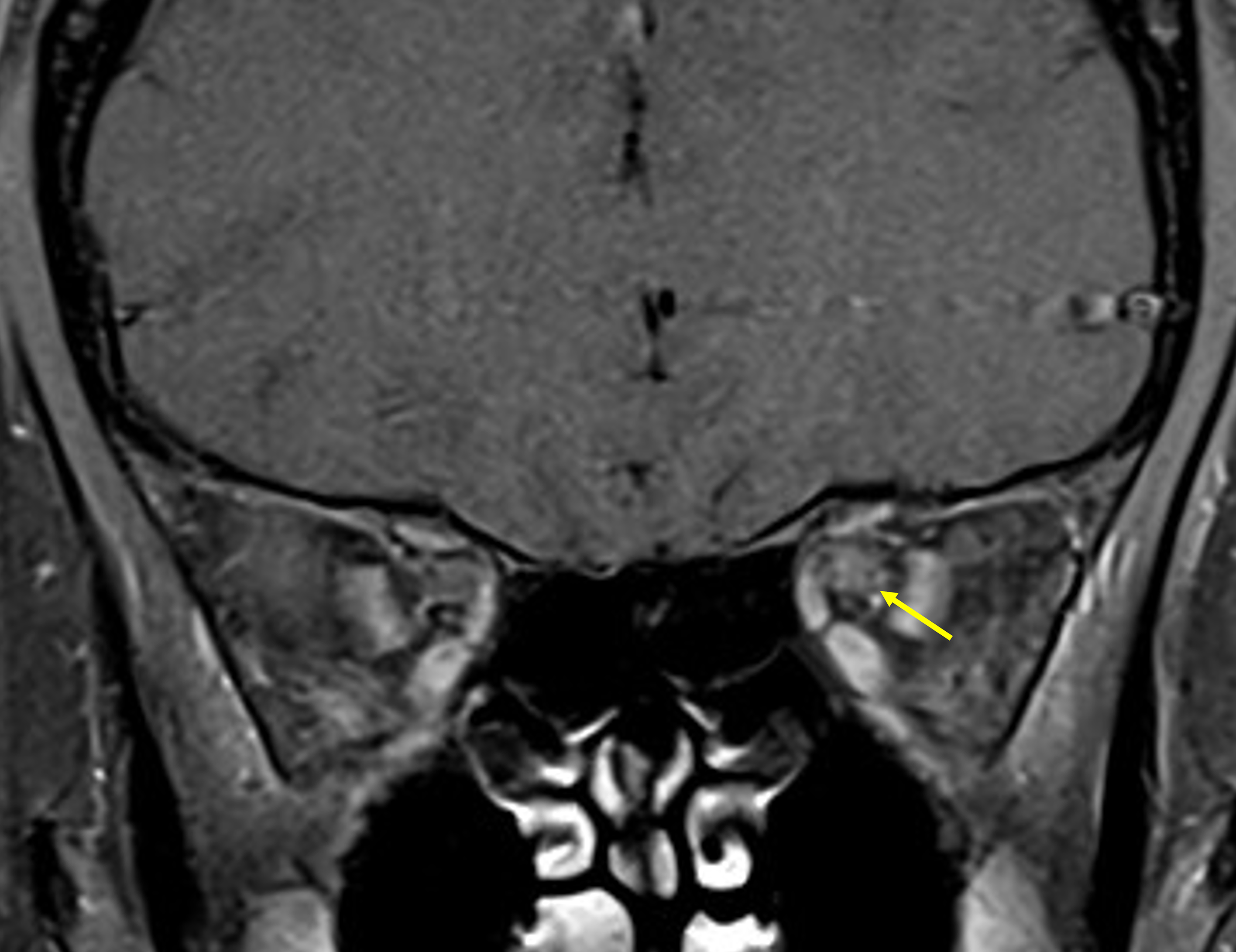
Case 2: A 28-year-old woman (G3P2) with unremarkable past medical history presented 10 weeks postpartum with a 6-week history of left retrobulbar pain followed by progressive vision loss over 3–5 days, which subsequently plateaued. The pain resolved after 4 weeks, but the visual loss persisted. She did not have symptoms in the right eye. Visual acuity was 20/20 OD and counting fingers OS, with a left RAPD. Fundoscopy revealed temporal optic disc pallor in OD, suggestive of a prior subclinical or unrecognized episode of ON, and optic disc edema in OS. OCT demonstrated temporal, superior and inferior RNFL thinning OD and circumferential RNFL thickening OS, with average RNFL thickness 64 μm OD and 195 μm OS. Macular GCL + IPL OCT demonstrated bilateral circumferential atrophy (average thickness 60 μm OD and 63 μm OS). HVF revealed an inferior arcuate defect in OD and complete loss in OS with mean deviation −6.71 OD and −33.77 OS. MRI showed T2 hyperintensity, thickening and enhancement of the orbital, intracanalicular and prechiasmatic segments of the left optic nerve (Figure 2). Investigations for alternative etiologies were negative. MOG antibodies were confirmed by cell-based assay (1:10 and 1:100). She received 1gm/day intravenous methylprednisolone for 5 days followed by oral prednisone 1 mg/kg. Due to persistent visual impairment at 14 days, plasmapheresis was initiated. At the time of plasmapheresis, mycophenolate therapy was also planned, and breastfeeding was held. After 6 weeks, her vision recovered to 20/200 OS with resolution of the optic disc edema. At 2-month follow-up, she remained stable without relapse. She declined repeat MRI and mycophenolate therapy.
Imaging findings in Case 2, the postpartum patient with unilateral ON. Coronal postcontrast T1-weighted fat-saturated MRI demonstrates enhancement of the left intraorbital optic nerve (arrow). ON = optic neuritis.

Pregnancy exerts a well-recognized modulatory effect on autoimmune demyelinating diseases.Reference Krysko, Dobson and Alroughani2 In multiple sclerosis, relapse rates decline during pregnancy and increase postpartum.Reference Schubert, Steinberg and Peper3,Reference Wang, Su and Zhou4 Although relapse activity in MOGAD is thought to be less common during pregnancy, these cases highlight the potential for pregnancy-related immunological adaptations to coincide with the clinical presentation of MOG-associated ON. Bilateral ON, marked optic disc edema and longitudinally extensive optic nerve involvement on MRI are characteristic features that raised clinical suspicion before serologic confirmation. In Case 2, contralateral optic disc pallor and RNFL thinning suggest a prior subclinical episode, indicating that the postpartum presentation may represent the first recognized manifestation of MOGAD rather than true disease onset.
Pregnancy induces a dynamic immunological shift, characterized by distinct phases of immune modulation. The first trimester exhibits a pro-inflammatory Type 1 T helper (Th1)-dominant response that facilitates embryo implantation and early placental development. This transitions in the second trimester toward immune suppression, where Type 2 T helper (Th2) and regulatory T cells (Tregs) dominate to promote fetal tolerance.Reference Robinson and Klein5 However, despite this immune suppression, persistently elevated estrogen levels throughout pregnancy can also enhance B-cell activation and antibody production, potentially overriding pregnancy’s immunosuppressive effects and facilitating the activation of dormant B-cell-driven autoimmune conditions such as MOGAD, as observed in the first case. These immunological adaptations persist into the postpartum period, where a rapid decline in Tregs and anti-inflammatory hormones, combined with a surge in Th1 cytokines, promotes a state of immune reactivation and heightened susceptibility to autoimmune flares, as observed in the second patient. No fetal or neonatal complications occurred in these cases.
Therapeutic management during pregnancy requires balancing maternal disease control with fetal safety. Although the FDA’s historical pregnancy risk categories have been replaced by the Pregnancy and Lactation Labeling Rule, the former classification remains a useful clinical reference. Category A and B drugs are generally low risk, while Category C may be used when benefits outweigh potential harm. Category D indicates known fetal risk but may be justified in severe disease, and Category X drugs are contraindicated (Supplemental Table 1). In MOG-associated ON, this framework aids risk stratification and guides treatment decisions to optimize maternal neurological outcomes while minimizing fetal exposure.
Prompt treatment with corticosteroids, which are permitted during pregnancy and breastfeeding as category C drugs, is critical to preserve vision.Reference Kemp, Newnham, Challis, Jobe and Stock6 Non-fluorinated corticosteroids such as methylprednisolone and prednisone are preferred as they undergo rapid metabolism by placental enzymes, minimizing fetal exposure. Failure of corticosteroids to improve vision in the postpartum patient may reflect a heightened inflammatory state related to immune rebound. Immunosuppressive therapy to prevent relapse must be tailored to balance disease control with maternal-fetal safety. Mycophenolate mofetil should be avoided during pregnancy due to teratogenic side effects as a category D drug.Reference Hoeltzenbein, Elefant and Vial7 Therapeutic plasma exchange may be considered for steroid-refractory cases, with reported safety during both pregnancy and breastfeeding.
Glucocorticoids have been used during breastfeeding for various autoimmune disorders, as the infant only ingests <0.1% of the total prednisone dose. The exposure to the infant is also minimized if breastfeeding is avoided for 4 hours after ingesting a high dose of prednisone.
Imaging decisions during pregnancy must also consider fetal safety. Gadolinium-based contrast agents, classified as category C, cross the placenta and may release free gadolinium ions that accumulate in fetal tissues, posing potential developmental risks.Reference Ray, Vermeulen, Bharatha, Montanera and Park8 In these cases, non-contrast MRIs were performed to facilitate diagnosis while minimizing fetal exposure.
Supplementary material
The supplementary material for this article can be found at https://doi.org/10.1017/cjn.2026.10658.
Author contribution
T.M. conceived and designed the study, collected and analyzed the data, interpreted the results and drafted the manuscript. E.I. contributed to data acquisition, interpretation of the findings and critical revision of the manuscript. R.A.S. contributed to data acquisition, interpretation of the findings and critical revision of the manuscript. M.M. contributed to data acquisition, interpretation of the findings and critical revision of the manuscript. A.N.E.S. supervised the study, contributed to study conception and design, interpretation of the findings and critical revision of the manuscript. All authors reviewed and approved the final manuscript.
Funding statement
None.
Competing interests
The authors declare no conflicts of interest.
Data sharing statement
Data related to this study are available upon reasonable request.



 Open access

