Introduction
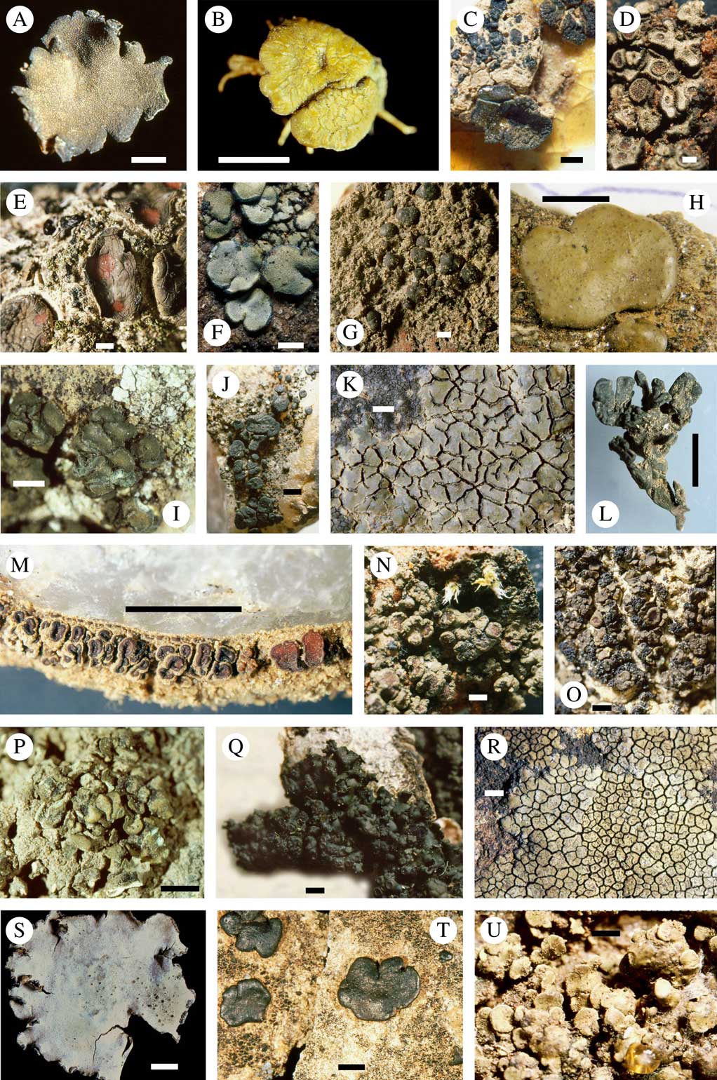
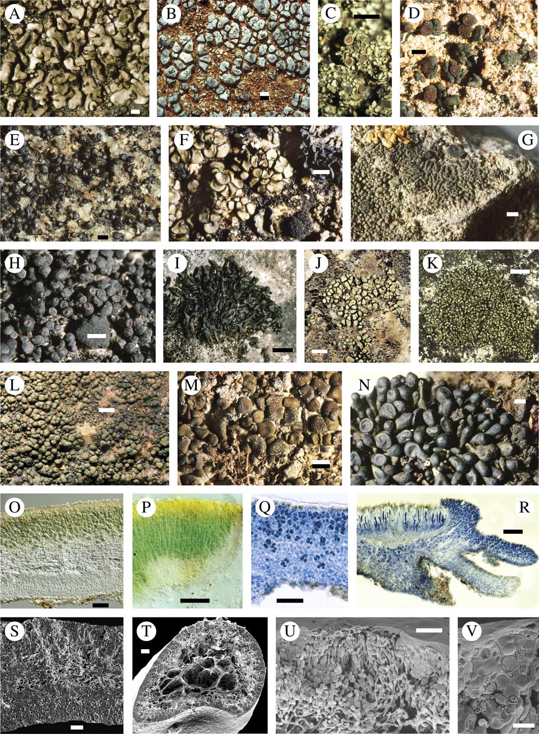
The lichen family Peltulaceae is exclusively associated with unicellular cyanobacterial photobionts, mostly of the genus Chroococcidiopsis, and presently comprises the three genera Peltula, Phyllopeltula and Neoheppia with a total of c. 47 species (Wetmore Reference Wetmore1970; Bubrick & Galun Reference Bubrick and Galun1984; Büdel 1987 Reference Büdela , Reference Büdelb , Reference Büdel1995; Egea Reference Egea1989; Büdel & Elix Reference Büdel and Elix1997; Kalb Reference Kalb2001; Donner Reference Donner2013; Marques et al. Reference Marques, Schultz and Paz-Bermúdez2013; Makryi Reference Makryi2016). However, the pantropical species Phyllopeltula corticola (Büdel & R. Sant.) Kalb was recently found to form chimeras with unicellular green algae incorporated into the thallus (Aptroot & Schumm Reference Aptroot and Schumm2010). Members of the Peltulaceae occur worldwide in arid and semi-arid climatic regions. However, under humid conditions they can exist in microclimatic arid islands (Büdel et al. Reference Büdel, Becker, Follmann and Sterflinger2000; Becker Reference Becker2002) as far north or east as Sweden or Baikal Siberia. The majority of species belong to the genus Peltula. Species from this genus have squamulose-compound (Figs 1P & U, 2A & R) and squamulose-semifruticose thalli (Fig. 2E, H–K), or exhibit singular peltate thalli with an umbilicus or central hyphal strand (Fig. 1A–I). All squamules of compound thalli are minute and usually grow tightly together (Büdel 1987 Reference Büdela ); they are not gelatinous (homoiomerous) but usually stratified (heteromerous) with an upper epinecral layer, a photobiont layer followed by a medulla with numerous air spaces of various sizes and typically a lower cortex (Fig. 2O & Q). An upper cortex is mostly absent but the epinecral layer serves as a protective layer for the photobiont and is often yellowish to brown pigmented (Büdel Reference Büdel1990; Büdel & Lange Reference Büdel and Lange1994). Soredia and isidia are rare but not unknown in a small number of species. Apothecia occur frequently in the Peltulaceae, with the exception of P. lingulata (Vain.) Swinscow & Krog and P. crispatula (Nyl.) Egea. Additionally, lichen substances are lacking in the Peltulaceae with the exception of P. langei Büdel & Elix, from which a yellow pigment similar to Myeloconon C could be extracted (Büdel & Elix Reference Büdel and Elix1997).
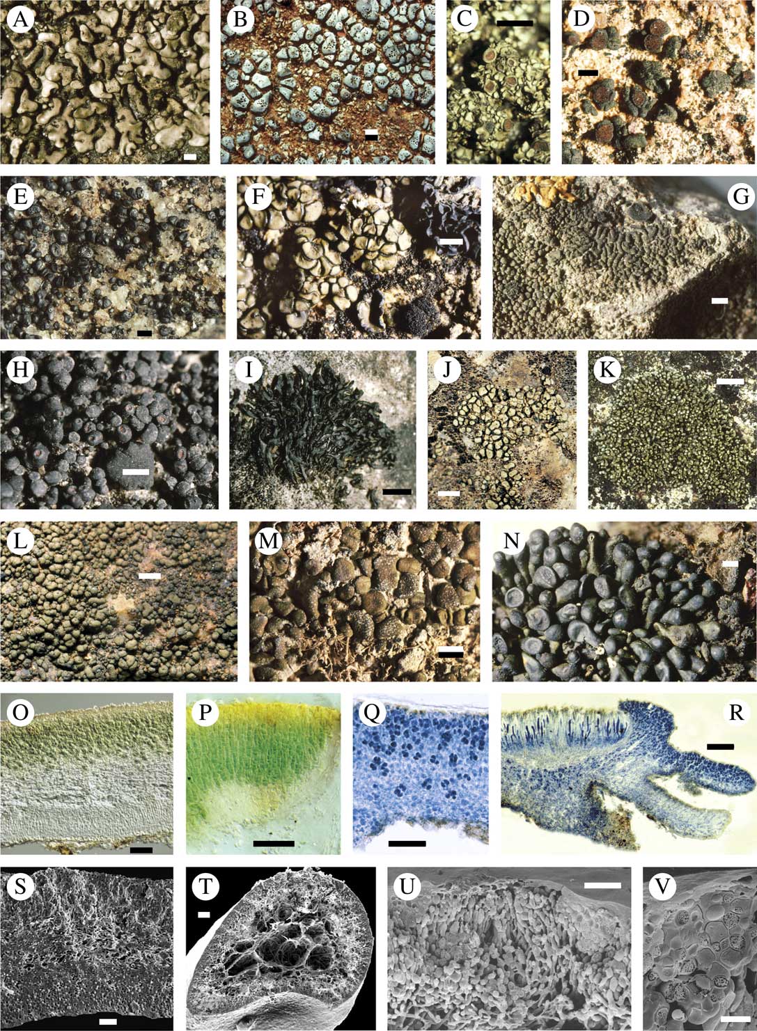
Peltula morphology. A, P. africana, Bu 14304b, South Africa; B, P. radicata, type species of the genus, isotype PC, North Africa; C, P. psammophila, lectotype, H-Nyl 30904, Algeria; D, P. patellata, Bu 18058a, Central Australia; E, P. richardsii, Schultz, Sonoran Desert, Arizona, USA; F, P. euploca, Central Australia; G, P. omphaliza, Vainio 11551; H, P. sonorensis, holotype ASU, Arizona, USA; I, P. bolanderi, Bu 14119b, South Africa; J, P. boletiformis, Bu 14117, South Africa; K, P. cataractae, topotype, Zaire BR 1330; L, P. capensis, isotype, LD, South Africa; M, P. inversa, Bu 15058, Namibia; N, P. imbricata, holotype MEL, Australia; O, P. hassei, lectotype FH, California; P, P. michoacanensis, FH, isotype, Mexico; Q, P. corticola, holotype UPS, Santesson 20745, Kenya; R, P. brasiliensis, holotype W, Brazil; S, P. farinosa, holotype M, Bu 14327b, South Africa; T, P. coriacea, Bu 14095a, South Africa; U, P. rodriguesii, isolectotype BM 2333, Island of Rodriguez. Scales: A, B, E, N, P, S & T=2 mm; C, D, G, I–K, O, Q, R & U=1 mm; F & L=5 mm; H=3 mm; M=10 mm.

Peltula morphology and anatomy. A, P. congregata, Ivory Coast; B, P. umbilicata, South Africa; C, P. auriculata, holotype M, Gröger 18, Venezuela; D, P. deserticola, Bu 18073, Australia; E, P. santessonii, holotype UPS, Santesson 23620; F, P. zahlbruckneri, MB Nash 8772, USA; G, P. placodizans, lectotype W, Blumer 112, Arizona, USA; H, P. clavata, Bu 18047, Australia; I, P. lingulata, Ivory Coast; J, P. marginata, isotype Bu 14017a, South Africa; K, P. tortuosa, Venezuela; L, P. impressa, MEL Brownlie 37806, Australia; M, P. impressula, Golubkova 1036, Mongolia; N, P. kofleri, holotype LD, Kofler 31010, South Africa; O, P. euploca, cross-section of peltate thallus with upper epinecral layer, cyanobiont layer, medulla and lower cortex; P, P. cataractae, topotype, cross-section of crustose thallus with cyanobiont arranged in vertical parallel rows; Q, P. corticola, cross-section of leaf-like thallus with compact cyanobiont layer and medulla without cavities; R, P. obscurans var. hassei, cross-section of crustose-leafy thallus; S, P. farinosa, cross fracture of peltate thallus with numerous cavities in cyanobiont layer and medulla, LT-SEM; T, P. tortuosa, cross fracture of subfruticose thallus with large medullary cave, LT-SEM; U, P. coriacea, cross fracture of upper thallus layer with pycnidium, LT-SEM; V, P. coriacea, cross fracture of upper cyanobiont and epinecral layer, LT-SEM. Scales: A, B, D, E, H, L & N=1 mm; C & I=5 mm; F & G=3 mm; J=2 mm; K=10 mm; M=4 mm; O & S–U=20 µm; P & R=100 µm; Q=50 mm; V=5 µm. LT-SEM=Low temperature scanning electron micrograph.
The remaining two genera, Neoheppia (Zahlbruckner Reference Zahlbruckner1909; Büdel Reference Büdel1995) and Phyllopeltula, comprise only two species each. Neoheppia cataractae Büdel & Sérus. (Fig. 1K) and N. brasiliensis Zahlbr. (Fig. 1R) differ from the remaining Peltulaceae by lacking a lower cortex (Fig. 2P) with their thalli being secondarily cracked, giving the impression of compound thalli (Fig. 1K & R). Phyllopeltula steppae Kalb and P. corticola (Büdel & R. Sant.) Kalb(Fig. 1Q) develop compound thalli with subfoliose squamules and, in contrast to rock- or soil-inhabiting Peltula and Neoheppia species, colonize bark (Büdel 1987 Reference Büdela ; Kalb Reference Kalb2001). Their thalli do not show internal air spaces in cross-section (Fig. 2P & Q).
Reproductive structures are uniform across all members of the family: a polysporous ascus with a gelatinous sheath and simple, single-celled, colourless spores. Pycnidia, conidiophores and pycnospores are also uniform among the species. The separation of the three genera necessarily relies on morphological characters only: thallus morphology and anatomy, substratum attachment and habitat, characters that have been shown to be of varying taxonomic use in cyanolichens (Wedin et al. Reference Wedin, Wiklund, Jørgensen and Ekman2009; Otálora et al. Reference Otálora, Aragón, Martínez and Wedin2013, Reference Otálora, Jørgensen and Wedin2014) and many other lichen groups (Printzen Reference Printzen2010; Ekman et al. Reference Ekman, Wedin, Lindblom and Jørgensen2014). For example, the lichen genera Coenogonium and Dimerella have identical apothecia but strikingly different thallus morphologies (filamentous vs. crustose, with intermediate forms), although molecular phylogenetic studies have shown that they are congeneric (Kauff & Lutzoni Reference Kauff and Lutzoni2002). Within the Peltulaceae, the limited number of growth forms (such as fruticose vs. subfruticose or crustose-squamulose) are often hard to distinguish, and intermediate forms exist, which further complicate the use of such characters.
The few morphological characters, their presence/absence, and the many modifications and intermediate forms make it difficult to characterize putative monophyletic lineages based on morphological data, and to date there is no comprehensive phylogenetic hypothesis for the Peltulaceae. Egea (Reference Egea1989) made a first attempt to find assemblies of putatively closely related species. He identified two major evolutionary lineages, mainly according to substratum fixation: one lineage containing species attached to the substratum with an umbilicus (e.g. P. euploca (Ach.) Poelt or P. africana (Jatta) Swinscow & Krog; Fig. 1A & F), the other lineage comprising species forming rhizohyphae (e.g. P. patellata (Bagl.) Swinscow & Krog or P. psammophila (Nyl.) Egea; Fig. 1C & D). Subfruticose thalli were considered as the most advanced traits for the Peltulaceae. Büdel (1987 Reference Büdela ) described two groups of closely related species: the so-called ‘form group’ comprising P. congregata (Nyl.) Swinscow & Krog, P. capensis (Brusse) Büdel, P. cylindrica Wetmore and P. tortuosa (Nees) Wetmore (Figs 1L, 2A & K), three of which develop squamulose-semifruticose thalli, and the ‘critical group’ (P. impressa (Vain.) Swinscow & Krog, P. marginata Büdel, P. placodizans (Zahlbr.) Wetmore, P. umbilicata (Vain.) Swinsc. & Krog and P. zahlbruckneri (Hasse) Wetmore; Fig. 2B, F, G, J & L), comprising species with squamulose-compound thalli.
Thus, the question arises whether the subdivision of Peltulaceae into three genera, based on thallus morphology and anatomical variation, reflects the evolutionary history of this family. Are there morphological characters that can be used to delimit natural groups of related taxa in the Peltulaceae? Neither the monophyly of any of the above-mentioned lineages, nor the monophyly of the three genera Peltula, Phyllopeltula and Neoheppia, have yet been explicitly verified with molecular data. Although the reproductive features of the Peltulaceae are too uniform and unsuitable for phylogenetic analysis, the usefulness of the remaining morphological characters (thallus anatomy, thallus growth form, substratum) currently used for genus and species delimitation has yet to be shown.
The aim of this study is to test the current morphology-based genus delimitation within Peltulaceae and the monophyly of Phyllopeltula and Neoheppia using a molecular phylogenetic analysis based on six gene loci.
Materials and Methods
Taxon sampling
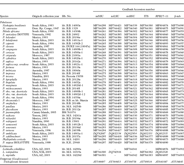
Thirty-seven of the 47 species classified within Peltulaceae were included in this study (Table 1). No specimens were available for P. applanata (Zahlbr.) J. C. Wei, P. minuta (H.Magn.) Golubk., P. tenuis Büdel & Henssen and P. zabolotnoji (Elenk.) Golubk., and the material for P. koflerae Henssen & Büdel and P. langei was too poor for a molecular approach. The new species P. lobata J. Marques et al., P. pannarioides Makryi and P. rosulata Makryi were not available at the time of the analyses (Marques et al. Reference Marques, Schultz and Paz-Bermúdez2013; Makryi Reference Makryi2016). The monophyly of the Peltulaceae has been shown previously (Schultz et al. Reference Schultz, Arendholz and Büdel2001). Three species of Lichinella and Trichoglossum hirsutum (Pers.) Boud. were chosen as outgroup. Six genetic loci (nuclear ribosomal small subunit RNA gene (nrSSU), nrLSU, nuclear ribosomal internal transcribed spacer region (ITS), mitochondrial ribosomal small subunit RNA gene (mtSSU), and the protein coding genes RNA polymerase II second largest subunit (RPB2) and beta-tubulin (β-tub) were analyzed. Voucher data and GenBank Accession numbers are given in Table 2.
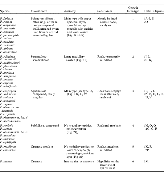
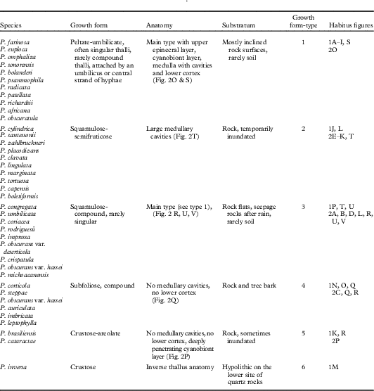
Growth forms of the lichen genus Peltula and their morphological and anatomical characteristics
Voucher information and GenBank Accession numbers for the species of Peltulaceae used in this study. Accession numbers are newly generated for this study with the exception of those in italics
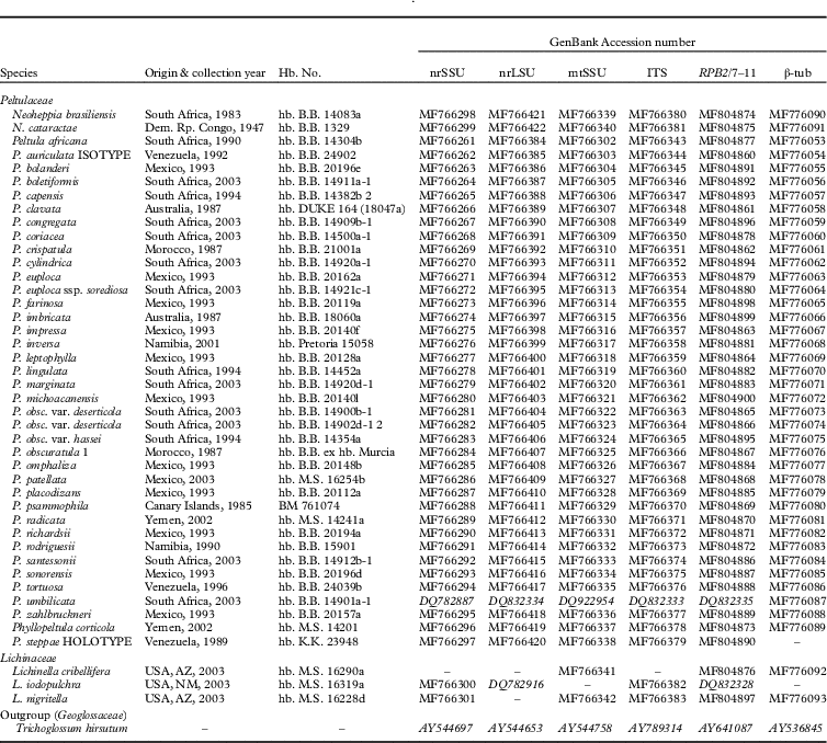
hb. B.B.: B. Büdel, Kaiserslautern, Germany; hb. M.S.: M. Schultz, Hamburg, Germany; hb. K.K.: K. Kalb, Neumarkt, Germany; hb. DUKE: Duke University, Durham, NC, USA.
DNA isolation and sequencing
DNA isolation followed the protocol of Hofstetter et al. (Reference Hofstetter, Clémençon, Vilgalys and Moncalvo2002). Polymerase chain reactions (PCR) were performed to amplify parts of six loci. The reaction was prepared to a final volume of 25 µl containing 4 µl dNTPs, 2·5 µl buffer, 1 µl BSA (bovine serum albumin, 10 mg ml−1, New England Biolabs, #B9001S), 1·25 µl of each primer (10 µM), 0·3 µl TAQ DNA polymerase (Abgene, Rochester, NY, USA), 10 µl DNA extraction solution at different dilution stages, and PCR buffer N (Abgene, #AB-0288). Half of the reactions were prepared using 12·5 µl PCR mastermix (Promega, Mannheim, Germany, #M7505), 0·625 µl BSA (20 mg/ml, Fermentas, St.-Leon-Rot, Germany, #B14), 2·5 µl of each primer (10 µM), 1·875 µl nuclease-free water (Promega), and 5 µl template DNA at different dilution stages. Several combinations of forward/reverse primers were used for amplification: nsu131/NS24, nsu131/nsu1088, nsu1088R/NS24 (nrSSU), LR0R/LR7, LR0R/LR5, LR5R/LR7 (nrLSU), mrSSU1/mrSSU3R, mrSSU1/mrSSU2R (mtSSU), ITS1/LR5 (ITS), RPB2-7F/RPB2-11aR, RPB2-7F/RPB2-11bR, fRPB2-7cF/fRPB2-11aR (RPB2/7–11), Bt3-LM/Bt10-LM, T1/T22, T1/Bt2b, Bt2a/T22 (β-tub) (lutzonilab.org/primer-sequences). All amplification primers were also used for sequencing (at a concentration of 5 µM) plus a variety of other primers: nssu634, nssu897R, SR6, SR7, SR7R, SR10, SR10R, SR11, SR11R, NS6 (nrSSU), LR2, LR2R, LR3, LR3R, LR6, LR6R (nrLSU), ITS4 (ITS), T2, T10, T12, T222, Bt698F, Bt705R (β-tub) (lutzonilab.org/primer-sequences). The primers Bt698F (5′-TCC GTT CGG TCA ACT CTT C-3′) and Bt705R (5′-TCA ACT TTT CCG TCC CGA-3′) were newly designed for this study. PCR was performed as follows: one initial cycle of 3 min at 95 °C, followed by 34 cycles of 1 min at 95 °C, 45 s at a variable annealing temperature depending on the targeted region, 2 min at 72 °C (1 min 30 s for RPB2/7–11 and β-tub). After completion of the 34 cycles, samples were held for 10 min at 72 °C. Gel electrophoresis was conducted on a TAE 1% agarose gel. PCR products were stained either with SYBRGreen I (Cambrex, East Rutherford, NJ, USA, #50513) or GelStar (Biozym, Hess. Oldendorf, Germany). A quantity of 0·5 µl MgCl2 (25 mM) was added to some reactions, in combination with a raised number of cycles. All ribosomal RNA genes (nrSSU, nrLSU, mtSSU) and the ITS region were annealed at 50 °C, RPB2/7–11 at 52 °C, and β-tub primers Bt3-LM and Bt10-LM at 60 °C or 52 °C. PCR reactions showing multiple bands or a weak signal on the gel were cloned using the TOPO TA cloning kit (Invitrogen, Carlsbad, CA, USA and Karlsruhe, Germany, #K4500). Cloning PCR reactions were prepared to a volume of 25 µl as for the standard PCR, replacing the template DNA solution with the same amount of nuclease-free water. At least four clone colonies were picked and added to the PCR reaction. PCR from cloning plasmids was performed as follows: one cycle of 10 min at 94°C, followed by 34 cycles of 1 min at 94 °C, 1 min at 55°C and 1 min 30 s at 72°C. The Montage single-column kit (Millipore, Billerica, MA, USA and Schwalbach, Germany, #UFC7PC) was used for PCR product cleaning. Sequencing reactions were prepared to a total volume of 10 µl, containing 1 µl of Big Dye (Big Dye Terminator Cycle Sequencing Kit, Abi Prism, Perkin-Elmer, Applied Biosystems), 2 µl of 5 µM primer, 3 µl sequencing buffer (5 ml 1M MgCl2, 200 µl 1M Tris at pH 9, 795 µl sterile double-distilled H2O), and 4 µl purified PCR product as template DNA. Sequencing PCR was performed as follows: one cycle of 2 min at 96°C, followed by 24 cycles of 30 s at 96°C, 15 s at 50°C and 4 min at 60°C. All samples were sequenced on an ABI automated sequencer (Perkin-Elmer, Applied Biosystems, Foster City, CA, USA).
Data set assembly
Sequence fragments were assembled using Sequencher 4.2 and 4.5 (Gene Codes, Ann Arbor, MI, USA). Previous to this study, sequences of only two species of Peltula were available from GenBank (www.ncbi.nlm.nih.gov/blast). BLAST searches (Altschul et al. Reference Altschul, Madden, Schäffer, Zhang, Zhang, Miller and Lipman1997) resulted in ambiguous results and the identity of newly sequenced Peltulaceae sequences could not be verified prior to phylogenetic analyses. Ribosomal sequences were aligned using SAM 3.5 (Karplus et al. Reference Karplus, Barrett and Hughey1998) with a model obtained by training with an automatic ClustalW alignment from BioEdit 7.04.1 (Hall Reference Hall1999). The single locus alignments were manually adjusted, and ambiguous regions (Lutzoni et al. Reference Lutzoni, Wagner, Reeb and Zoller2000) and introns were excluded from phylogenetic analyses.
Test for topological incongruence
To test for topological incongruence among loci, we used the program compat3 (available at lutzonilab.org/downloadable-programs). For each individual locus of the combined data set, 500 bootstrap replicates (Felsenstein Reference Felsenstein1985) were generated with RAxML 7.2.7 (Stamatakis Reference Stamatakis2006; Stamatakis et al. Reference Stamatakis, Hoover and Rougemont2008) and all pairwise comparisons between the six loci were performed. A conflict between two loci was assumed when a clade was supported as monophyletic with a bootstrap frequency ≥75% in one tree, but supported, using the same bootstrap threshold, as non-monophyletic in another (Mason-Gamer & Kellogg Reference Mason-Gamer and Kellogg1996). By comparing the significant topological differences, individual sequences causing conflicts were identified. Conflicting gene sequences were removed from alignments and the test was repeated until no further conflicts could be detected. Constraint-based filters (Ihlen & Ekman Reference Ihlen and Ekman2002) were used to check for congruence between different data sets. Constraints were defined for the whole topology as well as for single relationships in a phylogenetic tree. Congruence between data partitions was assumed if a constraint was present in at least 5% of the trees of a BMCMC sample.
Phylogenetic analyses
Phylogenetic searches were carried out by implementing maximum likelihood (ML) and Bayesian analyses. All analyses were run on the computer cluster of the Nano+Bio Center (University of Kaiserslautern, Germany).
For the ML analyses, the combined data set was split into eight partitions: mtSSU, nrLSU, nrSSU, ITS (excluding unalignable spacer regions), RPB2 1st, RPB2 2nd, RPB2 3rd codon position, β-tub 1st, β-tub 2nd, and β-tub 3rd codon position. ML analyses were carried out using RAxML 7.2.7 and 7.2.8 (Stamatakis Reference Stamatakis2006), implementing a GTR model of nucleotide substitution (Rodriguez et al. Reference Rodriguez, Oliver, Marin and Medina1990) with a gamma shaped distribution and a proportion of invariable sites, and searching for the most likely tree with 500 heuristic replicates. Bootstrap frequencies (Felsenstein Reference Felsenstein1985; Stamatakis et al. Reference Stamatakis, Hoover and Rougemont2008) were estimated with 500 replicates.
For the Bayesian analyses, the combined data sets were partitioned as described above. Three independent runs with 10 000 000 generations and four independent chains each were started with MrBayes 3.1.2 (Ronquist & Huelsenbeck Reference Ronquist and Huelsenbeck2003), sampling every 500th tree. The burn-in fraction of sampled trees was estimated both by eye with ln-likelihood plots and using AWTY (Nylander et al. Reference Nylander, Wilgenbusch, Warren and Swofford2008) to ensure that the runs reached stationarity.
Only nodes that received posterior probabilities (PP) ≥0·95 and ML-bootstrap support values (ML-BS) ≥70% were interpreted as strongly supported.
Results
Morphology
Ascomatal features as well as ascoma ontogeny are largely uniform within the Peltulaceae and cannot be used to separate species (Büdel 1987 Reference Büdela , Reference Büdelb ; Egea Reference Egea1989). On the basis of thallus morphology and vegetative thallus structure, six different growth types are distinguished. Two types share a common thallus anatomy while the remaining groups possess differing anatomical features (Table 1, Figs 1 & 2).
Phylogenetic analyses
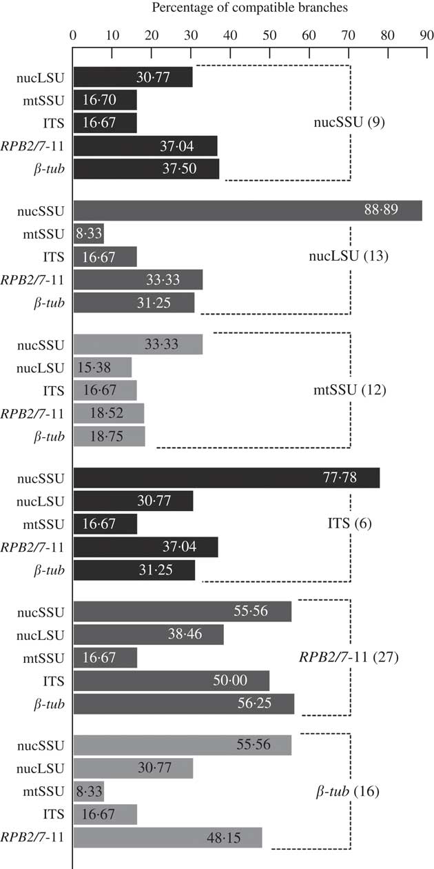
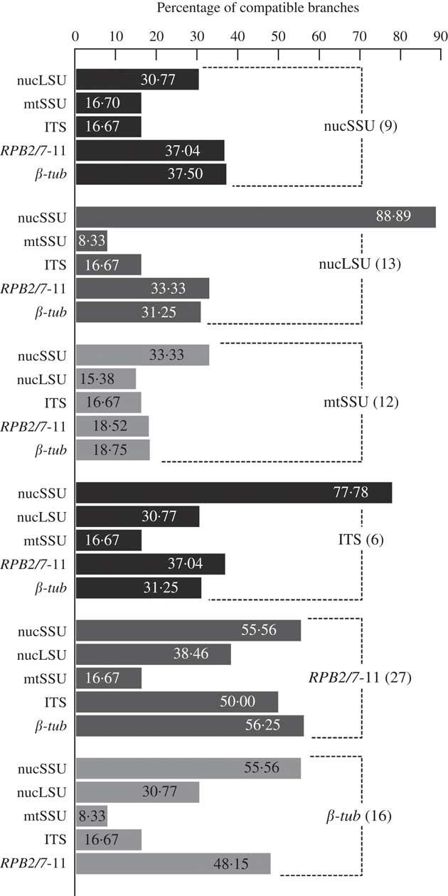
The concatenated alignments included a total of 10 212 characters. The alignment of the nrSSU contained a total of eight introns, and the nrLSU contained only one intron. After excluding hypervariable regions and introns, the final data set length for phylogenetic analyses was 5583 characters (nrLSU: 1321, nrSSU: 1635, ITS: 238, mtSSU: 703, RPB2: 876, β-tub: 810).The pairwise topological comparisons showed large amounts of conflict among clades supported with bootstrap ≥75%, especially between the mtSSU and the other loci (Fig. 3). After removing one sequence from the nrLSU alignment, one nrSSU sequence, three β-tub sequences and nine mtSSU sequences, all topological conflicts were resolved.
Pairwise topological comparisons of the six gene loci used in this study of Peltulaceae using constraint-based filtering. Bars represent the percentage of compatible branches. The presence of all supported branches (i.e. constraints) of each gene (single bars) was tested in the BMCMC tree sample of each other gene (bars grouped by dashed line). The number of constraints tested for each gene is given in brackets after the name of the gene.
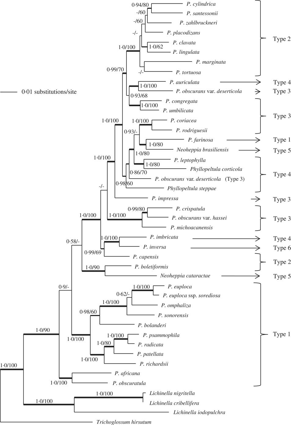
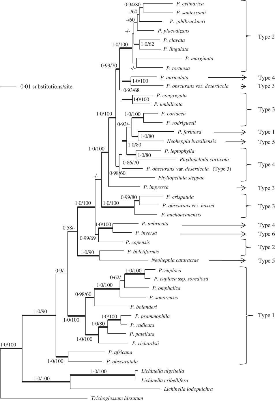
When analyzed in combination, both the original data set and the conflict-free data set produced very similar tree topologies, where only a few single taxa or small monophyletic groups of two or three taxa changed position. However, none of these alternative phylogenetic relationships were well supported by bootstrap frequencies or posterior probabilities. Alternatively, a complete removal of the mtSSU alignment did not alter the topology but resulted in minor changes of branch support. Therefore, we decided to use the conflict-free data set including the mtSSU for further discussion of the results.The results of the combined phylogenetic analyses, after the removal of conflicting sequences, are shown in Fig. 4. The Peltulaceae form a well-supported (90% ML-BS, 1·0 PP) monophyletic clade. The three outgroup species of the genus Lichinella also form a monophyletic clade with high support (100% ML-BS, 1·0 PP).
Best phylogenetic tree for the family Peltulaceae based on the six-locus data set calculated with RAxML, using a GTR+I+G model (likelihood value=−34 009·803617). Thicker branches indicate PP≥0·95 (before slash) or ML-BS≥70% (after slash) support. If one of the values is absent (denoted by a dash), the respective branch was not present in the associated analysis. Growth types based on thallus morphology and vegetative thallus structure are indicated by ‘Type 1–6’. For explanation, see Table 1. Genus is Peltula unless otherwise indicated.
Growth form versus phylogeny
Both Phyllopeltula and Neoheppia, two genera that were separated from Peltula due to their crustose or foliose growth (Büdel 1987 Reference Büdela , Reference Büdelb , Reference Büdel1995; Kalb Reference Kalb2001), are non-monophyletic (Fig. 4) and the representative species of these two genera are interspersed among Peltula species. Phyllopeltula corticola shares a most recent common ancestor with Peltula leptophylla (80% ML-BS, 1·0 PP), whereas Phyllopeltula steppae is sister to a clade comprising Phyllopeltula corticola, Neoheppia brasiliensis and five other Peltula taxa (60% ML-BS, 0·98 PP). Neoheppia brasiliensis is monophyletic with Peltula farinosa (80% ML-BS, 1·0 PP), and N. cataractae is a sister taxon to Peltula boletiformis (90% ML-BS, 1·0 PP). The polyphyly of Phyllopeltula and Neoheppia is consistent with the results of the single-gene analyses (see Supplementary Material Figs S1–S6, available online) and is well supported in both ML and Bayesian phylogenetic trees.
The different types of growth form in Peltula (peltate, squamulose, subfruticose and subfoliose) are scattered across the tree, underlining that growth form as a character appears to be unsuitable to track phylogenetic relationships within the Peltulaceae. The two smaller clades that resulted from the first split within the genus Peltula, both supported with 100% ML-BS and PP, exhibit peltate growth (type 1, Table 1); however, other peltate taxa are present across the rest of the tree (Fig. 4).
The same is true for the few remaining characters that are usually used to delimit species groups within the genus, such as the epinecral layer or anatomy of the medulla and presence/absence of the lower cortex. No other morphological and anatomical characters, individually or in combination, show any discernible pattern for the characterization of any of the clades present in the tree. None of the presumed evolutionary lineages described by Büdel (1987 Reference Büdela ) or Egea (Reference Egea1989) is in accordance with the results presented in this paper.
Peltula obscurans var. hassei and P. obscurans var. deserticola are not part of a monophyletic species. The two representatives of Peltula obscurans var. deserticola are not monophyletic either. While one is a well-supported sister to P. auriculata (100% ML-BS, 1·0 PP), the other forms a paraphyletic group with Phyllopeltula steppae (Fig. 4). Peltula obscurans var. hassei is sister with P. crispatula (80% ML-BS, 0·99 PP) in a well-supported clade with P. michoacanensis, with high support (100% ML-BS, 1·0 PP).
New Combinations and Synonyms
Peltula brasiliensis (Zahlbr.) Büdel, Kauff & Bachran comb. nov.
MycoBank No.: MB 821278
Basionym: Neoheppia brasiliensis Zahlbr., Denkschr. kaiserl. Akad. Wiss. Wien math.-naturw. Kl. 83: 144 (1909); type: Brazil, São Paulo, Salto Grande do Rio Paranapanema, 1901, V. Schiffner 20702 (W—holotype, not seen; SI—isotype).
(Fig. 1R)
Peltula cataractae (Büdel & Sérusiaux) Büdel, Kauff & Bachran comb. nov.
MycoBank No.: MB 821279
Basionym: Neoheppia cataractae Büdel & Sérusiaux, Mycotaxon 54: 140 (1995); type: Zaïre, Tshopo Waterfalls near Kisangani, 1947, J. Léonard 1329 (BR—holotype).
(Fig. 1K)
Peltula corticola Büdel & R. Sant.
MycoBank No.: MB 130538
Biblioth. Lichenol. 23: 79 (1987); type: Kenya, Santesson 20745 (UPS—holotype).—Phyllopeltula corticola (Büdel & R. Sant.) Kalb, Biblioth. Lichenol. 78: 158 (2001).
(Fig. 1Q)
Peltula hassei (Zahlbr.) Büdel, Kauff & Bachran comb. nov.
MycoBank No.: MB 821280
Basionym: Heppia hassei Zahlbr., Beih. Bot. Centralbl. 13: 157 (1902); type: California, Palm Springs, H. E. Hasse (W—lectotype).
Peltula obscurans var. hassei (Zahlbr.) Wetmore, Ann. Missouri. Bot. Gard. 57: 191 (1971).
(Fig. 1O)
Peltula leptophylla (Vain.) Büdel & M. Schultz comb. nov.
MycoBank No.: MB 822531
Basionym: Heppia leptophylla Vain., Acta Soc. Flora Fauna Fenn. 7(1): 216 (1890); type: “supra rupem graniticam litoralem, Rio de Janeiro, n. 135b, leg. Edw. Vainio, 1885”; Lich. Bras. Exs. 1891 (TUR-VAIN 12474—isotype!).
Peltula steppae (Kalb) Büdel, Kauff & Bachran comb. nov.
MycoBank No.: MB 821277
Basionym: Phyllopeltula steppae Kalb, Biblioth. Lichenol. 78: 159 (2001); type: Venezuela, Lara, Distr. Torres, c. 35 km E of Barquisimeto, 20 Aug. 1989, Kalb (M—holotype).
Discussion
Peltula, Phyllopeltula and Neoheppia
The single-locus tree topologies are not identical when compared to each other or to the combined analysis (Supplementary Material Figs S1–S9, available online). However, they all concur that Phyllopeltula and Neoheppia are not monophyletic. Since reproductive features cannot be used in species delimitation (see Introduction), the prior generic concept is exclusively based on morphological characters. Phyllopeltula and Neoheppia were segregated from Peltula based on the morphology of their cortex and the substratum, features that are likely to adapt quickly to environmental factors. Phenotypic plasticity in lichens is not necessarily reflected in a corresponding genetic variability (Purvis Reference Purvis1997). In the Peltulaceae, the creation of an epinecral layer is dependent on the light intensity of the existing habitat (Büdel Reference Büdel1990; Büdel & Lange Reference Büdel and Lange1994). Growth types of Peltula (peltate, squamulose and subfruticose) are connected to characteristic habitats with different water availabilities.
The results from the combined molecular phylogenetic analyses, confirmed by the analyses of the single-locus data sets, clearly highlight that the separation of Neoheppia and Phyllopeltula from Peltula into distinct genera does not reflect the evolutionary history of this family. Consequently, the two genera Phyllopeltula and Neoheppia are subsumed here within Peltula.
The genus Phyllopeltula was originally established by Kalb (Reference Kalb2001) based on Peltula corticola, together with Phyllopeltula steppae as a new species. Thus, Phyllopeltula corticola becomes a synonym of Peltula corticola, whereas Peltula steppae is proposed as a new combination for Phyllopeltula steppae. The genus Neoheppia, originally erected by Zahlbruckner (Reference Zahlbruckner1909), is a synonym of Peltula. Consequently, Peltula brasiliensis and P. cataractae are proposed as new combinations for Neoheppia brasiliensis and N. cataractae, respectively.
The two varieties Peltula obscurans var. deserticola (Zahlbr.) Wetmore and P. obscurans var. hassei (Zahlbr.) Wetmore were established due to their differences in thallus morphology by Wetmore (Reference Wetmore1970), based on the two species Heppia deserticola Zahlbr. and H. hassei Zahlbr. Both species were treated as synonyms of P. obscurans and ranked as varieties of this species by the author. As neither are supported as varieties of P. obscurans, and cluster independently with other species of Peltula in this study, P. obscurans var. hassei is resurrected here as a species while P. obscurans var. deserticola is not a monophyletic entity and needs further investigation.
Species delimitation in Peltula
The anatomical uniformity of structures associated with sexual reproduction in the Peltulaceae leaves only morphological and anatomical characters of vegetative thalli for delimiting species within the genus Peltula. Main characters, among others, are the different growth forms (e.g. peltate, subfruticose, squamulose), attachment to the substratum (rhizines, umbilicus), presence or absence of apothecia, isidia, and characters associated with thallus medulla and cortex. The different Peltula species are currently distinguished by unique combinations of these features. It is thus not unexpected that the few attempts to establish phylogenetic lineages within the genus (or family) based on morphology have remained controversial (Büdel 1987 Reference Büdela , Reference Büdelb ; Egea Reference Egea1989). Our molecular analyses presented here confirm the difficulties with identifying characters that could play a role in characterizing presumably monophyletic evolutionary lineages. Morphological characters exhibit lability and, consequently, finding unique character states or combinations of characters for any of the clades has not been possible (results not shown). Despite the apparent lack of alternative characters, it is still not known whether phenotypic features can be diagnostic at the species level within the genus Peltula. The taxon sampling of this study is insufficient to answer this question. Printzen (Reference Printzen2010) emphasized that the phylogenetic usefulness of specific characters varies among families and orders. However, certain taxa do have specific traits. For example, some Peltula species are characterized by shield-like (peltate) thalli, either with an umbilicus or a strand of hyphae (rhizines), occurring as many individuals (compound) or as single thalli (e.g. P. euploca, P. bolanderi, P. radicata, P. patellata, P. africana; Fig. 1A, B, D, F & I) and the squamulose to semifruticose group is characterized by large medullary cavities in the thalli that can occur in temporarily inundated habitats (e.g. P. tortuosa, P. zahlbruckneri, P. clavata, P. lingulata; Fig. 2F, H, I & K). It seems that there is a considerable amount of intraspecific variation in the thallus morphology of several species. This needs to be investigated systematically with a considerably enlarged taxon sampling. Considering the poor taxonomic value of morphological and anatomical features of vegetative thalli in the family Peltulaceae, scientists should refrain from describing new species until a clearer connection between genetically and morphologically defined “species” is established.
This study was supported by the grant BU 666/10-1 from the German Research Foundation (Deutsche Forschungsgemeinschaft DFG). Part of this work was supported by the ‘Assembling the Fungal Tree of Life’ (AFTOL) project, which itself is supported by the National Science Foundation (NSF) ‘Assembling the Tree of Life’ (ATOL) grants DEB-9615542 and DEB-0133891 to FL. We acknowledge Klaus Kalb’s (Neumarkt, Germany) kind provision of the P. steppae material.
Supplementary Material
For supplementary material accompanying this paper visit https://doi.org/10.1017/S0024282918000105