

INTRODUCTION
Iron deficiency is a common agricultural problem in many soils such as in North African countries (Ksouri et al., Reference Ksouri, Gharsalli and Lachâal2005) as the majority of arable land consists of calcareous and alkaline soils (Mtimet, Reference Mtimet, Zdruli, Steduto, Lacirignola and Montanarella2001). This phenomenon cannot be easily overcome by the use of iron-containing fertilizers because iron availability problem is due to its low solubility rather than to a low concentration (Guerinot, Reference Guerinot2001). Therefore, imbalance between plant need and iron solubility in the soil is the primary cause of iron-related chlorosis. The exploration of phytogenetic resources to identify species and varieties tolerant to iron-related chlorosis could help improve crop productivity on calcareous soils (Mahmoudi et al., Reference Mahmoudi, Labidi, Ksouri, Gharsalli and Abdelly2007). In fact, a large variability in responses to Fe deficiency has been found either among plant species or among varieties (Ali et al., Reference Ali, Ahmad, Riaz-ul-Haque and Tufail1995; Krouma et al., Reference Krouma, Gharsalli and Abdelly2003).
As an abiotic stress, iron deficiency affects plant morphology and metabolism (Briat, Reference Briat, Kader and Delseny2007). Iron-deficient plants are characterized by the development of a pronounced interveinal chlorosis occurring first in the youngest leaves. This symptom is accompanied by a decrease in leaf chlorophyll and carotenoid concentration and a reduction in fresh and dry weights of plants (Rabhi et al., Reference Rabhi, Barhoumi, Ksouri, Abdelly and Gharsalli2007; Zaharieva et al., Reference Zaharieva, Gogorcena and Abadıa2004; Zocchi et al., Reference Zocchi, De Nisi, Dell'Orto, Espen and Gallina2007). In addition, Fe deficiency reduces photosynthetic rate, light absorption and Rubisco activity of several species (López-Millán et al., Reference López-Millán, Morales, Andaluz, Gogorcena, Abadía, de las Rivas and Abadía2000; Rombolà et al., Reference Rombolà, Gogorcena, Larbi, Morales, Balde, Marangoni, Tagliavini and Abadía2005). However, plants induce a coordinated set of responses that maximizes iron mobilization and uptake from the soil, utilizes its stores and prioritizes its allocation to crucial cell processes. All angiosperms except grasses use what we call ‘Strategy I’ to solubilize and transport iron into roots when it is limiting (Römheld, Reference Römheld1987). This response includes: (i) proton release into the soil via an ATPase localized at the plasma membrane of root cells to acidify the surrounding solution and thus increase Fe3+ solubility (Dell'Orto et al., Reference Dell'Orto, Santi, De Nisi, Cesco, Varanini, Zocchi and Pinton2000; Rabotti and Zocchi, Reference Rabotti and Zocchi1994; Zocchi and Cocucci, Reference Zocchi and Cocucci1990); (ii) an induction of the activity of a ferric chelate reductase (FCR) to convert Fe(III)-chelates to Fe(II) (Schmidt, Reference Schmidt1999) and phosphoenolpyruvate carboxylase (PEPC) activity in root tissues, thus accumulating organic acids (mainly citrate) and accelerating the translocation of solubilized Fe to shoots (De Nisi and Zocchi, Reference De Nisi and Zocchi2000; López-Millán et al., Reference López-Millán, Morales, Andaluz, Gogorcena, Abadía, de las Rivas and Abadía2000). These responses are associated to gene induction involved in the mechanism of iron reduction and uptake. First of all, the induction of a specific transporter for Fe uptake from soil, named IRT1. IRT1 is expressed in the epidermal cells of Fe-deficient roots and localizes to the plasma membrane (Kim and Guerinot, Reference Kim and Guerinot2007).
In addition, phenolic compounds are frequently reported to be the main components of root exudates in response to Fe deficiency (Curie and Briat, Reference Curie and Briat2003; Hell and Stephan, Reference Hell and Stephan2003; Römheld and Marschner, Reference Römheld and Marschner1986; Susín et al., Reference Susín, Abián, Sánchez-Beyes, Peleato, Abadia, Gelpi and Abadia1996). It has been shown that iron deficiency in peanut plant increased the release of caffeic acid in culture medium (Römheld and Marschner, Reference Römheld and Marschner1983). In addition to their functions including antioxidant and antimicrobial activities, it has been reported that phenolic compounds reduce Fe in the apparent free space of the roots and/or in the external solution.
Lactuca sativa is one of the most important commercial plants belonging to the family Asteraceae. It is also known as a source of phytonutrients including vitamins, carotenoids and antioxidants (Kang and Saltveit, Reference Kang and Saltveit2002; Nicolle et al., Reference Nicolle, Cardinault, Gueux, Jaffrelo, Rock, Mazur, Amouroux and Rémésy2004). In this work, two varieties of L. sativa were compared in terms of their tolerance to iron deficiency using several physiological parameters.
MATERIALS AND METHODS
Plant growth
Lettuce (L. sativa) seeds from cultivars ‘Romaine’ and ‘Vista’ were provided by the Tunisian Ministry of Agriculture. After seed germination in Petri dishes with distilled water for 7 days (25 °C in the dark), the plantlets were transferred into pots, of 4 plants, containing 2 l of Hoagland's nutrient solution (Hoagland and Arnon, Reference Hoagland and Arnon1950). For each variety, a total of 48 plants were kept for 21 days in control solution containing 20 μM Fe(III)-ethylenediaminetetraacetic acid (EDTA) then divided into two treatments: 24 plants were used as control (20 μM Fe(III)-EDTA) and 24 as deficient (0 μM Fe(III)-EDTA). Nutrient solutions (with pH initially adjusted to 6.5) were renewed weekly and continuously aerated with an air-inflating pump linked to capillaries introduced each into a pot. Then, during the last 6 days of treatment, a follow-up of the medium pH was realized. Values were determined everyday using a Hanna pH 210 pH-meter. Plants were cultivated in a growth room at 22/18 °C of day/night temperature regime under a photoperiod of 16/8 h light/dark and a light intensity of 300 μmol m−2 s−1. The plants were harvested after 15 days of treatment and fresh weight was determined. Total leaf area and number were measured using a scanner (Epson Perfection 640U) and Optimas software.
Nutrient extraction and analysis
Mineral nutrients were extracted following the method of Liorente et al. (Reference Liorente, Lèon, Torrecillas and Alcaraz1976). Plant materials (8 plants per treatment) were washed with distilled water, dried at 70 °C for 72 h, weighed then ground with a mortar. The obtained powder samples were digested using 1 N HCl solution. K, Mg and Fe concentrations were determined by atomic absorption spectrophotometry.
Chlorophyll and carotenoid concentrations
For each treatment, 6 plants were used, and individually treated. Fresh leaves of each plant (second leaves from the apex) was separately incubated in the dark for 72 h at 4 °C in acetone 80% (v/v). Absorbance of each of the acetone extracts was measured with a DU 640 Beckman spectrophotometer. Concentrations of chlorophyll and total carotenoids were calculated using the equations proposed by Lichtenthaler (Reference Lichtenthaler1988).
Chlorophyll fluorescence parameters
Fluorescence induction measurements were performed in intact leaves (6 plants per treatment) of each varieties using Plant Efficiency Analyser (Hansatech, King’ Lynn, Norfolk, UK). Dark-adapted leaves were excited with saturating red actinic light (655 nm and an intensity of 3000 μmol m−2 s−1) provided by light emitting diodes. The signal at 40 μs was taken as F 0, the initial fluorescence. Variable fluorescence, F v (the difference between F 0 and the maximal fluorescence, F m in dark-adapted samples) was used to calculate F v/F m ratio.
RuBP carboxylation assay
For each variety and each treatment, 6 individual plants were treated separately. Extracts for measuring RuBP carboxylation activity were prepared by grinding approximately 200 mg of fresh tissue material with 1 ml of extraction buffer (100 mM Tris-bicine, pH 8.0) containing 1 mM EDTA, 5% glycerol (v/v), 5 mM MgCl2, 1% mercaptoethanol (v/v), 1 mM phenylmethylsulfonylfluoride (PMSF) and 5% polyvinylpyrrolidone (PVP) (w/v). The homogenate was centrifuged for 20 min at 14 000 g and 4 °C and the supernatant was collected and used for enzyme assays. The activity of Rubisco (EC 4.1.1.39) was measured in crude leaf extracts following Sato et al. (Reference Sato, Nishida and Yamada1980). The assay reaction mixture consisted of 100 mM Tris-bicine (pH 8.0), 10 mM MgCl2, 0.2 mM EDTA, 5 mM dithiothreitol (DTT), 40 mM NaHCO3, 4 mM ATP, 0.2 mM NADH, 0.2 mM ribulose 1,5-biphosphate (RuBP) and one enzyme unit of 3-phosphoglycerate kinase (PGK) and glyceraldehyde 3-phosphate dehydrogenase (3-PGADH) in a final volume of 1 ml. The change in absorbance was measured at 340 nm.
Root medium acidification
Six different pots were used as replicates for root acidification follow-up; everyday, the pH of the nutrient solution was measured with a Hanna pH 210 pH-meter.
In vivo FCR activity
Fe(III)-reductase activity was measured in roots by using bathophenanthrolinedisulphonate (BPDS) (Chaney et al., Reference Chaney, Brown and Tiffin1972). Excised root (1 g from apical region) of 6 plants of each treatment were placed in a bottle filled with 5 ml of an assay solution consisting of: 0.1 mM Fe(III)-EDTA, 0.3 mM BPDS and 10 mM 2-(N-morpholino) ethanesulfonic acid (MES) at pH 6 adjusted by NaOH, 1 M. The bottles were kept in the dark at 25 °C. After 30 min, 2 ml of the solution were withdrawn and the absorbance at 535 nm was determined spectrophotometrically. The amount of reduced Fe was calculated by the concentration of the formed Fe(II)- (BPDS) complex, using an extinction coefficient of 22.14 mM−1 cm−1.
PEPC assay
PEPC was measured in crude root extracts according to Ouerghi et al. (Reference Ouerghi, Cornic, Roudani, Ayadi and Brulfert2000). Three plants per treatment were used. Extracts were prepared in the same way as RuBP carboxylation assay. PEPC reaction mixture contained 100 mM Tris-bicine (pH 8.0), 5 mM MgCl2, 1 mM DTT, 5 mM NaHCO3, 0.2 mM NADH, 4 mM PEP, 5 enzyme units of malate dehydrogenase (MDH). The crude extract was added to the reaction medium then the activity was monitored at 340 nm for 15 min.
Phenolic compounds analysis
Roots were dried at room temperature for 2 weeks. Organ extracts were obtained by magnetic stirring for 30 min of 2.5 g of dry organ powder with 25 ml of pure acetone. Then, extracts were kept at 4 °C for 24 h, filtered through a Whatman No. 4 filter paper and stored at 4 °C until analysis.
Total phenolics concentrations
Total phenolics were assayed using the Folin–Ciocalteu reagent, following Singleton and Rosi's (Reference Singleton and Rosi1965) method, based on the reduction of a phosphowolframate–phosphomolybdate complex by phenolics to blue reaction products and slightly modified by Dewanto et al. (Reference Dewanto, Wu, Adom and Liu2002). An aliquot of diluted sample extract was added to 0.5 ml of distilled water and 0.125 ml of the Folin–Ciocalteu reagent. The mixture was shaken and allowed to stand for 6 min, before addition of 1.25 ml of 7% Na2CO3. The solution was then adjusted with distilled water to a final volume of 3 ml and mixed thoroughly. After incubation in dark, the absorbance at 760 nm was read versus the prepared blank. Total phenolic concentration in roots was expressed as milligrams of gallic acid equivalents per gram of dry weight (mg GAE g−1 DW) through the calibration curve with gallic acid. All samples were analysed in three replications.
Total flavonoid concentration
Total flavonoids were measured using a colorimetric assay developed by Dewanto et al. (Reference Dewanto, Wu, Adom and Liu2002). An aliquot of diluted sample or standard solution of (+)-catechin was added to 75 μl of NaNO2 solution (7%), and mixed for 6 min, before adding 0.15 ml AlCl3 (10%). After 5 min, 0.5 ml of 1 M NaOH solution was added. The final volume was adjusted to 2.5 ml, thoroughly mixed and the absorbance of the mixture was determined at 510 nm. Total flavonoids were expressed as mg (+)-catechin equivalent g−1 DW (mg CE g−1 DW), through the calibration curve of (+)-catechin (0–400 μg ml−1 range). All samples were analysed in three replications.
Total condensed tannins
Procyanidins were measured using the modified vanillin assay described by Sun et al. (Reference Sun, Richardo-da-Silvia and Spranger1998). Three millilitres of 4% methanol vanillin solution and 1.5 ml of concentrated H2SO4 were added to 50 μl of suitably diluted sample. The mixture was allowed to stand for 15 min, and the absorbance was measured at 500 nm against methanol as a blank. The amount of total condensed tannins was expressed as mg (+)-catechin equivalent g−1 DW. All samples were analysed in three replications.
Statistical analysis
Statistical analysis was performed with Statistica™ software, using one-way analysis of variation (ANOVA) and Newman–Keuls test for post hoc mean comparison.
RESULTS
Morphological effects and dry matter yield
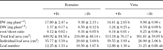
The primary symptom of iron deficiency is interveinal chlorosis in young leaves; it was initially observed in ‘Romaine’ (after 5 days of treatment) and later in ‘Vista’. These effects were intensified with time (data not shown). The inhibitory effects of iron deficiency on plant growth in both varieties were reflected in the fresh weight of plants measured after 15 days that was significantly lower as compared to the control. This growth decline was more pronounced in ‘Romaine’ than in ‘Vista’: 47% and 31%, respectively (Table 1). In addition, we found that iron deprivation had a similar effect on plant dry weights. The increased root/shoot ratio of treated plants especially observed in ‘Vista’ shows that shoots were more affected than roots (Table 1).
Plant growth parameters of two lettuce varieties after 15 days of growth under Fe-sufficient (+Fe) or Fe-deficient (–Fe) conditions.
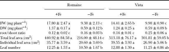
Data are means of 3 replicates ±standard error. Means followed by the same letter are not statistically different according to the ANOVA test at p ≤ 0.05.
Total and individual leaf areas were also significantly reduced by Fe deficiency as compared to the values measured in Fe-sufficient lettuce. The decreases were in total and individual leaf area (in ‘Romaine’ and ‘Vista’) was 51 and 41% and 42 and 32%, respectively (Table 1). However, the effect of Fe-deficiency was less pronounced in leaf number.
Mineral nutrition
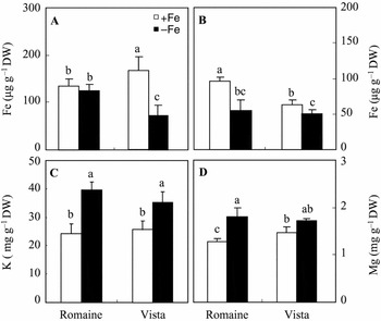
Mineral concentrations were measured in Fe-sufficient and Fe-deficient plants after 15 days of treatment. As compared to ‘Vista’, ‘Romaine’ Fe-sufficient plants showed higher amounts of Fe in shoots and lower amounts in roots. But, in Fe-deficient plants of both varieties, shoot Fe amounts were diminished by iron deficiency (Figure 1). The larger diminution (–43%) was observed in ‘Romaine’. In roots, Fe concentration decreased only in ‘Vista’ under iron deficiency conditions.
Root A) and shoot B) Fe concentrations and shoot K C) and Mg D) concentrations in two lettuce varieties after 15 days of growth under Fe-sufficient (+Fe) or Fe-deficient (–Fe) conditions. Data are means of 8 replicates ±standard error. Bars labelled by the same letter are not statistically different according to the ANOVA test at p ≤ 0.05.

In both varieties, shoot K concentration of plants grown without Fe was significantly higher than that of plants grown with Fe (Figure 1). However, Mg concentration was significantly increased only in ‘Romaine’ shoots.
Chlorophyll and carotenoid concentration
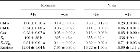
Noticeable differences between the two varieties appeared when leaf chlorophyll and carotenoid concentrations were measured. Under control conditions, ‘Romaine’ (dark green leaves) showed more pigment concentration than ‘Vista’ (light green leaves). Differences were also observed under Fe-limiting conditions; iron deficiency had more marked influence on chlorophyll a, chlorophyll b and carotenoid concentrations in ‘Romaine’ than in ‘Vista’. In fact, the reductions of total chlorophyll and carotenoid concentrations observed in ‘Romaine’ and ‘Vista’ was 81 and 82% and 54 and 62%, respectively (Table 2).
Leaf pigment concentrations (mg g−1 FW), fluorescence parameters and Rubisco activity (μmol h−1 g−1 FW) in two lettuce varieties after 15 days of growth under Fe-sufficient (+Fe) or Fe-deficient (–Fe) conditions.
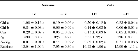
Data are means of 6 replicates ±standard error. Means followed by the same letter are not statistically different according to the ANOVA test at p ≤ 0.05.
Photosystem II (PSII) activity
A significant decrease in F v/F m values, that reflects the quantum yield of PSII, was found with Fe deficiency. In control leaves, F v/F m was higher than 0.8 for both varieties. At the end of the treatment period, F v/F m ratio decreased to 0.71 and 0.44 for ‘Vista’ and ‘Romaine’, respectively. In chlorotic leaves of ‘Romaine’, an increase of F 0 was observed, a phenomenon that was not observed in ‘Vista’ (Table 2). In addition, a significant reduction of Rubisco activity was found in leaves of ‘Romaine’ plants exposed to Fe deficiency (Table 2).
Root acidification capacity and FCR activity
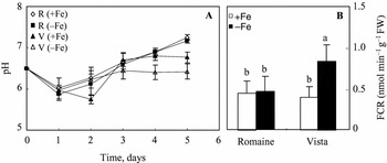
The time courses of the medium acidification by roots of hydroponically grown lettuce in the presence and absence of iron are shown in Figure 2A. A consistent but relatively weak acidification of the medium occurs during the first 2 days of the experiment. Then, pH values increased in both varieties. However, we noted that ‘Vista’ deficient plants retained lower pH values during the last 5 days of treatment.
pH and Fe-chelate reductase activity in two lettuce varieties after 15 days of growth under Fe-sufficient (+Fe) or Fe-deficient (–Fe) conditions. Data are means of 6 replicates ±standard error. Bars or points labelled by the same letter are not statistically different according to the ANOVA test at p ≤ 0.05.

Fe3+ reduction activity did not change significantly in ‘Romaine’ with iron deficiency. However, the stress significantly enhanced Fe3+-reducing capacity of the roots, from 0.46 to 0.98 nmol Fe2+ min−1 g−1 FW, in ‘Vista’ and this capacity was thus about twice that of ‘Romaine’ grown in the absence of iron (Figure 2B).
PEPC activity
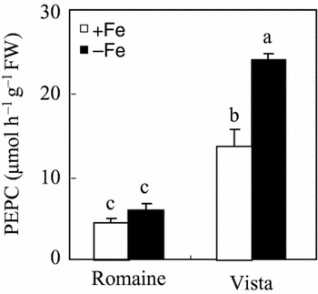
PEPC activity was also measured in root extracts from the two varieties. Iron deficiency induced an increase in the activity of this enzyme in ‘Vista’, whereas it did not significantly affect it in ‘Romaine’ (Figure 3).
PEPC activity in two lettuce varieties after 15 days of growth under Fe-sufficient (+Fe) or Fe-deficient (–Fe) conditions. Data are means of 3 replicates ±standard error. Bars labelled by the same letter are not statistically different according to the ANOVA test at p ≤ 0.05.

Total phenol, flavonoid and condensed tannin concentrations
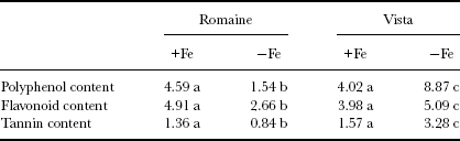
The total phenolic, flavonoid and condensed tannin concentrations in roots of two varieties are given in Table 3. Results showed that phenolic compound accumulation in roots was stimulated by Fe deficiency in ‘Vista’ variety. However, iron deficiency condition induced a decrease of phenolic concentration in ‘Romaine’ extract. Moreover, flavonoid and condensed tannin concentrations are decreased in deficient root of ‘Romaine’ variety.
Total polyphenol (mg GAE g−1 DW), Flavonoid and condensed tannin content (mg CE g−1 DW) in roots of two Lactuca sativa varieties after 15 days of growth under Fe-sufficient (+Fe) or Fe-deficient (–Fe) conditions.
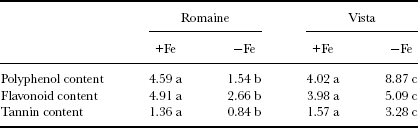
Data are means of 6 replicates ±standard error. Means followed by the same letter are not statistically different according to the ANOVA test at p ≤ 0.05.
DISCUSSION
Differences in tolerance to Fe-deficiency chlorosis between varieties have been described for many plant species (Alcántara et al., Reference Alcántara, Cordeiro and Barranco2003). In this investigation, the two lettuce varieties showed different physiological responses to the stress. As shown here, ‘Vista’ experienced slighter chlorosis symptoms than ‘Romaine’ under Fe deficiency. Iron stress resulted in decreased fresh weights in both lettuce varieties. Total leaf area also decreased significantly. This restriction concerned mainly leaf expansion (individual leaf area) as leaf number was much less affected by iron deficiency, especially in ‘Vista’. The differences between the two varieties could be attributed to (i) an inhibition of leaf induction in ‘Romaine’, practically absent in ‘Vista’, and (ii) a restriction of leaf expansion, present in both varieties but more important in ‘Romaine’. This is consistent with other findings in several species (Ksouri et al., Reference Ksouri, Debez, Mahmoudi, Ouerghi, Gharsalli and Lachaal2007; Mahmoudi et al., Reference Mahmoudi, Koyro, Debez and Abdelly2008; Zocchi et al., Reference Zocchi, De Nisi, Dell'Orto, Espen and Gallina2007). In the present study, iron deficiency affected dry matter distribution leading to a higher root/shoot ratio. This effect was more pronounced in ‘Vista’. The increased root/shoot ratio in deficient plants is in consonance with earlier observations in Spathiphyllum (Yeh et al., Reference Yeh, Lin and Wright2000) and Vitis vinifera (Gruber and Kosegarten, Reference Gruber and Kosegarten2002). It may be a response mechanism to the lack of Fe, associated with an increase in the number of secondary roots and the differentiation of transfer cells in the root apex. In contrast to lettuce, other species such as sugar beet showed a larger growth restriction in roots than in aerial parts as subjected to Fe deficiency (Rombolà et al., Reference Rombolà, Gogorcena, Larbi, Morales, Balde, Marangoni, Tagliavini and Abadía2005).
Fe concentration was more reduced in aerial organs of ‘Romaine’ than in those of ‘Vista’, which could explain the more pronounced decrease in biomass production in the former. However, its concentration in roots was reduced only in ‘Vista’. According to these data, it appears that ‘Vista’ is more able to transport iron towards its aerial parts than ‘Romaine’. Our findings showed also that iron deficiency increased shoot potassium concentration in both varieties; whereas it increased that of magnesium only in ‘Romaine’. These results agree with the findings of other authors in spinach (Assimakopoulou, Reference Assimakopoulou2006) and peach trees (Belkhodja et al., Reference Belkhodja, Morales, Sanz, Abadía and Abadía1998). A higher potassium concentration in the shoots of the Fe-deficient plants has been shown to correlate with a fall in leaf calcium concentrations in some plant (DeKock et al., Reference Dekock, Hall and Mcdonald1960). López-Millán et al. (Reference López-Millán, Morales, Andaluz, Gogorcena, Abadía, de las Rivas and Abadía2000) reported that in iron-deficient leaves of sugar beet, the apoplastic concentration of potassium and magnesium increased in comparison with the control. They also showed that the increase of cation concentrations was associated to an increase of organic acid concentration in leaves (Abadía et al., Reference Abadía, López-Millán, Abadía and Rombolà2002). This supports that an increase in cation uptake by roots accounted for the increase in malate concentrations under Fe deficiency, as proposed by Welkie and Miller (Reference Welkie, Miller, Barton and Hemming1993).
The severe effect of iron deficiency on total dry weight in ‘Romaine’ may be due to the considerable decrease in PSII efficiency. In fact, it is known that a low Fe supply negatively affects chlorophyll concentration and other chloroplast components, which reduces photosynthesis and growing capacity in plants (De La Guardia and Alcántara, Reference De La Guardia and Alcántara2002). A more pronounced effect of the stress on chlorophyll and carotenoid concentrations was observed in ‘Romaine’ than in ‘Vista’. PSII activity was also affected by iron deficiency as shown by the declined F v/F m ratio often used as indicators of PSII sensitivity to many kinds of stress. This decline was more pronounced in ‘Romaine’ leaves and was accompanied by an increase of the minimal fluorescence F 0 in this variety. Similar observations were previously reported in cyanobacteria (Guikema, Reference Guikema1985; Guikema and Sherman, Reference Guikema and Sherman1983; Riethman and Sherman, Reference Riethman and Sherman1988) and in some higher plants (Larbi et al., Reference Larbi, Abadía, Abadía and Morales2006; Morales et al., Reference Morales, Abadía and Abadía1991; Timperio et al., Reference Timperio, D'Amici, Barta, Loreto and Zolla2007) affected by iron deficiency. Such a reduction of PSII efficiency can be attributed to a structural modification particularly a destruction/dissociation of antennae pigments (Morales et al., Reference Morales, Abadía and Abadía1991), as borne out by the results of chlorophyll concentration assays in our study. The activity of Rubisco, one of the major enzymes of the carbon fixation cycle, was declined in ‘Romaine’ plants exposed to Fe deficiency. Similar reduction in the activity of this enzyme was shown by Bertamini et al. (Reference Bertamini, Nedunchezhian and Borghi2001) in Fe-deficient grapevine. Winder and Nishio (Reference Winder and Nishio1995) also reported a decline in Rubisco activity with severe Fe deficiency in sugar beet.
The fact that the two varieties investigated in the present work exhibited some contrasting physiological responses may help evaluate the relative importance of these properties in terms of whole plant response. Rizosphere acidification has been recognized for many years as an important response to iron deficiency for Strategy I plants (Zocchi et al., Reference Zocchi, De Nisi, Dell'Orto, Espen and Gallina2007). However, in our case, acidification capacity was weak in both varieties. A similar result was found by Landsberg (Reference Landsberg1982) who showed that acidification capacity is strongly limited even in the efficient genotypes of soybean. Unlike acidification capacity, a significant varietal difference was observed at the end of treatment in FCR and PEPC activities and polyphenol accumulation that were higher in ‘Vista’ than in ‘Romaine’. These findings suggest that the induction of FCR and PEPC activities in roots in response to Fe deficiency constitutes an important physiological adaptation enabling ‘Vista’ plants to tolerate the stress. On the other hand, polyphenol accumulation coincides with a higher PEPC and FCR activities in Fe-deficient roots of ‘Vista’ variety. It has been suggested that phenolics compounds enhance the Fe availability in the rhizosphere soil, through chelating and reducing insoluble Fe (Dakora and Phillips, Reference Dakora and Phillips2002). In contrast, Jin et al. (Reference Jin, You, He, Tang, Wu and Zheng2007) demonstrated that enhanced FCR activity was not involved by phenolics secretion. However, the secreted phenolics played a critical role in facilitating the reutilization of the apoplastic Fe in roots.
In conclusion, these data demonstrates that ‘Vista’ was more tolerant to iron deficiency than ‘Romaine’. The capacity of ‘Vista’ to prevent iron deficiency effect was due to its ability to induce its root responses including polyphenol accumulation, FCR and PEPC activities and then to reduce and transport iron towards its aerials parts.
Acknowledgement
The authors wish to thank Professor R. Carpentier for careful reading of the manuscript and stimulating discussions on this work.