Introduction
Nasal obstruction is an important health problem; it can be congenital or due to acquired anatomical abnormalities or various disorders affecting the nasal airway passage. A frequent cause of nasal obstruction is turbinate hypertrophy. This has been one of the most comprehensively studied pathologies by rhinologists since the late nineteenth century.Reference Passali, Laureiello, Bellussi and Anselmi1, Reference Myrthe, Hol and Huizing2
At present, nasal turbinate hypertrophy has various medical and surgical treatments. Topical or systemic decongestant drugs and topical steroids are common treatment modalities in primary healthcare. However, medical treatments are not always successful and surgery is often required. The many surgical treatments for turbinate hypertrophy include corticosteroid injection into the turbinate, cryotherapy, electrocauterisation, out-fracture, total or partial turbinectomy, submucosal resection, laser-assisted turbinoplasty, and radiofrequency tissue ablation. The large number of surgical methods indicates that there is no single ideal method of therapy for turbinate hypertrophy. Therefore, new alternative methods are required to treat this disorder. The ideal therapeutic method should reduce turbinate volume while retaining physiological function, with no complications.Reference Lippert and Werner3–Reference Hol and Huizing7
The drug, OK-432 (Picibanil; Chugai Pharmaceutical, Tokyo, Japan), has been widely used in recent years. It is a mixture of freeze-dried avirulent group A Streptococcus pyogenes and penicillin G,Reference Ogita, Tsutu and Tokiwa8 which has been used as an immunotherapeutic agent for malignancies. OK-432 is currently used to treat sclerotherapy because of its antitumoral action. It is also frequently used to treat cystic lesions in the head and neck region: it destroys the cyst wall by causing inflammation of endothelial cells, which leads to fibrosis and thus lesion shrinkage.Reference Ogita, Tsuto, Deguchi, Tokiwa, Nagashima and Iwai9, Reference Herrera, Di Segni, Kaye, Castaneda-Zuniga and Tadavarthy10 This effect led us to hypothesise that OK-432 could also be used to reduce the size of hypertrophic turbinates.
This study aimed to assess the histopathological effect of injected OK-432 (Picibanil) on nasal turbinates and to determine whether it could form an alternative therapy for nasal turbinate hypertrophy.
Materials and methods
This prospective, controlled, experimental study was carried out using 21 New Zealand adult male rabbits, each weighing 2500–3500 g (average 3000 g). The study plan was approved by the Ethics Committee for Animal Experiments of Firat University (4 April 2013; approval number 43). Standard experimental reagents were supplied by the Scientific Research Projects Unit of Firat University (project number TF13.41). All applicable international, national and/or institutional guidelines for the care and use of animals were followed.
Nasal cavities of the rabbits were examined using a 2.7-mm endoscope (Karl Storz, Tuttlingen, Germany), and rabbits with normal-looking turbinate ventralis (i.e. without hypertrophy or increased mass) were included in the study. Animals were randomly divided into three groups of seven and the same procedure was performed on all by the same surgeon. Animals were anaesthetised with an intramuscular injection of 2 mg/kg xylazine hydrochloride (Rompun, Bayer, Leverkusen, Germany) and 5 mg/kg ketamine hydrochloride (Ketalar, Eczacıbaşı İlaç, Turkey). OK-432 or saline (0.2 ml OK-432, 0.5 ml OK-432 or 0.6 ml saline per animal) was then injected twice into the submucosa of the one-third anterior, middle and posterior parts of both nasal turbinates ventralis using a 27-gauge needle, with 3 weeks between each injection.
Six weeks after the first injection, all rabbits were sacrificed under ketamine hydrochloride anaesthesia according to the Guide for the Care and Use of Laboratory Animals.11 The turbinates nasalis ventralis were then completely excised using the lateral rhinotomy technique. Excised specimens were fixed in 10 per cent formaldehyde and then embedded in paraffin. Paraffin blocks were then cut into 5 μm-thick sections using Ultratome III glass blades (Shandon Finesse, Thermo Scientific, Hemel Hempstead, UK). Sections were stained with trichrome Masson and hematoxylin and eosin stains and examined by the same pathologist under light microscopy (Olympus, BX51, Tokyo, Japan) at ×40, ×100, ×200 and ×1000 magnification.
Ciliary loss, increased submucosal vascularisation, goblet cell loss, inflammatory cell infiltration, fibrosis and epithelial damage were scored as follows: none, –; mild, +; intermediate, ++; severe, +++. An eyepiece graticule (1 × 1 mm ocular micrometer containing 100 equal squares) attached to the light microscope was used as a measuring scale. Four microscope fields from four tissue sections from each rabbit were scored (at ×40, ×100, ×200 and ×1000 magnification), and the mean score for all fields was calculated for each group.
Statistical analysis of histopathological data from the test animals was performed using IBM SPSS Statistics for Windows, Version 21.0. (IBM Corp. Armonk, New York, USA). The Mann–Whitney U test and the Kruskal–Wallis test were used for between-group comparisons. A p value less than 0.05 was considered significant.
Results
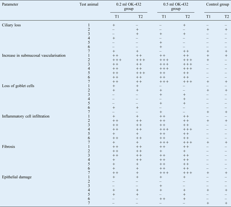
The histopathological scoring parameters for all groups are shown in Table I. Ciliary loss was most severe in the 0.2 ml OK-432 (mild in 6) and 0.5 ml OK-432 (mild in 5, intermediate in 1) treatment groups. In the control group, mild ciliary loss was seen in only 2 turbinates (Table I). There were no significant between-group differences related to ciliary loss (p > 0.05).
Histopathological scoring of parameters in the groups

T = turbinate; – = no effect; + = mild or low effect; ++ = intermediate effect; +++ = high or severe effect
Intense submucosal vascularisation was mostly observed in the 0.5 ml OK-432 treatment group (intermediate in 6 and intense in 8), followed by the 0.2 ml OK-432 treatment group (intermediate in 10 and intense in 4). In the controls, a mild increase in submucosal vascularisation was seen in only six turbinates (Table I). There was a significant increase in submucosal vascularisation in both OK-432 treatment groups compared with controls (p < 0.05; Fig. 1). However, there was no significant difference in submucosal vascularisation between both OK-432 treatment groups (p > 0.05).
Photomicrograph showing a strong increase in submucosal vascularisation in 0.5 ml OK-432 treated turbinate tissue. (H&E ×400)
Intermediate or severe goblet cell loss did not occur in any group; mild goblet cell loss was observed in six turbinates in the 0.2 ml OK-432 treatment group, seven turbinates in the 0.5 ml OK-432 treatment group and four turbinates in the control (Table I). There was no significant between-group difference in goblet cell loss (p > 0.05).
Inflammatory cell infiltration was mainly observed in the 0.5 ml OK-432 treatment group (low in two, intermediate in eight, high in four), followed by the 0.2 ml OK-432 treatment group (low in six, intermediate in eight) and the control group (low in four; Table I and Fig. 2). There was a significant difference in inflammatory cell infiltration between both OK-432 treatment groups and the control group (p < 0.05). However, there was no significant difference in this parameter between OK-432 treatment groups (p > 0.05).
Photomicrograph showing severe inflammatory cell infiltration in 0.5 ml OK-432 treated turbinate tissue. (H&E ×400)
The lowest degree of fibrosis was observed in the control group (mild in 2) and the highest degree was observed in the 0.5 ml OK-432 treatment group (mild in 2, intermediate in 10, severe in 2), followed by the 0.2 ml OK-432 treatment group (mild in 6 and intermediate in 8; Table I and Fig. 3). There was a significant difference in the degree of fibrosis between the control group and each OK-432 treatment group (p < 0.05). There was also a significant difference in the degree of fibrosis between OK-432 treatment groups (p < 0.05).
Photomicrograph showing severe fibrosis in 0.5 ml OK-432 treated turbinate tissue. (H&E ×400)
The lowest level of epithelial damage was observed in the control group (mild in four) and the highest was observed in the 0.5 ml OK-432 treatment group (mild in 7, intermediate in 1; Table I). There was no significant difference in epithelial damage between the OK-432 treatment groups (p > 0.05).
Discussion
The medical and surgical treatment modalities used for nasal obstruction due to nasal turbinate hypertrophy do not always yield successful results. This has led scientists to seek new therapeutic techniques. In recent years, the injection of chemicals into turbinates has been widely practised.Reference Lippert and Werner3–Reference Hol and Huizing7
There are a limited number of studies on sclerosing substance injection into the turbinate. Swift and Siegler reported a marked reduction in turbinate size after ethanolamine oleate injection into the inferior turbinate.Reference Swift and Siegler12 Likewise, Lafayette injected sodium salicylate and mercuric chloride solutions as sclerosing agents into the submucosa of inferior turbinates, resulting in marked turbinate shrinkage.Reference Lafayette13 However, after these sclerosing agents were reported to cause pyemia, renal dysfunction and pulmonary embolism, their use for turbinate reduction was stopped.Reference Miyoshi, Ohshiba, Matsumoto and Takada14, Reference Woods15
OK-432 is a sclerosing drug that is used as an immunotherapeutic agent for malignant tumours. No serious complication of OK-432 treatment has been reported, other than local inflammation.Reference Ogita, Tsutu and Tokiwa8, Reference Brewis, Pracy and Albert16–Reference Smith, Burke, Sato, Poust, Kimura and Bauman22 In recent years, OK-432 safety has been supported by case reports describing its in utero application.Reference Ogita, Suita, Taguchi, Yamanouchi, Masumoto and Tsukimori23 Hamamoto et al. used radiofrequency ablation combined with OK-432 to treat experimental lung tumours in rabbits and reported the development of systemic antitumoral immunity at the end of treatment.Reference Hamamoto, Okuma, Yamamoto and Kageyama24 Many studies have described the use of OK-432 for treating ranula.Reference Parekh, Stewart, Joseph and Lawson25–Reference Kim, Sung and Roh28 Chang et al. reported the successful use of OK-432 to treat recurrent or progressive thyroid nodules.Reference Chang, Yoon, Chung and Park29 To the best of our knowledge, no study has reported the histopathological effects of OK-432 on turbinates. This, as well as the known fibrosing effect of OK-432, led us to investigate whether OK-432 can reduce the size of hypertrophic turbinates. In the present study, OK-432 caused minimal changes in ciliary loss, goblet cell loss and epithelial damage in rabbit turbinates, but caused significant changes in increased submucosal vascularisation, inflammatory cell infiltration and fibrosis. In particular, the severe fibrosis observed in both test groups compared with the control group led us to conclude that OK-432 can achieve turbinate reduction.
Coste et al. reported that turbinate reduction by radiofrequency ablation does not cause epithelial damage, ciliary loss or goblet cell loss, and that surgical methods, except for submucosal resection of the turbinates, are not as effective in protecting the nasal epithelium.Reference Coste, Yona and Blumen30 Hol and Huizing reported that laser therapy for hypertrophic turbinates provides the optimal volume reduction but cannot protect mucociliary function, thus leading to irreversible functional damage to the turbinate.Reference Hol and Huizing7 Our study showed mild ciliary loss and epithelial damage in both OK-432 test groups and control group (p = 0.101), suggesting that OK-432 does not damage mucociliary function. Further detailed studies are needed to verify this result.
Berger et al. compared the histopathology of tissue that had been treated or not treated with radiofrequency ablation and found dispersed infiltrates of inflammatory cells, fibrosis and many sites of vascularisation along the submucosal stroma underlying the epithelium of radiofrequency-ablated tissue.Reference Berger, Ophir, Pitaro and Landsberg31 Kaplama et al. studied the effect of radiofrequency ablation and submucosal diathermy on the turbinates and found mild inflammatory cell infiltration and mildly increased submucosal vascularisation but marked fibrosis in radiofrequency-ablated turbinates.Reference Kaplama, Kaygusuz, Akpolat, Karlıdağ, Keleş and Alpay32 Similarly, we found a significant degree (p = 0.000) of inflammatory cell infiltration, increased submucosal vascularisation and fibrosis in both OK-432 treatment groups. There was a higher degree of fibrosis in the 0.5 ml OK-432 treatment group than in the 0.2 ml OK-432 treatment group (p = 0.019). The finding that a higher degree of fibrosis is associated with a higher OK-432 dose in rabbits suggests that a high dose of OK-432 could be used to treat hypertrophic turbinates in humans. Further experimental and clinical studies are necessary to verify this result.
• Turbinate hypertrophy is a frequent cause of nasal obstruction
• The effect of OK-432 (Picibanil) on nasal turbinate histopathology in rabbits was assessed microscopically
• The marked fibrosis after OK-432 injection may lead to size reduction in human hypertrophic turbinates
Zborayova et al. compared histopathological changes in the inferior turbinate induced by radiofrequency and laser ablation.Reference Zborayova, Ryska, Miroslav, Petr, Januskova and Vokurka33 They observed intact superficial epithelium and fibrosis in radiofrequency-ablated specimens but necrosis, ulcerations, fibrin aggregation in the mucosa and damage to the superficial epithelium in laser-ablated specimens. In this study, we observed a significant degree of fibrosis only in OK-432 treated specimens and no necrosis or ulceration in any experimental group. This finding suggests that OK-432 therapy may be safer than laser ablation for turbinate reduction.
Conclusion
Slight ciliary loss, goblet cell loss and epithelial damage were observed in turbinates treated with OK-432, but these changes were not significantly different than in the control group. This finding suggests that OK-432 may not affect nasal mucociliary transport. However, the OK-432 use was associated with a significant increase in submucosal vascularisation, inflammatory cell infiltration and fibrosis. In particular, the increased degree of fibrosis suggests that OK-432 may reduce turbinate size, thus facilitating air passage through the nose. Despite these positive results, more experimental and clinical studies are needed to verify OK-432 as an alternative therapy for turbinate reduction. Furthermore, clearance studies to show the status of the mucociliary transport and rhinomanometric studies to more objectively determine the degree of nasal airflow should form the next step in future investigations.