Introduction
The design of the conventional alloys was primarily based on one or two principal alloying elements along with the addition of a few other minor elements to tailor the microstructures and their properties. The understanding of alloy design has been advanced by the seminal work of Cantor et al. [Reference Cantor, Chang, Knight and Vincent1] and Yeh et al. [Reference Yeh, Chen, Lin, Gan, Chin, Shun, Tsau and Chang2] by advocating a new class of materials, known as high-entropy alloys (HEA). The name “high-entropy alloy” is justified due to higher mixing entropy compared with the conventional alloys for stabilizing the solid solution phase. This new class of alloys has received considerable attention due to its interesting novel properties compared with the conventional alloys [Reference Mukhopadhyay3]. HEAs generally should contain at least five principal alloying elements whose atomic concentration varies in the range of 5–35% [Reference Yeh4]. Due to the presence of multi-principal elements, these alloys exhibit high-entropy (configurational) effect, favoring the formation of simple solid solution (body centered cubic (BCC)/face centered cubic (FCC)/hexagonal close packed (HCP)) phases [Reference Joo, Kato, Jang, Moon, Kim, Hong and Kim5]. A solid solution phase is understood to conform to the crystal structures resemble one of their parent structure in which the metal atoms are present in proportion to their concentration. However, the stability of the simple solid solution phases over a wide range of temperature is a major concern. It has been found that these simple solid solution phases can decompose into the other phases, including intermetallic phases, when exposed to prolonged heat treatment [Reference Mohanty, Maity, Mukhopadhyay, Sarkar, Gurao, Bhowmick and Biswas6, Reference Moravcik, Cizek, Gavendova, Sheikh, Guo and Dlouhy7]. HEAs possess good properties, i.e., high hardness, high strength, excellent thermal stability, magnetic properties, and good resistance to wear, oxidation, and corrosion due to their unique composition design concept [Reference Zhang, Zuo, Cheng and Liaw8, Reference Chou, Yeh and Shih9, Reference Kai, Li, Cheng, Chu, Huang, Tsay and Kai10, Reference Fu, Chen, Jiang, MacDonald, Lin, Chen, Zhang and Lavernia11, Reference Zhang and Fu12].
The inspection of the literature on HEAs reveals that most of the HEAs have been synthesized through melting and casting route, i.e., vacuum arc melting, vacuum induction melting, and injection casting. One of the major drawbacks that is inevitable with this route is the formation of inhomogeneous microstructure due to segregation of the alloying elements during solidification. Therefore, subsequent heat treatment is required to homogenize these microstructures. Alternatively, the solid-state processing route, i.e., mechanical alloying (MA), is used extensively to prepare the HEAs with homogenized and refined microstructures [Reference Varalakshmi, Kamaraj and Murty13, Reference Wang, Li, Ren, Yang and Fu14, Reference Chen, Hu, Hsieh, Yeh and Chen15, Reference Fu, Chen, Chen, Wen and Lavernia16, Reference Suryanarayana, Ivanov and Boldyrev17, Reference Suryanarayana18, Reference Fu, Chen, Wen, Zhang, Chen, Zheng, Zhou and Lavernia19, Reference Fu, Chen, Wen, Chen and Lavernia20]. MA has the ability to synthesize the elements with different melting point and vapor pressure [Reference Koch21]. The MA followed by spark plasma sintering (SPS) ensured the formation of bulk nanostructured materials with nanocrystalline and uniform structure and high density [Reference Mohanty, Maity, Mukhopadhyay, Sarkar, Gurao, Bhowmick and Biswas6]. SPS has the ability to consolidate the alloy powders by applying pressure and passing electric current for a very short soaking time. This short consolidation time is beneficial to retain the fine microstructure and to achieve the better mechanical properties with nanocrystalline grains [Reference Joo, Kato, Jang, Moon, Kim, Hong and Kim5]. Zhang et al. [Reference Zhang, Fu, Zhang, Wang, Lee and Niihara22] have reported the formation of supersaturated BCC and FCC solid solution phase in equiatomic AlCoCrFeNiTi HEA after MA. They have observed that this phase transformed into two BCC solid solution phases after annealing the milled powder at 600 °C for 1 h. Zhang et al. [Reference Zhang, Fu, Zhang, Wang, Wang, Wang, Zhang and Shi23] in their another work on non-equiatomic CoCrFeNiTiAlx alloy, prepared through vacuum arc melting, have observed that CoCrFeNiTi alloys have the FCC crystal structure. The phase composition transformed to stabilize the BCC solid solution phase after the addition of Al to the alloy. From these studies, it could be inferred that HEAs are affected by processing routes and the addition of alloying elements for controlling the evolution of phases. Although equiatomic AlCoCrFeNiTi HEA has been reported earlier, there is further scope to investigate the alloying behavior and thermal stability in a systematic manner to remove the apparent inconsistencies.
In view of the above-mentioned findings, the present work was motivated mainly by two factors. First, there are fewer reports on the thermal stability of the hexanary AlCoCrFeNiTi HEA. Second, the systematic studies of the alloying behavior and the phases evolved during the milling of AlCoCrFeNiTi HEA have rarely been investigated. Thermal stability of the alloy has been studied with X-ray diffraction (XRD), electron microscopy, and differential scanning calorimetry. Finally, attempts have been made to consolidate the HEA powder through microwave sintering. Microwave sintering is the new kind of sintering route (in the context of HEAs) to sinter the HEA powder. Contrary to the conventional sintering, in the microwave sintering, both direct heating of metallic powder and indirect heating by an auxiliary SiC absorber are associated [Reference Veronesi, Colombini, Rosa, Leonelli and Rosi24]. Compared with the conventional sintering, microwave sintering has various advantages, such as rapid heating rate, time-saving, and retaining fine microstructure [Reference Thostenson and Chu25]. Here, microwave sintering was used as an exploratory method to sinter the HEA. It has been found that best results with increased density can be obtained when pressure is applied during sintering.
Results
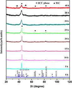
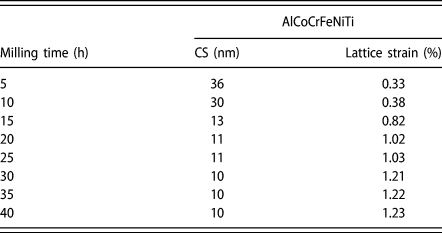
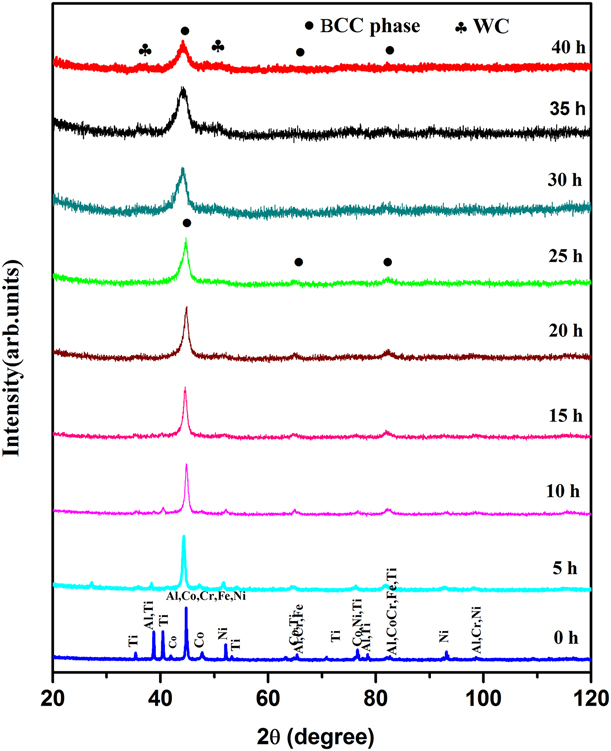
XRD patterns of AlCoCrFeNiTi HEA milled powder with various milling time are displayed in Fig. 1. Before milling (0 h), diffraction signals corresponding to all the constituent elements were observed. After 5 h of milling, it has been found that diffraction intensities of few peaks corresponding to Ti (d 101 = 2.228 Å), Ti (d 103 = 1.32 Å), and Ti, Al, Ni (d 112/311/220 = 1.23 Å) start losing their intensity. Further, on increasing the milling time up to 10 h, it has been noted that the diffraction intensities of low-angle peak corresponding to Co start reducing earlier to other cubic structures. Earlier reports on mechanically alloyed HEAs suggest that the melting point of the element is related to the cause of disappearance of the peaks corresponding to the elements [Reference Chen, Fu, Fang, Xiao and Zhu26]. Chen et al. reported that the HEAs of lower melting point elements go to the solid solution phase earlier than those of the high melting point elements due to faster diffusivity. Here, it has been found that in addition to the melting point, the structure of the elements plays an important role in the formation of the solid solution phase [Reference Chen, Hu, Hsieh, Yeh and Chen15]. Similar behavior was observed in our previously reported AlCoCrFeNi and AlCoCrFeNiMn HEAs [Reference Shivam, Basu, Shadangi, Singh and Mukhopadhyay27, Reference Shivam, Basu, Pandey, Shadangi and Mukhopadhyay28]. However, it is also well established that alloying behavior of elements during MA is a function of many parameters. Further increase in milling for next 5 h gives more support to the above-mentioned statement. After 25 h of milling, peaks corresponding to the constituent elements disappeared and the formation of single-phase BCC structure was observed. To understand the alloying behavior and phase stability of the alloy, milling was extended further. On increasing the milling time, it has been noted that along with the existing BCC (a = 2.85 ± 0.01 Å) phase, a new peak corresponding to an unexpected phase appears. After careful investigation, it has been found that these signals conform to the existence of hexagonal tungsten carbide (WC) (a = 2.90 Å, c = 2.83 Å) phase. This indication becomes more prominent on further increase in milling time up to 40 h. Throughout the diffraction patterns analysis, it has been noted that the intensity of the major peaks decreases with increase in broadening. This behavior was expected because the increase in milling time decreases the crystallite size and increases the lattice strain. It also supports the formation of nanocrystalline structure. The pseudo-Voigt function was used for fitting XRD peak profile analysis after eliminating the instrumental broadening contribution by using standard silicon sample. Crystallite size and lattice strain were calculated by using Williamson–Hall method with X’Pert HighScore Plus software (Malvern PANalytical, Malvern, UK). The variation in crystallite size (nm) and lattice strain (%) of AlCoCrFeNiTi HEA with milling time is reported in Table I. It could be observed that at initial milling time the decrease in crystallite size is prominent with significant induction of lattice strain. This increase in lattice strain is mainly due to the introduction of elements in the host lattice of the HEA and mechanical working of powder during MA processing [Reference Fu, Chen, Chen, Wen and Lavernia16, Reference Wang, Ji and Fu29]. After 40 h of milling, it has been found that the diffraction intensities of the higher angle peaks are very weak in nature. This weak nature of the peaks (intensity) might be the reason for atomic mismatches (due to the constituent alloying elements), which destroyed the corresponding parent lattices.
XRD patterns of AlCoCrFeNiTi HEA powders with different milling time. Evolution of BCC and WC phase was evident with the progress of milling.
The calculated values of the crystallite size (nm) and lattice strain (%) in AlCoCrFeNiTi HEA at different milling schedules.

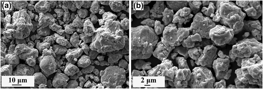
The morphology and size of the powder particles were investigated using scanning electron microscopy (SEM). Figures 2(a) and 2(b) show the secondary electron micrographs of 40 h milled AlCoCrFeNiTi HEA powder. It is observed that nanosized particles got agglomerated and formed micron-sized particles. The size of these micron range particles varies in the range of 3–18 μm. This is happening because during MA, the powder particles get flattened, cold-welded, and fractured repeatedly. The process of welding and fracturing reaches to a saturation value after a certain period.
(a, b) SEM micrographs of 40 h milled AlCoCrFeNiTi HEA powders at different magnifications. In both the cases, fractured particles with agglomerated form can be observed.

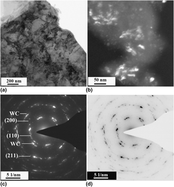
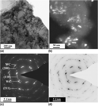
Phase evolution and alloying effects have also been confirmed through transmission electron microscopy (TEM). Bright-field image, corresponding selected area diffraction (SAD), and dark-field image are shown in Fig. 3. In the bright-field image, highly strained regions can be seen. Formation of ring patterns in the SAD confirms that the particles are strained and randomly oriented. Indexing of the ring pattern further corroborates the formation of the BCC phase along with WC. The dark-field imaging on the BCC reflections shows that the phase is in the nanometer ranges confirming to the evolution of nano BCC phase.
TEM images of 40 h milled AlCoCrFeNiTi HEA powder. (a) Bright-field image, (b) dark-field image, (c) SAD pattern, and (d) inverted SAD pattern for better visibility. Indexing of ring pattern confirmed the formation of a solid solution phase of BCC crystal structure along with WC phase.

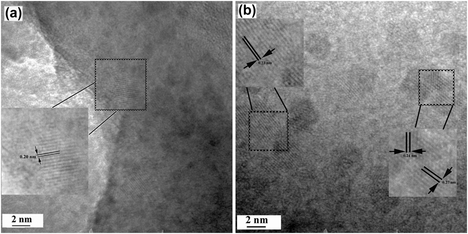
High-resolution imaging of the 40 h milled powder sample is shown in Fig. 4. It has been ascertained that similar “d” spacings of the BCC phase are observed in Fig. 4(a). From indexing of the lattice fringes in Fig. 4(b), it can be confirmed that some intermetallic phases have been formed after MA. Careful investigation shows that few fringes are due to the WC phase.
High-resolution imaging of the 40 h milled AlCoCrFeNiTi HEA powder. (a) Lattice fringes of similar “d” spacing of BCC phase were observed. (b) Formation of WC phase was also observed.

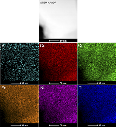
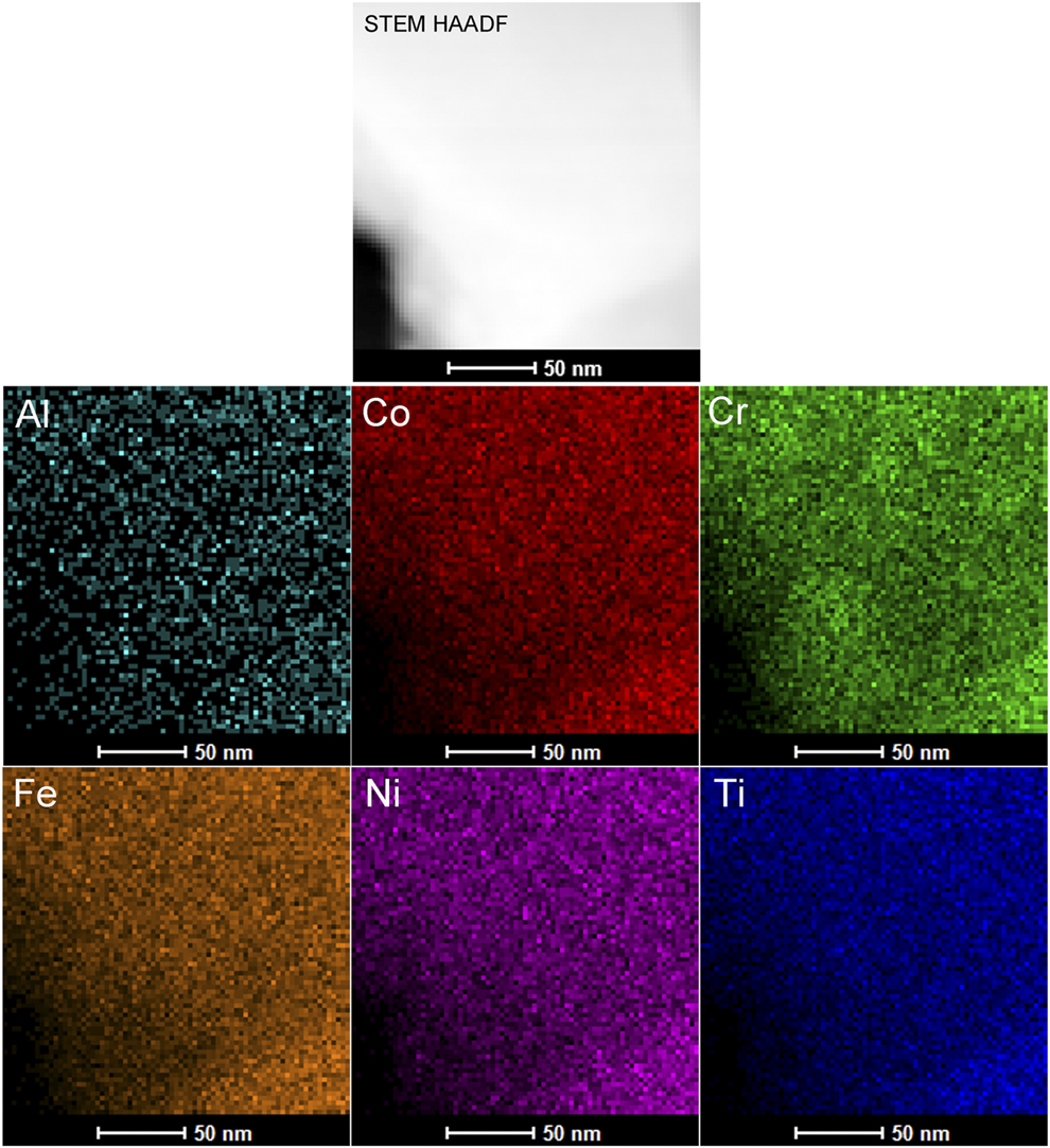
The chemical compositions of the constituent elements were analyzed using high angle annular dark field (HAADF) and scanning transmission electron microscopy-energy dispersive spectroscopy (STEM-EDS) mapping of the 40 h milled AlCoCrFeNiTi HEA and are displayed in Fig. 5. It could be visualized that all the constituent elements are homogenously distributed in the matrix of the AlCoCrFeNiTi HEA. It confirms that all the constituent elements are present in the equal participation, leading to high entropy of the alloy.
The mapping of equiatomic AlCoCrFeNiTi hexanary HEA after 40 h of milling. No segregation of elemental composition was observed.
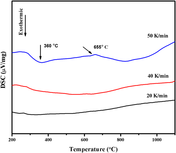
The differential scanning calorimetry (DSC) thermogram of the 40 h milled AlCoCrFeNiTi HEA is shown in Fig. 6. This thermogram is the plot of the energy evolved during the heating of the alloy powder up to 1200 °C (1473 K) with three heating rates 20, 40, and 50 K/min. It has been found that there was no sign of phase change at 20 and 40 K/min heating rates. Further increase in the heating rate (50 K/min) showed the presence of two thermodynamic heat events. First heat event that is exothermic in nature shows its signal at around 360 °C (633 K) and second event that is associated with the endothermic shows its signal at around 655 °C (928 K). The first exothermic event is related to the release of the internal stresses, such as structural deformation and lattice strain [Reference Zhuang, Xue, Chen, Hu and He30, Reference Tariq, Naeem, Hasan, Akhter and Siddique31]. The second peak (endothermic in nature) at around 928 K in the DSC thermogram might be the evolution of B2 phase. In the DSC thermograms, it has been observed that no peak (in 50 K/min heating rate) shows the sharp nature during the heat evolution. It can be elucidated that in HEAs due to the presence of the several elements in the alloy matrix, the kinetics of the phase change is very slow in nature and time dependent. Therefore, the phase transformations in all these alloys are diffusional in nature and depend on the range of temperature and time.
DSC thermogram of 40 h milled nanocrystalline equiatomic AlCoCrFeNiTi HEA heated up to 1200 °C. First heat event is related to the release of the internal stresses and the second might be related to the speculation of the B2 phase.
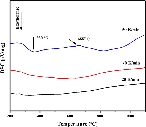
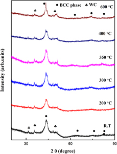
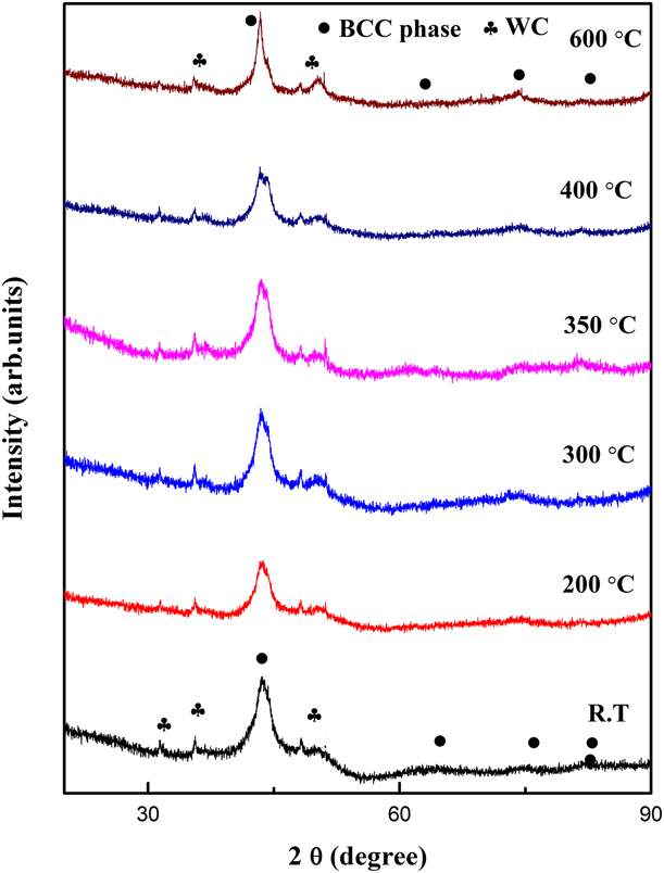
Figure 7 shows the in situ high-temperature XRD plots of the 40 h milled AlCoCrFeNiTi HEA powder. These temperatures were selected based on heat events evolved in the DSC thermogram. It has been noted that the first heat event in DSC thermogram was at around 360 °C (633 K), which might be related to the release of the internal stress. There was no change in phase constituents observed at this temperature (633 K). Further selected temperature (600 °C) also falls in the same line and confirmed that there is no transformation. Therefore, from this study, it can be concluded that AlCoCrFeNiTi alloy is stable up to 600 °C (873 K).
In situ XRD patterns of 40 h milled AlCoCrFeNiTi HEA powder heated up to 600 °C (873 K). No phase transformation is observed, which signifies that the alloy is stable up to 600 °C (873 K).
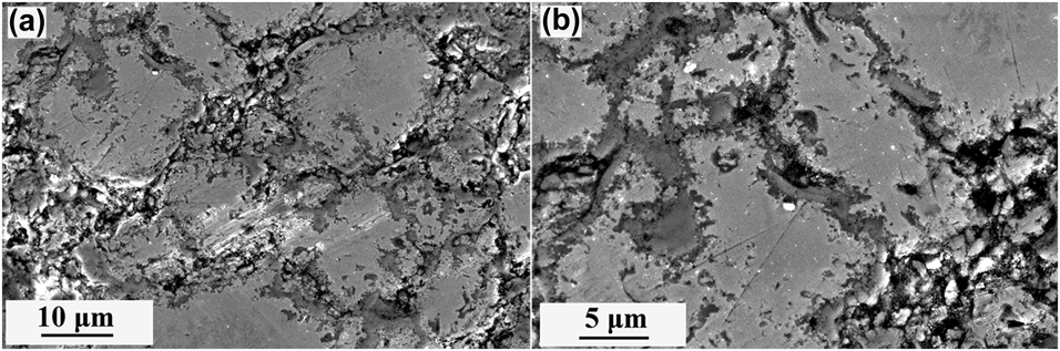
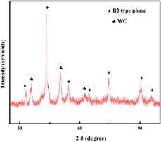
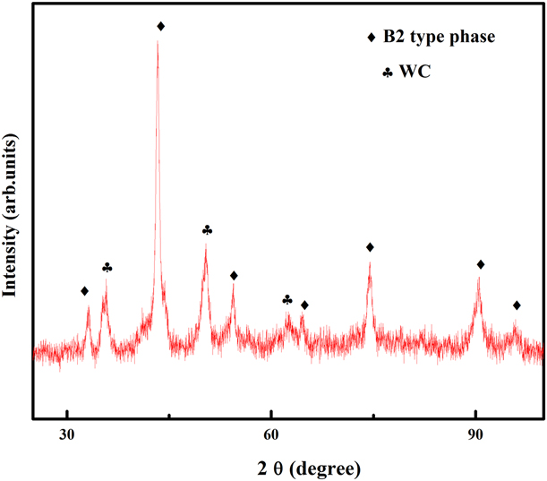
The secondary electron imaging of the microwave-sintered pellet is given in Fig. 8 (Supplementary material) at different magnifications. Two types of the particle could be visualized from electron micrographs. It has been found that particles of sizes in the range of 7–12 μm are formed after microwave sintering. Some amount of porosity could also be observed from the SEM micrographs. Therefore, from this study, it could be concluded that density and porosity are the major concern in the microwave-sintered samples. High density with better homogeneity can be achieved through powder metallurgy route if pressure is applied during the sintering likewise SPS and hot pressing. The XRD pattern of microwave-sintered AlCoCrFeNiTi HEA compacted at 1000 °C (1273 K) is illustrated in Fig. 9. It has been found that after microwave sintering, the BCC phase transforms into the B2 (a = 2.87 ± 0.03 Å) type phase co-existing with the hexagonal phase. The increase in diffraction intensity results from the coarsening of the crystal and release of the internal stresses.
Secondary electron (SE) imaging of the microwave-sintered pellet of AlCoCrFeNiTi HEA at 1000 °C at two different magnifications (a&b).

XRD patterns of microwave-sintered AlCoCrFeNiTi alloy compacted at 1000 °C (1273 K). The evolution of B2-type phase along with formation of WC phase can be discerned.
Discussion
Solid solution formation criteria in AlCoCrFeNiTi HEA
HEAs are getting more attention over more than a decade due to their simple solid solution phase forming ability. An important question related to the design of these multi-principal alloys is not clearly understood. It has been reported that these alloys form simple solid solution phases due to high mixing entropy (ΔS mix). This high mixing entropy lowers the Gibbs free energy (ΔG mix), which promotes the different elements to be randomly distributed in the crystal lattice. This reduces the tendency of ordering and segregation in the alloy. In addition to that, thermodynamically it has been found that the effect of high mixing entropy (ΔS mix) becomes more prominent as the temperature increases. Zhang et al. [Reference Zhang, Zhou, Lin, Chen and Liaw32] have proposed phase formation criteria in HEAs using parameters, such as enthalpy of mixing (ΔH mix) and delta parameter (δ), to form simple solid solution phase. In the same approach, Yang et al. [Reference Yang and Zhang33] have suggested that solid solutions with simple crystal structure are formed when Ω ≥ 1.1 and δ ≤ 6.6%. The parameters Ω and δ are defined in the following two equations:
 $$\Omega = {{{T_{\rm{m}}}\Delta {S_{{\rm{mix}}}}} \over {\left| {\Delta {H_{{\rm{mix}}}}} \right|}}\quad ,$$
where T m is the melting point of the ith component of the alloy and can be calculated as 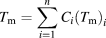 ${T_{\rm{m}}} = \sum\limits_{i = 1}^n {{C_i}{{\left( {{T_{\rm{m}}}} \right)}_i}}$. The enthalpy of mixing (ΔH mix) for multicomponent alloy system can be calculated using the Miedema’s macroscopic model for binary alloys as
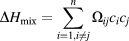 $\Delta {H_{{\rm{mix}}}} = \sum\limits_{i = 1,i \ne j}^n {{\Omega _{ij}}{c_i}{c_j}}$, where
 ${\Omega _{ij}} = 4\Delta H_{{\rm{AB}}}^{{\rm{mix}}}$ is the regular solution interaction parameter between ith and jth elements, c i and c j are the at.% of the ith and jth component, respectively, and
 $\Delta H_{{\rm{AB}}}^{{\rm{mix}}}$ is the enthalpy of mixing of binary equiatomic alloy [Reference Miedema, de Châtel and de Boer34]. Configurational entropy in a multicomponent system is given by the equation
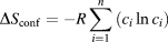 $\Delta {S_{{\rm{conf}}}} = - R\sum\limits_{i = 1}^n {\left( {{c_i}\ln {c_i}} \right)}$, where R represents the universal gas constant and c i the mole fraction of the ith component in the alloy. The effect of the atomic size difference (δ) in the n-element alloy is normally expressed as follows:
 $$\delta = \sqrt {\mathop \sum \limits_{i = 1}^n {c_i}{{\left( {1 - {{{r_i}} \over {\bar{r}}}} \right)}^2}} \quad ,$$
where c i is the concentration of ith component and  $\bar{r}$ is the average value of atomic radii.
The calculated results of the all these parameters are reported in Table II. The Ω of this alloy system is ∼1.19, which is in good agreement to form solid solution phase (Ω > 1). Along with Ω factor, delta factor (δ) is also in range for the AlCoCrFeNiTi HEA to form solid solution phase. These two factors imply that simple solid solution phase can be formed theoretically and getting validation from the experimental results.
Calculated thermodynamic parameters for AlCoCrFeNiTi HEA.

The MA behavior of AlCoCrFeNiTi HEA
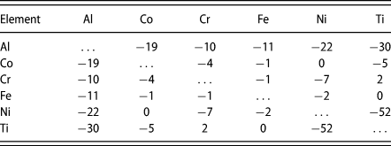
Alloying behavior and the phase evolutions in the AlCoCrFeNiTi HEA were monitored at a specific time schedule shown in Fig. 1. It has been found that Ti, Co, and Al are the elements, which go to the lattices of the solid solution first compared to other constituent alloying elements. It could be concluded that apart from the melting point, the structure of the constituent elements is also an important factor. Throughout the milling process, peak intensity decreases with an increase in broadening, mainly due to refined crystal sizes, the evolution of high lattice strain, and a decrease in crystallinity (Table I). A single-phase solid solution of BCC phase was observed after 25 h of milling, and milling was extended up to 40 h of milling to understand if there any further changes in phases occur or not. It has been found that there are some extra peaks appeared as the milling proceeds. Careful investigation suggests that these peaks are corresponding to the WC. It helps in understanding that milling powder has been contaminated. Noticeably, it is happening after a definite time period. The formation of carbide is due to the wet atmosphere of milling using toluene for mechano-chemical reaction. Here, toluene reacted with the WC vials and balls, and led to the formation of the carbide phase. From this study, it can be concluded that high-energy ball milling in a wet atmosphere after a certain period of time started contaminating the powder. However, it may be emphasized that the formation of WC does not change the composition of HEAs formed as the content of all these six elements does not get affected. The presence of these phases has been ascertained through TEM. The mixing enthalpies of the binary atomic pairs in Table III of AlCoCrFeNiTi HEA show that this alloy possess limited solid solubility under an equilibrium condition. Here, the solid solubility extension is mainly governed by high mixing entropy (in HEAs) and nonequilibrium state of the MA process. This high mixing entropy of alloy lowers the tendency to form the intermetallic ordered phases and supports to form stable solid solution phases. It can be concluded that the formation of simple solid solution phases after MA is dependent on composition and processing routes.
The chemical mixing enthalpy [ΔH mix (kJ/mol)] of atomic pairs of AlCoCrFeNiTi HEA calculated by the Miedema’s approach.

Thermal stability of the phases evolved in AlCoCrFeNiTi HEA
From the differential scanning calorimetry thermogram, it has been found that there were two separate heat events evolved when the powder is heated up to 1200 °C (1473 K). First heat event is exothermic in nature at around 360 °C (633 K) and related to the release of the internal stresses induced during the mechanical working of the alloy. Second heat event is endothermic in nature at around 650 °C (923 K), which might be the indication of the phase transformation. To ascertain these two events, in situ high-temperature XRD was done at the selective temperatures. It is observed that this alloy is stable up to 600 °C (873 K), because there was no phase change found in the XRD plot. Similar kinds of phases observed were present in the 40 h milled powder alloy. Microwave-sintered AlCoCrFeNiTi HEA at 1000 °C (1273 K) showed that the BCC phase transformed to ordered B2 (a = 2.87 ± 0.03 Å) type phase. It has been reported in the literature that the formation of intermetallics is somehow empirically related to the heat of mixing [Reference Basu, Murty and Ranganathan35]. For current alloy, the heat of mixing is higher for the Ni–Ti and Al–Ni among all the other alloying elements. Therefore, the formation of intermetallic phases in the AlCoCrFeNiTi HEA can be justified through their heat of mixing.
Conclusions
The following conclusions can be drawn from the present study.
(1) The equiatomic composition of AlCoCrFeNiTi (hexanary) HEA leads to the formation of a single-phase BCC structure with minor WC phase after 40 h of MA. The evolution of WC phase is due to the contamination of the milling media and process controlling agent.
(2) Phase stability of the AlCoCrFeNiTi has been confirmed through differential scanning calorimetry and in situ high-temperature XRD and found that this alloy is stable up to ∼600 °C (873 K).
(3) The semi-empirical thermodynamic analysis reveals that the ratio of TΔS/ΔH (Ω) is ∼1.19, which is well within the range of the solid solution phase formation criterion of HEA.
(4) Consolidated and sintered pellets of AlCoCrFeNiTi HEA show the evolution of a B2-type phase along with minor WC phase arising from the milling media.
Experimental details
Elemental powders of Al, Co, Cr, Fe, Ni, and Ti with a purity of ≥99 wt% and a particle size of ∼45 μm (∼325 mesh) were used as the starting material. The equiatomic composition of powder was milled in a high-energy planetary ball mill (Retsch PM 400, Retsch GmbH, Haan, Germany) using WC vials and balls (diameter: 10 mm). The powder was milled in the wet atmosphere using toluene and with a rotation speed of vials at 200 rpm. Ball-to-powder weight ratio (BPR) of 10:1 was maintained during milling experiment. The powder was extracted at a regular interval of 5 h to investigate the phase evolution through XRD. Milling machine was stopped for half an hour (at every one-hour interval time) to avoid any phase change due to overheating of the powder. Structural and microstructural characterizations were carried out by XRD [Rigaku Mini flex-600 (40 kV–15 mA); Rigaku, Tokyo, Japan] and high-temperature XRD by Rigaku SmartLab (45 kV–200 mA) with Cu Kα radiation (Kα = 0.154 nm), SEM (FEI Quanta 200F; Thermo-Fisher - FEI, Eindhoven, Netherlands) operated at 20 kV, and TEM (TECNAI G2T20; Thermo-Fisher - FEI, Eindhoven, Netherlands) operated at 200 kV). Thermal analysis of 40 h milled powder was performed by using NETZSCH DSC 404 F3 Pegasus apparatus (NETZSCH GmbH, Selb, Germany) in a nitrogen atmosphere with constant heating rates of 20, 40, and 50 K/min. Subsequently, 40 h milled powder was consolidated in the form of green pellets of 10 mm diameter. The normal load of 637 MPa was applied to form these pellets in the 30-ton capacity hydraulic press. These green pellets were then sintered in the microwave heating furnace (with a frequency of 2.45 GHz and power of 3 kW) at a temperature of 1000 °C (1273 K) for 1 h and allowed to furnace cooling.
Supplementary material
To view supplementary material for this article, please visit https://doi.org/10.1557/jmr.2019.5.