Introduction
The endoscopic management of hypopharyngeal diverticula is well established. Division of the cricopharyngeal bar using an endoscopic stapling device has largely replaced the use of laser and diathermy as described in the classical Dohlman's procedure.Reference Martin-Hirsch and Newbegin1
Complications are rare but include bleeding, perforation and mediastinitis.2
Here, we describe a technique for accurately applying the endoscopic stapling device so as to increase the number of successful procedures and reduce complications.
When performing an endoscopic diverticulotomy, it is necessary to ensure that the distal tip of the stapling device reaches the floor of the pouch, so as to completely divide the cricopharyngeal bar. Although it is possible to check the position of the stapling device with a Hopkins rod, the cricopharyngeal bar may still move before the staples are deployed. This can result in incomplete division of the cricopharyngeal bar, necessitating a second application of the stapling device.
As well as the obvious financial implications of using more than one set of staples, there is evidence to suggest that this may increase the risk of bleeding and perforation.Reference Roth, Sigston and Vallance3
Failure to engage the stapling device over a small cricopharyngeal bar has also been cited as a reason why endoscopic diverticulotomy is not suitable for small pouches.Reference Jaramillo, McLay and McAteer4 The procedure described below directly addresses these issues.
Method
General anaesthesia is induced and an endotracheal tube sited. The patient is positioned supine with a pillow half under the shoulders, half under the neck, allowing for head and neck extension.
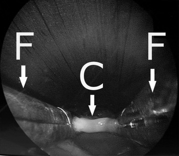
A double-lipped Weerda endoscopic stapling diverticuloscope (Karl Storz, Tuttlingen, Germany) is inserted in the standard fashion, with appropriate dental protection. The primary surgeon uses two oesophageal forceps to grasp the cricopharyngeal bar as shown in Figure 1.
Endoscopic view of the cricopharyngeal bar (C) held with two oesophageal forceps (F).

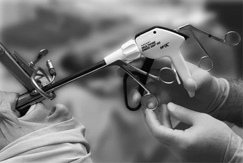
The assistant then takes hold of the right oesophageal forceps. Next, the primary surgeon inserts the endoscopic stapling device between the two oesophageal forceps as shown in Figure 2. At this point, the assistant can check the position of the instruments by introducing a 0° Hopkins rod into the diverticuloscope.
Intra-operative photograph showing relative positions of the oesophageal forceps and Endo GIA 30 stapler (Covidien, Mansfield, MA, USA) within the Weerda pharyngoscope.

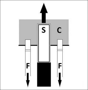
The stapling device is advanced over the cricopharyngeal bar while the bar itself is pulled into the stapler by the oesophageal forceps (Figure 3). The stapling device is then fired and the result is checked with the Hopkins rod.
Diagram showing how the endoscopic stapler (S) is advanced over the cricopharyngeal bar (C) while counter-traction is provided by the forceps (F).

Discussion
This technique enables the surgeon to accurately place the stapling device over the cricopharyngeal bar. This increased accuracy reduces the need to apply a second set of staples. This has the benefit of reducing potential complications, as well as keeping the costs of the procedure to a minimum.
The use of a Hopkins rod further improves the safety of this procedure; the surgeon is able to confirm there is no perforation, as well as to exclude a carcinoma of the pharyngeal pouch. Furthermore, by using the method described above the surgeon is able to accurately engage the stapling device over a small cricopharyngeal bar, which previously would have been regarded as too small for stapling.