According to the American Diabetes Association, 8% of the general population and 25% of people over the age of 65 are afflicted with diabetes in the USA (Ref. 1). One of the most outward and debilitating complications of diabetes is the development of chronic, nonhealing foot ulcerations, which occur in 15% of diabetics. In the worst cases, lower-limb amputation is required; indeed, diabetic foot ulceration leads to 82 000 amputations per year and is the leading cause of nontraumatic (disease-associated) amputation in the USA (Refs 1, 2). The national economic burden of diabetic foot ulceration and amputation is correspondingly staggering: it was estimated at near US$11 billion in the year 2001 (Ref. Reference Boulton3).
The pathophysiological trail from hyperglycaemia to foot ulceration traverses a complex interplay between multiple dysfunctioning systems. Traditionally, ischaemia, neuropathy, trauma and infection were considered the culprits of the recurring chronic wound, and treatment revolved largely around wound management and revascularisation (Refs Reference Edmonds4, Reference Thomson5). More recently, impairment of the cutaneous wound-healing process itself has been recognised as a major contributor to the failure to heal, and wound healing has been appreciated as yet another biological system hindered by the metabolic, vascular, neurological and inflammatory alterations present in diabetes (Ref. Reference Falanga6). In light of this recent understanding, numerous investigations are pursuing therapies for diabetic foot ulceration that act to restore the molecular and cellular processes required for successful wound healing.
Wound healing requires the well-orchestrated integration of the complex biological and molecular events of cell migration, cell proliferation, and extracellular matrix (ECM) deposition (Ref. Reference Falanga6). Normal wound healing can be divided into four overlapping phases: haemostasis, inflammation, proliferation and remodelling. The cell types involved in each phase are responsible for mediating specific events that lead to wound closure. Haemostasis lasts for 2–3 h, during which a fibrin plug is formed and aggregated platelets release pro-inflammatory mediators such as cytokines and growth factors. Cytokines recruit neutrophils and monocytes to the wound area triggering the inflammatory phase of wound healing, which lasts from hours to days. The proliferation phase involves several different cell types such as epithelial cells, endothelial cells and fibroblasts. Migration of these cells to the wound site is again the result of cytokine stimulation. ECM deposition coupled with angiogenesis and re-epithelialisation leads to wound contraction and closure. Remodelling, the last step in wound healing, can last several weeks, and is marked by ECM remodelling and formation of scar tissue. Whereas acute wounds progress linearly through the phases of wound healing, chronic nonhealing wounds become stalled in different phases at the same time and do not follow an orderly and reliable progression of healing (Ref. Reference Falanga6).
The first observations regarding the impact of diabetes on wound healing focused on an impaired leukocyte function related to hyperglycaemia (Refs Reference Bagdade, Root and Bulger7, Reference Nolan, Beaty and Bagdade8). However, additional factors common to other chronic wounds, such as decubitus ulcers and venous ulcers, were later found to participate in impaired diabetic wound healing (Ref. Reference Falanga9). These abnormalities included the development of pericapillary fibrin cuffs (Ref. Reference Claudy10), impaired expression of ECM (Ref. Reference Loots11), aberrant cellular infiltration (Ref. Reference Rosner12), insufficient macrophage activation (Ref. Reference Moore, Ruge and Harding13), impaired re-epithelialisation and impaired angiogenesis (Refs Reference Herrick14, Reference Duraisamy15).
Traditionally, research in the field of diabetic wound healing focused on the role of growth factors and their therapeutic potential. However, to date only one growth factor – platelet-derived growth factor (PDGF) – has been approved by the US Food and Drug Administration for treating diabetic wounds. Mediators of inflammation participate in all four phases of wound healing and represent an alternative therapeutic option for promoting proper healing. Numerous investigations have focused on the systemic implications of inflammation in diabetes yet few have examined the local role of inflammation in the wound microenvironment. The mechanism by which local inflammatory pathways are affected in diabetes is poorly understood, but an emerging hypothesis suggests the involvement of the neuroendocrine system in the inflammatory impairment present in diabetic wounds (Ref. Reference Elenkov16). Recently, the role of nerves and their interaction with immune and cutaneous cells in wound healing has become evident, and neuropathy, an early and ubiquitous complication of diabetes, has been shown to impair wound healing by influencing inflammatory pathways. This review summarises the role of neuroinflammatory mediators – principally neuropeptides and cytokines – in diabetes and wound healing.
Inflammation and wound healing
Tissue injury triggers an acute inflammatory response, beginning with the arrival of neutrophils, monocytes/macrophages and mast cells to the site of injury (Refs Reference Engelhardt17, Reference Hebda, Collins and Tharp18, Reference Trautmann19). These cells produce inflammatory cytokines and growth factors that co-ordinate wound repair (Refs Reference Riches and Clark20, Reference Sunderkotter21). Cytokines are a heterogeneous group of pleiotropic signalling proteins and glycoproteins that can generally be classified as pro-inflammatory or anti-inflammatory and allow an organism to respond rapidly to an immune challenge (Ref. Reference Guest22). Proper wound healing requires a sequential, self-limited, cytokine–immune-cell interaction in order to achieve an adequate immune response necessary for bacterial clearance, organised tissue breakdown, and subsequent regeneration. A study of adult human wound healing showed that the cytokines interleukin (IL)-8, CXCL1 (growth-related oncogene α; GRO-α), CCL2 (macrophage chemoattractant protein 1; MCP-1) CXCL10 [interferon (IFN)-γ-inducible protein 10; IP-10] and CXCL9 (mitogen-inducible gene; MIG) are sequentially and differentially expressed during phase-specific infiltration of leukocyte subsets (Ref. Reference Engelhardt17). Tumour necrosis factor (TNF)-α and IL-1β are early regulators of the immune response and both induce the release of secondary cytokines, such as IL-6 and IL-8 (Ref. Reference Hildebrand, Pape and Krettek23). Released cytokines, predominantly produced by monocytes and macrophages, activate and selectively guide various leukocyte subsets to specific microanatomical sites of skin wounds during the phases of tissue repair. Keratinocytes and dermal endothelial cells are additional cell types that respond to and release molecular mediators that orchestrate wound healing. These cytokines mediate a variety of overlapping effects, and their actions can be additive. More recently, inflammatory mediators have been found to regulate angiogenesis, another critical process for wound repair (Ref. Reference Belperio24).
Inflammation and diabetes
Both type 1 and type 2 diabetes are characterised by the chronic upregulation of pro-inflammatory cytokines. In type 2 diabetes, several mediators, such as the pro-inflammatory cytokines IL-1, TNF-α, IL-6 and CCL5 (RANTES), released from various cells, including immune cells and adipocytes, have been implicated in the development of insulin resistance (Refs Reference Tilg and Moschen25, Reference Bogdanski26). In addition, inflammatory markers correlate significantly with insulin resistance indices, such as haemoglobin A1c, lipid profile, hypertension, positive family history of type 2 diabetes, low physical fitness, and mixed high-fat and high-carbohydrate diet (Ref. Reference Syrenicz27). In children with type 1 diabetes, cytokines such as IL-1α, IL-4 and IL-6 are acutely elevated during hyperglycaemia, and these elevations persist for hours after hyperglycaemia has been corrected (Ref. Reference Rosa28). In a similar study of monocyte function in type 1 diabetes, increased levels of IL-6, IL-α and superoxide anion were identified (Ref. Reference Devaraj29).
Furthermore, a study of lean young individuals found that even high-normal physiological increases in blood glucose after meals can aggravate inflammatory processes by activation of the pro-inflammatory transcription factor nuclear factor κB (NF-κB) (Ref. Reference Dickinson30). This study reveals how inflammatory pathology is not simply a consequence of long-standing diabetes but can be triggered acutely by the hyperglycaemic environment.
Inflammation and diabetic wound healing
Altered immune-cell function
It has been questioned whether chronic systemic inflammation present in diabetes translates into chronic peripheral inflammation. In patients with metabolic syndrome, oral glucose tolerance testing was associated with increased levels of intercellular adhesion molecule 1 (ICAM-1), TNF-α and IL-6 in peripheral blood leukocytes, compared with patients without metabolic syndrome, 2 h after the test was performed. Similar results were obtained in vitro after 72 h of cultivation in a high-glucose medium. Hence, it is thought that metabolic syndrome may support peripheral inflammation by causing leukocytes to release pro-inflammatory mediators in response to glucose (Ref. Reference Kempf31). In diabetes there is a pronounced imbalance of pro- and anti-inflammatory cytokines, which are released in a nonsequential manner leading to impaired tissue repair and weakened cellular and humoral immune defence mechanisms (Refs Reference Fisman, Adler and Tenenbaum32, Reference Hatanaka33).
These immune cells show abnormal activity and cytokine-release profiles (Ref. Reference Hand, Hand and Vasquez34). Neutrophils, which act as first-line-of-defence cells, show reductions in functional activity, contributing to the high susceptibility and severity of infections in diabetes (Ref. Reference Stegenga35). Clinical investigations in diabetic patients and experimental studies in diabetic rats and mice consistently demonstrate defects of neutrophil chemotactic, phagocytic and microbicidal activities (Refs Reference Alba-Loureiro36, Reference Mastej and Adamiec37, Reference Marhoffer38). Lowering of blood glucose levels by insulin in diabetic patients or experimental animals correlated significantly with improvement of neutrophil functional activity (Ref. Reference Alba-Loureiro39). A prospective clinical study that examined the phagocytic activity index and intracellular killing activity of neutrophils as well as IL-1β levels in a group of patients with healing and nonhealing diabetic foot infections found a dysfunction of neutrophils and IL-1β regulation in patients with diabetic foot infections, possibly indicating ineffective immunological responses in diabetic patients (Ref. Reference Oncul40). It has been shown in diabetes that immune cell infiltration is decreased in early-stage wounds, followed by a persistence of neutrophils and macrophages in chronic, nonhealing wounds. These changes in inflammatory cell recruitment are consistent with alterations in growth factor expression (Ref. Reference Ochoa, Torres and Shireman41).
Advanced glycation, glycosylation and cytokine production
Metabolic routes by which hyperglycaemia is linked to neutrophil dysfunction include the advanced protein glycosylation reaction, the polyol pathway, oxygen-free-radical formation, the nitric-oxide–cGMP pathway, and the glycolytic and glutaminolytic pathways. Another consequence of diabetes is the formation of advanced glycation end (AGE) products, which act via the receptor RAGE (AGER) and are implicated in various diabetic complications including inflammation. S100b, an inflammatory protein as well as a specific RAGE ligand, significantly increased CXCL10 mRNA and protein levels in THP-1 monocytes as well as peripheral blood monocytes (Ref. Reference Shanmugam, Ransohoff and Natarajan42). Similar to AGE products but less characterised are advanced lipoxidation end (ALE) products, which are also formed as a consequence of diabetes. A synthetic ALE product significantly increased the expression of CXCL10, β1 and β2 integrins, cyclooxygenase 2 (COX2), CCL2, IL-6 and -8, and inducible nitric oxide synthase (NOS), which are all associated with monocyte dysfunction (Ref. Reference Shanmugam43).
Altered cytokine production by nonimmune cells
Cytokine production in diabetes is not limited to inflammatory cells but also comes from skin cells, including keratinocytes and microvascular endothelial cells (Ref. Reference Ochoa, Torres and Shireman41). A study analysing the expression of various chemotactic and growth factors in the margin of diabetic foot ulcers found elevations of TGF-β1 and its receptor TGFβR1 in keratinocytes and significantly increased expression of CCL2, CXCR1 and TGFβR1 in ulcer dermal endothelial cells. There was decreased expression of IL-10, IL-15 and TGF-β1 in the ulcer dermal endothelial cells and blunted upregulation of IL-8, CCR2A and IL-10 receptor in both keratinocytes and endothelial cells. Finally, there was a lack of upregulation of IL-10 and IL-15 in keratinocytes in the ulcer margins. The blunting of certain leukocyte chemotactic factors along with the reduced influx of immune cells led to poor formation of granulation tissue and chronicity of ulcer epithelialisation (Ref. Reference Galkowska, Wojewodzka and Olszewski44). Similarly, it is now recognised that the crosstalk between adipocytes and adipose tissue stromal cells such as macrophages contributes to local and systemic inflammation. This interaction is mediated by adipocyte-derived free fatty acids that enhance macrophage-induced inflammation via CXCL10, IL-8, CCL2, COX2 and CXCL9, and active NF-κB (Ref. Reference Laine45).
Oxidative stress
In addition to cytokines, nitrosative and oxidative stress contributed by monocytes and leukocytes also significantly add to the wound-healing problem. Nitric oxide, a known scavenger of superoxide (O2−), is the main component of oxidative stress and also enhances angiogenesis and proliferation (Ref. Reference Maulik and Das46). However, under conditions of excessive and prolonged production of O2− in the wounds, nitric oxide in fact leads to a significant increase in the production of peroxynitrite (ONOO−) and peroxynitrous acid (ONOOH), both of which are highly reactive and damaging radicals, significantly impairing wound healing (Ref. Reference Soneja, Drews and Malinski47). In a diabetic rat model of wound healing, lipoic acid, a known scavenger of several free oxygen radicals, accelerated wound healing and this effect of lipoic acid was attributed to its protection of endothelial cells from oxidant damage (Ref. Reference Lateef48).
Summary
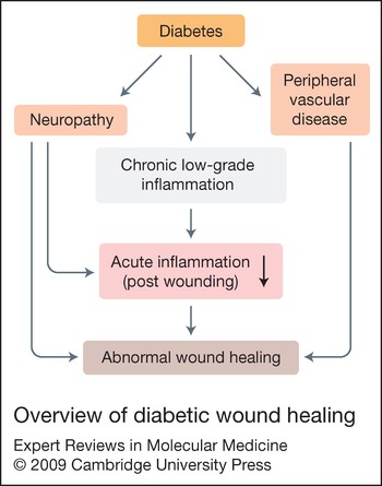
The aforementioned reports demonstrate how the chronic inflammatory environment of diabetes disturbs the interaction of pro- and anti-inflammatory cells and mediators at several levels to produce a persistent increase in pro-inflammatory cytokines by various immune and nonimmune cells. It can therefore be hypothesised that the chronic inflammatory state of diabetes blunts the focused acute immune/cytokine response needed for the second phase of wound healing (Refs Reference Straino49, Reference Corrales50, Reference Maruyama51) (Fig. 1). Rapid and effective leukocyte migration is pivotal during this early phase of wound healing. However, later observations show that leukocytes persist locally in poorly healing wounds of diabetic patients. These partly dysfunctional leukocytes (mainly neutrophils and macrophages) are believed to additionally promote the chronic inflammatory state. Further understanding of how to augment chronic low-grade inflammation, the lack of acute focused immune response, and the behaviour of certain subpopulations of dysfunctional immune cells will aid the development of new approaches for treating nonhealing wounds in diabetic patients.
Overview of diabetic wound healing. Diabetes leads to neuropathy, peripheral vascular disease and chronic low-grade inflammation, both systemically and peripherally. Neuropathy and chronic low-grade inflammation together blunt the acute focused inflammatory response to injury and together with peripheral vascular disease lead to impaired wound healing.

Neuropathy and diabetic wound healing
Diabetic neuropathy is an insidious, oftentimes unnoticed, long-term complication of diabetes characterised by the progressive loss of somatic and autonomic nerve fibres (Ref. Reference Urbancic-Rovan52). Autonomic neuropathy results in anhidrosis, leading to altered neurogenic regulation of cutaneous blood flow and rendering the skin dry and vulnerable to cracks and fissures (Ref. Reference Urbancic-Rovan52). Somatic diabetic neuropathy, reported in about 30–50% of diabetic patients, is the most common and sensitive predictor of foot ulceration (Refs Reference Sheehan53, Reference Young, Breddy, Veves and Boulton54, Reference Adler55). The consequent vulnerability to physical and thermal trauma increases the risk of foot ulceration by sevenfold; however, sensory loss must be profound before resulting in the loss of protective sensation (Refs Reference Reiber56, Reference Yao57). Peripheral sensory neuropathy is considered to be a major contributor to foot amputations and increases risk of foot amputations by 1.7-fold alone and by 36-fold if there is a prior history of ulceration (Ref. Reference Lindberger58). It has been shown that loss of peripheral sensory and autonomic nerves along with diminished neuropeptide production precedes clinical symptoms of neuropathy (Ref. Reference Vinik59). Skin biopsies from type 2 diabetic patients with and without peripheral sensory neuropathy were found to have severe denervation of the skin with active foot ulcers, irrespective of clinically identifiable sensory neuropathy (Ref. Reference Galkowska60).
Cutaneous neuroimmunology
The contribution of peripheral nerves and cutaneous neuroimmunology to normal wound healing is of emerging importance. A bidirectional connection between the nervous and immune systems, together with signalling between the central nervous system (CNS) and peripheral nervous system (PNS) with endocrine feedback, coalesce to produce complex immunomodulation (Ref. Reference Quattrini, Jeziorska and Malik61). Both cutaneous nerve fibres and inflammatory cells are able to release neuromediators activating specific receptors on target cells in the skin or transient immunocompetent cells (Ref. Reference Luger62). Mediators of the neuroimmune axis include neuropeptides, neurotransmitters and cytokines. Neuropeptides commonly involved in immune regulation include substance P [SP; also known as tachykinin 1 (TAC1)], calcitonin-gene-related peptide (CGRP), neuropeptide Y (NPY), vasointestinal peptide (VIP) and pro-opiomelanocortin (POMC)-derived peptides such as melanocyte-stimulating hormone (MSH); these may be released from sensory or autonomic nerve fibres and several epidermal as well as dermal cells. Neurotransmitters include catecholamines and acetylcholine. Cytokines that are commonly activated by these neurohormones include IL-1, IL-6, IL-8 and TNF-α – the same cytokines dysregulated in diabetes.
Sensory C nerve fibres are capable of inducing inflammation in response to a variety of irritants. This is achieved by the release of SP, CGRP and other mediators from nerve terminals, which in turn induce mast cell discharge and increase the permeability of blood vessels (Ref. Reference Berczi63). It is important to discuss the findings of the clinical study by Krishnan et al. showing that despite a significant reduction in the neurogenic flare response and dermal nerve fibre density, the rate of wound closure was identical between control subjects and diabetic patients (Ref. Reference Krishnan64). In this study, the wounds were acute and surgically induced on the dorsum surface in 12 well-screened diabetic and 12 healthy subjects who were not at risk of developing wound failure. The skin is different at the dorsum and plantar surface of the foot. In chronic diabetic ulcers, the wounds penetrate the underlying muscle and bone. Surgical wounds are known to heal even in patients with peripheral arterial disease following revascularisation procedures (Ref. Reference Pomposelli65). This further emphasises the differences between acute surgical wounds and diabetic foot ulcers; the latter are chronic wounds accompanied by chronic inflammation, subclinical infection and considerable tissue damage during the period the ulcer develops.
Not only do neuropeptides modulate the cytokine response, but, conversely, inflammation in the form of nitrosative and oxidative stress may affect nerve function. A recent study showed that peroxynitrite injures peripheral nerve and dorsal root ganglia, leading to development of peripheral diabetic neuropathy (Ref. Reference Vareniuk, Pavlov and Obrosova66). Interestingly, poly(ADP-ribose) polymerase (PARP), a downstream effector of free-radical- and oxidant-induced single-strand DNA breakage, was previously implicated in diabetic neuropathy (Ref. Reference Obrosova67). In a mouse study of experimental diabetic peripheral neuropathy, deletion of the COX2 gene, encoding another inflammatory pathway enzyme, protected the mice from functional and biochemical deficits of experimental diabetic peripheral neuropathy and against nerve fibre loss (Ref. Reference Kellogg68). Not just hyperglycaemia but a high-fat diet too has been shown to induce neuropathy in prediabetic and obese mice by increased sorbitol pathway activity, oxidative–nitrosative stress, PARP activation and 12/15-lipoxygenase activation (Ref. Reference Obrosova69).
Neuropeptides and diabetic wound healing
It might be argued that the occurrence of foot ulceration in those with neuropathy does not necessarily indicate a direct or mechanistic link between wound healing and neuropathy. One link that can directly connect neuropathy to wound healing is the neuropeptides. Neuropeptides act by binding to specific receptors found on various cells in the skin, including immune cells, Langerhans cells, endothelial cells, mast cells, fibroblasts and keratinocytes (Ref. Reference Roosterman70). They activate these cutaneous cells through high-affinity neuropeptide receptors, leading to direct activation of intracellular G-protein signalling cascades. The neuropeptides involved in wound healing are SP, NPY, CGRP, corticotropin-releasing factor (CRF), α-MSH and neurotensin (NT) (Refs Reference Nakamura71, Reference Movafagh72, Reference Ekstrand73, Reference Kuo, Abe and Zukowska74, Reference Delgado, McManus and Chambers75, Reference Zukowska, Grant and Lee76). Roles for α-MSH and NT in diabetic wound healing await more thorough investigation.
Substance P
SP is a 10 amino acid peptide widely distributed in the CNS and PNS (Refs Reference Hokfelt77, Reference Hokfelt78). In the PNS, SP is localised in primary sensory neurons and neurons intrinsic to the gastrointestinal, respiratory and genitourinary tracts (Ref. Reference Maggi79). There are three SP receptors – neurokinin (NK)1R, NK2R and NK3R – of which the predominant SP receptor, NK1R, is a G-protein-coupled receptor (Ref. Reference Maggi80). NK1R is present on a variety of cell types including immune cells, endothelial cells, neurons, epithelial cells and glial cells, and couples to Gq/11, Gs and G0 proteins. The interaction of SP with NK1R produces a net rise in intracellular [Ca2+] via phospholipase C, and in cAMP via adenylate cyclase (Refs Reference Khawaja and Rogers81, Reference Harrison and Geppetti82).
SP and diabetes
The first study investigating serum SP levels in type 1 diabetes showed a significant decrease in the diabetic group compared with control patients and even greater reductions in patients with diabetic neuropathy (Ref. Reference Kunt83). Skin biopsies from both type 1 and type 2 diabetics showed reduced density of SP-positive nerve fibres (Ref. Reference Vinik59). SP is metabolised by the enzyme enkephalinase, a zinc metalloprotease also known as neutral endopeptidase (NEP), present in the cell membrane of cells expressing NK1R. NEP is expressed in keratinocytes, dermal appendages, the microvasculature of the skin (Ref. Reference Olerud84), and cultured skin fibroblasts (Ref. Reference Bou-Gharios85). NEP expression and activity is increased in the skin of diabetic patients, leading to SP degradation (Ref. Reference Antezana86). In the mouse diabetic model, use of an NEP inhibitor improved wound healing (Ref. Reference Spenny87).
SP–inflammation interaction and wound healing
SP is released from peripheral neurons upon noxious stimuli such as a burn or pressure. Once released at the site of injury, SP binds to various cell types and initiates a cascade of events ensuring proper wound healing. SP binds to NK1R receptors on endothelial cells and elicits vasodilation via nitric oxide, altering vascular permeability and enhancing the delivery of leukocytes to tissues for the expression of the local immune response (Ref. Reference Pernow88). SP is not only produced by immune cells such as T cells, macrophages, dendritic cells and eosinophils but is also a strong chemoattractant for lymphocytes, monocytes, neutrophils and fibroblasts (Refs Reference Ho89, Reference Lai, Douglas and Ho90, Reference Lai91, Reference Lambrecht92, Reference Lambrecht93, Reference Weinstock94, Reference O'Connor95, Reference Schratzberger96). SP can stimulate the production of TNF-α, IL-1β, IL-2, IL-8 and IL-6 from T cells, macrophages and neutrophils (Ref. Reference Delgado, McManus and Chambers97). SP increases the expression of endothelial leukocyte adhesion molecule 1 on human microvascular endothelium and leukocyte-function-associated antigen 1 (LFA-1; also known as αLβ2 integrin) on murine endothelial cells and lymphocytes (Refs Reference Matis, Lavker and Murphy98, Reference Vishwanath and Mukherjee99). SP has been shown to activate coincident NF-AT- and NF-κB pathways to induce ICAM and vascular cell adhesion molecule (VCAM) gene expression in microvascular endothelial cells through intracellular [Ca2+] mobilisation (Ref. Reference Quinlan100).
By promoting vasodilatation, leukocyte chemotaxis and leukocyte–endothelial-cell adhesion, SP ensures the extravasation, migration and subsequent accumulation of leukocytes at sites of injury. Induction of cell adhesion molecules on endothelial cells is important for the recruitment and transmigration of immune cells through the endothelial monolayer. The accumulated leukocytes release cytokines, creating a pro-inflammatory microenvironment that leads to proliferation of endothelial cells and angiogenesis. In intestinal wound healing, SP modulates epithelial cell restitution via the release of TGF-β from fibroblasts (Refs Reference Bulut101, Reference Felderbauer102). Endogenous SP release in rat knee synovium, a model of neurogenic inflammation, showed enhanced angiogenesis (Ref. Reference Seegers103). In another study, exogenously administered SP stimulated fibroblast proliferation, angiogenesis and collagen organisation during Achilles tendon healing in rats (Ref. Reference Burssens104). Similar results were found in the diabetic mouse model (db/db) of wound healing, where fewer SP-positive nerves were found compared with control mice, and exogenous treatment of wounds with SP shortened wound closure time (Ref. Reference Gibran105). Although corneal wound healing cannot be equated with skin wound healing because of the avascular nature of the cornea, corneal epithelial wound healing in a study of diabetic rats was accelerated by the combination of an SP-derived peptide (FGLM-NH2) and insulin-like growth factor 1. This study suggests that these peptides may have an effect on epithelial cells in addition to the vascular cell types (Ref. Reference Nakamura71). It is evident SP plays a major role in the inflammatory and angiogenesis phases of wound healing, and dysregulation in the SP pathway in diabetes can significantly hinder wound repair.
Neuropeptide Y
NPY is a highly conserved 36 amino acid polypeptide, and is one of the most abundant neurotransmitters in the mammalian CNS and PNS. NPY elicits diverse biological functions, including hypothalamic control of food intake, anxiolysis and sedation (Ref. Reference Blomqvist and Herzog106). Non-neuronal cells also express NPY, including megakaryocytes, liver, heart, spleen and endothelial cells (Refs Reference Ericsson107, Reference Strand108). NPY activates members of the NPY receptor family of heptahelical G-protein-coupled receptors. Four NPY Y receptor cDNAs have been cloned: Y1, Y2, Y4 and Y5. All these receptors mediate their responses through pertussis-toxin-sensitive G proteins, resulting in inhibition of adenylate cyclase activity and increase in intracellular [Ca2+] (Ref. Reference Larhammar109). Although NPY was isolated and characterised 20 years ago, little is known about the molecular mechanisms that regulate NPY receptor activity and the biological significance of NPY in the periphery. NPY is also costored and coreleased with adrenaline and ATP in the sympathetic nerve terminal and in the adrenal medulla with adenaline. NPY regulates vascular tone by inducing contraction of blood vessels (Ref. Reference Franco-Cereceda, Lundberg and Dahlof110). It also stimulates growth of vascular smooth muscle cells and hypertrophy of ventricular cardiomyocytes (Refs Reference Erlinge, Brunkwall and Edvinsson111, Reference Zukowska-Grojec112).
NPY and diabetes
Most research related to NPY and diabetes is focused on the central effect of NPY on feeding and energy homeostasis, where NPY is known to induce feeding and conservation of energy (Ref. Reference Frankish113). Both insulin and the antiobesity peptide leptin inhibit NPY expression in the hypothalamus. Although NPY levels in the hypothalamus are increased in type 1 and type 2 diabetic and obese patients, NPY levels in the skin are reduced in patients with both type 1 and type 2 diabetes (Refs Reference Ahlborg and Lundberg114, Reference Wallengren115, Reference Levy116). Recently, NPY expression was also shown to be reduced in streptozotocin (STZ)-induced diabetic rats (Ref. Reference Kuncova117). The vascular smooth muscle contractile response to NPY was significantly reduced in circular segments of isolated vessels in vitro from rabbits with alloxan-induced diabetes mellitus compared with those from control rabbits (Ref. Reference Andersson118).
NPY–inflammation interaction and wound healing
NPY, acting through its Y1 receptor, has pleiotropic effects on both the innate and adaptive arms of the immune system, with effects ranging from the modulation of cell migration to macrophage and T helper (Th) cell cytokine release, antigen presentation, activation of natural killer cells and antibody production (Refs Reference Wheway, Herzog and Mackay119, Reference Groneberg120, Reference Bedoui121). In a study of rabbit ligament healing, NPY induced expression of IL-2 and TNF-α in specimens of injured ligament cultured two weeks after injury (Ref. Reference Salo122). Only recently, NPY has been reported to directly stimulate endothelial cell proliferation and migration through the Y2 and Y5 receptors, leading to angiogenesis (Refs Reference Ghersi123, Reference Marion-Audibert124, Reference Zukowska-Grojec125). Deletion of Y2 in mice resulted in blockage of NPY-induced angiogenesis and delayed wound healing (Ref. Reference Ekstrand73). NPY is also implicated in vascularisation of neural-crest-derived tumours (Ref. Reference Kitlinska126). The enzyme that activates NPY, dipeptidyl peptidase IV (DPP IV), is expressed in endothelial cells. It cleaves the 36 amino acid NPY to NPY3–36, the fragment that is agonistic to Y2 and Y5 angiogenic receptors (Ref. Reference Zukowska127). Endothelial cell Y2 and DPP IV expression was significantly reduced in ageing mice (Ref. Reference Kitlinska128). Although the receptors involved in NPY-mediated angiogenesis are known, the downstream signalling pathways are not yet clear. In conclusion, similar to SP, NPY is also important for both the inflammatory and angiogenic phases of wound healing.
Calcitonin-gene-related peptide
CGRP is a 37 amino acid peptide generated from the alternative splicing of the calcitonin gene, both in the CNS and the PNS (Ref. Reference van Rossum, Hanisch and Quirion129). In the PNS, CGRP is costored and coreleased with SP from capsaicin-sensitive peripheral afferent neurons (Refs Reference Holzer130, Reference Maggi131). It is also produced in non-neuronal tissues such as liver, lungs, kidneys, prostate and testis (Ref. Reference Russwurm132). CGRP receptors are found in the brain, including the cerebellum, as well as in various peripheral organs such as heart, vasculature, liver, spleen, skeletal muscle and lung, and in lymphocytes (Ref. Reference van Rossum, Hanisch and Quirion129). The data regarding CGRP receptors and their structure are not very clear. It has been proposed that there are two CGRP receptors: CGRP R1 and CGRP R2 (Ref. Reference Hay, Poyner and Quirion133). CGRP activates adenylate cyclase and/or increases intracellular cAMP, as seen in different tissues including liver, heart, skeletal muscle and astrocytes (Ref. Reference van Rossum, Hanisch and Quirion129). It is one of the most potent vasodilators.
CGRP and diabetes
Diabetes reduces CGRP in the hearts of mice and in the dorsal root ganglion and dura mater of rats (Refs Reference Song134, Reference Dux135, Reference Adeghate136). In diabetic rats, CGRP-mediated vasodilation was significantly reduced (Refs Reference Oltman137, Reference Oltman138, Reference Sheykhzade139). In a diabetic cardiomyopathy model, both CGRP and CGRP receptor expression were shown to be reduced (Refs Reference Chottova Dvorakova140, Reference Yorek141). It is clear that diabetes decreases the expression, release and action of CGRP.
CGRP–inflammation interaction and wound healing
CGRP not only induces inflammatory pathways, but inflammation too can induce CGRP release (Refs Reference Ambalavanar142, Reference Ambalavanar143). Similar to SP, CGRP is known to induce neurogenic inflammation via release of histamine from mast cells, leading to recruitment of neutrophils to the site of injury (Ref. Reference Hu, Xiang and Hu144). CGRP is also produced by macrophage-activated adipocytes and monocytes (Ref. Reference Linscheid145), and can act in an autocrine fashion, as seen in human adipocytes (Ref. Reference Linscheid146). CGRP increases the release of IL-1α and IL-8 from keratinocytes (Ref. Reference Dallos147), and is chemotactic towards T cells, modulates lymphocyte proliferation and inhibit IL-2 production (Refs Reference Wang148, Reference Foster149). In the airway epithelium, CGRP increases IL-1, IL-8 and ICAM-1 expression, leading to the accumulation of neutrophils (Ref. Reference Zhang150). In humans, CGRP induces IL-8 but not CCL2 or CCL5 in corneal epithelial cells (Ref. Reference Tran151). Both CGRP and SP increase the levels of IL-1β, IL-6 and TNF-α in dental pulp fibroblasts and IL-1β and TNF-α in macrophages (Refs Reference Yamaguchi152, Reference Yaraee153). Although the role of CGRP as a neuroinflammatory mediator is apparent this interaction is not yet directly linked to its effect on wound healing. More direct involvement of CGRP in wound healing is shown through its effect on angiogenesis via enhancement of vascular endothelial growth factor (VEGF) release and the cAMP pathway (Refs Reference Toda154, Reference Haegerstrand155).
Hypothalamic–pituitary–adrenal axis and corticotropin-releasing factor
The hypothalamic–pituitary–adrenal (HPA) axis, comprising the hypothalamus, the pituitary gland and the adrenal glands, controls reactions to stress and regulates digestion, the immune system, mood and emotions, sexuality, and energy storage and expenditure. CRF is a peptide of 41 amino acids, derived from a 191 amino acid chain. CRF, along with the other members of the CRF family – urocortin 1, urocortin 2 and urocortin 3 – is secreted by the paraventricular nucleus (PVN) of the hypothalamus in response to stress. In addition, CRF is synthesised and produced in multiple peripheral tissues and might be involved in many other biological functions, such as energy balance, metabolism, and regulation of the immune response (Refs Reference Richard, Huang and Timofeeva156, Reference Baigent157). CRF binds to and activates two known receptors: CRFR1 and CRFR2 (Ref. Reference Carlin, Vale and Bale158). These receptors belong to the family of G-protein-coupled receptors whose actions are mediated through activation of adenylate cyclase. CRFR1 is the predominant CRF receptor and is expressed in the brain, pituitary, gonads and skin (Ref. Reference Perrin and Vale159). CRF binding to the receptors in the anterior lobe of the pituitary gland causes release of adrenocorticotrophic hormone (ACTH); ACTH then activates the adrenal glands to release glucocorticoids such as cortisol.
HPA axis, CRF and diabetes
Diabetes has been shown to modulate the HPA axis, and type 2 diabetic patients have subclinical hypercortisolism (Refs Reference Chiodini160, Reference Ghizzoni161, Reference Roy162). In a study of diabetic rats, hyperactivation of the HPA axis was associated with an impaired stress response due to decreased pituitary and adrenal sensitivity, and basal hyperactivation of the diabetic HPA axis in the morning was found to be due in part to decreased glucocorticoid negative feedback sensitivity (Refs Reference Chan163, Reference Chan164). The impairment in the HPA axis was corrected by insulin treatment (Ref. Reference Chan165). In an in vitro study, CRF-induced ACTH release from cultured anterior pituitary cells of short-term diabetic rats was increased in part by the cAMP system (Ref. Reference Gohshi240). By contrast, the decrease in the CRF-induced ACTH release from cultured anterior pituitary cells of long-term diabetic rats indicated a change in the properties of the L-type Ca2+ channel coupled with the CRF receptor, or the CRF receptor itself.
Interaction of the HPA axis and CRF with inflammation, and wound healing
HPA-axis dysfunction is generally caused by stress. Given that immune dysfunction is a known manifestation of chronic stress, it is not surprising that alteration in the HPA axis leads to immune dysfunction. Also, cytokines have been shown to lead directly to HPA-axis dysfunction, suggesting a bidirectional communication between inflammatory mediators and the HPA axis (Ref. Reference Turnbull and Rivier166). Glucocorticoids such as cortisol are known to suppress inflammatory pathways and are known immunosupressants (Refs Reference Lozovaya and Miller167, Reference Chrousos168). Glucocorticoids secreted in response to acute, subacute and chronic stress suppress cytotoxic T cell (CD8+ T cell)-mediated immunological responses but enhance the T helper cell (CD4+ T cell)-mediated humoral immune responses (Refs Reference Elenkov169, Reference Elenkov and Chrousos170). Glucocorticoids also affect the balance of the CD4+ T cell subsets of Th1 and Th2 (Ref. Reference Webster, Elenkov and Chrousos171). Th1 cells produce cytokines associated with cell-mediated immune responses against intracellular pathogens and induce organ-specific autoimmune diseases; Th2 cells produce cytokines that are associated with atopic and allergic conditions (Ref. Reference Monney172). Thus, by affecting the Th1–Th2 balance, the hyperactivated HPA axis seen in diabetes can decrease resistance to viral and bacterial infections (Refs Reference Brown and Zwilling173, Reference Sheridan174).
Immune cells including lymphocytes and monocytes express receptors for CRF, ACTH and cortisol (Refs Reference Webster, Elenkov and Chrousos171, Reference Webster175). CRF inhibits IFN-α release from Th1 cells and IFN-γ from monocytes (Refs Reference Elenkov and Chrousos170, Reference Karalis176). Similar to SP and CGRP, CRF stimulates the activation and degranulation of mast cells via the CRFR1, leading to histamine release (Ref. Reference Theoharides177). Glucocorticoids suppress the activity of activator protein 1 (AP-1) and the transcription factor NF-κB (Ref. Reference Lozovaya and Miller167). Glucocorticoids inhibit IL-1, IL-6 and TNF-α, and upregulate IL-4 and IL-10 (Ref. Reference Lozovaya and Miller167). In the CNS, CRF is known to induce IL-1 and in peripheral tissues stimulates the proliferation of B and T cells, induces IL-2 receptor expression, and releases IL-1 and IL-2 from mononuclear cells (Ref. Reference Lozovaya and Miller167). Thus, predictably, CRF is implicated in the pathology of inflammatory diseases such as rheumatoid arthritis, osteoarthritis, thyroiditis and ulcerative colitis (Refs Reference Webster175, Reference Karalis178, Reference Crofford179).
Chronic stress delays wound healing in humans and rodents, which is mediated by the HPA axis and, in particular, increases in corticosteroids (Refs Reference Glasper and Devries180, Reference Detillion181). In a rhesus monkey model of wound healing, stress decreased the expression of IL-8 and CCL3 and impaired wound healing (Ref. Reference Kalin182). In a restraint stress model of mouse wound healing, the expression of IL-β and PDGF was decreased, accompanied by delayed wound healing. An antiglucocorticoid, androstenediol, inhibited this suppression of IL-β and PDGF and accelerated wound healing, suggesting glucocorticoids are responsible for stress-induced delayed wound healing (Ref. Reference Head183). In a similar study, cutaneous expression of IL-1β and keratinocyte growth factor 1 (KGF-1) was shown to be reduced and was responsible for delayed wound healing (Ref. Reference Mercado184). As mentioned earlier, activation of the HPA axis leads to immune suppression making wounds susceptible to opportunistic infections. Female mice subjected to restraint stress had delayed healing and a 102–105 times increase in opportunistic bacteria (e.g. Staphylococcus aureus) when compared with wounds from control animals. This effect was obliterated by the application of the glucocorticoid receptor antagonist RU486 (Ref. Reference Rojas185).
In diabetes, the neuropeptides SP, NPY and CGRP are downregulated, leading to immune dysfunction and impaired wound healing. But in the case of the HPA axis and CRF, there is hyperactivation, which can have similar deleterious consequences on the immune system and wound healing. Therefore, whereas the sensory neuropeptides need to be substituted, the peptides of the HPA axis need to be suppressed to ensure successful wound healing.
Melanocyte-stimulating hormone
Melanocortins, which are traditionally known to be responsible for the regulation of pigmentation and cortisol production, are structurally related peptides involved in processes such as food intake, energy homeostasis, sexual behaviour, exocrine gland function, and inflammatory responses (Ref. Reference Gantz and Fong186). The melanocortins ACTH and α-MSH are released from the anterior and pars intermedia region of the pituitary gland, respectively. These two melanocortins are derived from pro-opiomelanocortin (POMC), which is processed by distinct members of the prohormone convertase family (Ref. Reference Seidah187). Human skin is a potent source for α-MSH and contains all the necessary machinery for its production (Refs Reference Thody188, Reference Slominski189, Reference Mazurkiewicz, Corliss and Slominski190). CRF and inflammatory cytokines regulate expression of melanocortins including that of α-MSH. Different cell types including melanocytes, keratinocytes, fibroblasts and endothelial cells not only synthesise α-MSH but also express melanocortin receptors (MCRs), thus forming a complex endocrine, paracrine and autocrine network (Ref. Reference Bohm191). There are five known MCRs (MC1R to MC5R), which are seven-transmembrane-domain G-protein-coupled receptors. MC1R is present on human monocytes, macrophages, neutrophils, mast cells, dendritic cells, microglia and astrocytes (Ref. Reference Bhardwaj192). On binding to the MC1R, α-MSH activates adenylate cyclase, which, in turn, causes an increase in intracellular cAMP. This is the classical pathway by which α-MSH is believed to mediate its melanogenic effects on melanocytes (Ref. Reference Bohm191).
α-MSH and diabetes
POMC levels have been shown to affect glucose metabolism. Homozygous POMC-null mice lack central as well as peripheral α-MSH signalling in addition to lacking adrenal glands. This mouse was unable to recover from insulin-induced hypoglycaemia, suggesting that the regulation of glucose homeostasis requires the integration of both central and peripheral melanocortin signalling systems (Ref. Reference Hochgeschwender193). In mice, centrally administered melanocortin agonists inhibited basal insulin release and altered glucose tolerance. Furthermore, increased plasma insulin levels occurred in the young lean MC4R-knockout mouse (Ref. Reference Fan194). In diet-induced obesity, long-term activation of α-MSH reduced body weight, adiposity and hepatic fat accumulation, and improved glucose metabolism. These results suggest that long-term melanocortinergic activation could serve as a potential strategy for the treatment of obesity and its deleterious metabolic consequences (Ref. Reference Lee195). In a diabetic rat study, POMC mRNA in arcuate nucleus and pituitary was decreased and normalised after insulin treatment (Ref. Reference Kim196). In a similar study of diabetic rats, hypothalamic POMC mRNA expression was significantly reduced and was partially reversed by insulin treatment (Ref. Reference Havel197). These studies clearly demonstrate that α-MSH influences glucose metabolism and insulin sensitivity, and similarly diabetes itself can affect the POMC–α-MSH pathway.
α-MSH–inflammation interaction and wound healing
α-MSH is known to be anti-inflammatory and has been shown to attenuate inflammatory pathways that affect inflammatory bowel disease, heart transplantation, and brain inflammation (Refs Reference Abou-Mohamed198, Reference Rajora199, Reference Rajora200, Reference Catania201, Reference Gatti202, Reference Catania203). α-MSH modulates transcription factors AP-1 and NF-κB and regulates the expression of IL-8 in human dermal fibroblasts (Ref. Reference Bohm204). α-MSH downregulated CD86, a major T cell costimulatory molecule, in lipopolysaccharide (LPS)-stimulated monocytes (Ref. Reference Bhardwaj192). In human peripheral blood monocytes and cultured human monocytes, α-MSH increased the production and expression of the anti-inflammatory cytokine IL-10. In the THP-1 monocytic cell line, α-MSH inhibited LPS-stimulated release of TNF-α. In septic patients, small concentrations of α-MSH added to LPS-stimulated whole blood samples inhibited TNF-α and IL-1β production (Refs Reference Bhardwaj205, Reference Taherzadeh206, Reference Catania207). It inhibited nitric oxide production induced by LPS and IFN-γ in RAW264.7 mouse macrophages (Refs Reference Star208, Reference Mandrika, Muceniece and Wikberg209). α-MSH also affects production of inflammatory mediators by nonimmune cells. In endothelial cells, α-MSH downregulated LPS-induced expression of adhesion molecules VCAM-1 and E-selectin, caused an increase in the expression and release of IL-8 in resting endothelial cells, decreased IL-8 release in stimulated dermal fibroblasts, and in human keratinocytes increased expression of IL-10 (Refs Reference Bohm204, Reference Kalden210, Reference Hartmeyer211, Reference Redondo212). In rabbits, corneal wound healing was achieved by topical administration of the C-terminal tripeptide (KPV) sequence of α-MSH (α-MSH11–13) with a mechanism that may have involved NO disposition in corneal tissue (Ref. Reference Bonfiglio213). Thus, similar to the other neuropeptides, MSH too affects inflammatory pathways and, although MSH is abundantly found in cutaneous cells, the potential implication of this interaction needs a thorough investigation in cutaneous diabetic wound healing.
Neurotensin
NT, a tridecapeptide, is localised predominantly in the CNS (predominantly hypothalamus and pituitary) and in endocrine cells (N cells) of the jejunum and ileum. In the CNS, NT functions by inhibiting dopaminergic pathways. In the periphery, NT stimulates growth of various gastrointestinal tissues as well as adrenal gland, hepatocytes and fibroblasts (Ref. Reference Evers214). NT mediates its functions by binding to two specific G-protein-coupled receptors: NT receptor 1 (NTR1), which is the predominant NT receptor, and NT receptor 2 (NTR2) (Ref. Reference Gross and Pothoulakis215). There is a third receptor, NTR3, which is an intracellular receptor with a single transmembrane domain. All three receptors are found throughout the CNS. NTR1 is functionally coupled to the phospholipase C and the inositol phosphate signalling pathway and it is also known to function through cGMP, cAMP, arachidonic acid production, mitogen-activated protein (MAP) kinase phosphorylation and inhibition of AKT activity (Ref. Reference St-Gelais, Jomphe and Trudeau216). NTR2 receptors produce ligand-induced internalisation of receptor–ligand complexes, which is associated with activation of extracellular-signal-regulated kinases 1/2 (ERK1/2) but not Ca2+ mobilisation (Ref. Reference St-Gelais, Jomphe and Trudeau216). NTR3 may be involved in the NT-induced migration of human microglial cells via the stimulation of both MAP and phosphoinositide (PI) 3-kinase-dependent pathways (Ref. Reference St-Gelais, Jomphe and Trudeau216).
NT and diabetes
Similar to α-MSH, NT has been shown to play a role in the pathogenesis of diabetes. Raised concentrations and total contents of NT were observed in the pancreas of obese (ob/ob) mice and the intestine of both ob/ob and diabetic (db/db) mice (Ref. Reference Sheppard217). In a similar study of ob/ob and db/db mice, pancreatic NT concentration was regulated by insulin, with elevated levels occurring in association with insulin deficiency and its metabolic consequences but not with insulin resistance (Ref. Reference Berelowitz and Frohman218). Daily administration of insulin to diabetic rats completely reversed this effect, and pancreatic NT levels in these animals returned to control values. These findings suggest that elevated levels of pancreatic NT may contribute to some of the metabolic and hormonal disturbances occurring in diabetes (Ref. Reference Fernstrom219). However, these results were not consistent. In one study, NT levels were not different between lean and obese diabetic mice (Ref. Reference El-Salhy220). Moreover, the results of these animal studies were not supported by human studies in which there was no difference in NT levels between nondiabetic subjects and lean and obese diabetic patients pre- and post-prandially (Ref. Reference Service221).
NT–inflammation interaction and wound healing
NT has been reported to modulate cell functions of both innate and adaptive immunity (Refs Reference Goldman222, Reference Koff and Dunegan223, Reference Lemaire224, Reference Garrido225, Reference Evers226). NT-positive fibres and NT mRNA have been found in the skin where NT it is known to activate skin mast cells to release histamine (Refs Reference Hartschuh, Weihe and Reinecke227, Reference Donelan228). Since NT is implicated in numerous gastrointestinal tract diseases, most research focused on the effect of NT on inflammation is in the gastrointestinal tract, and investigations in the skin are limited. NT mediates acute intestinal inflammation in vivo and stimulates NF-κB-dependent IL-8 expression in nontransformed human colonocytes in vitro, by stimulating IκBα phosphorylation and degradation, p65 phosphorylation and transcriptional activity, and Rho-dependent pathways (Refs Reference Zhao229, Reference Zhao230). NT stimulates intestinal wound-healing following chronic intestinal inflammation via inducing the COX2 pathway (Ref. Reference Brun231). In a cerebral tissue wound-healing model and a chemotaxis assay, NT elicited the migration of the human microglial cell line C13NJ by a mechanism dependent on both PI 3-kinase and MAP kinase pathways via the receptor NTR3 (Ref. Reference Martin, Vincent and Mazella232). From these studies once again it is apparent that NT is an important immunomodulator. At this time inferences about the effect of diabetes on NT in the periphery, along with the potential role it may play in wound healing, are lacking. It could be expected that NT would enhance wound healing by increasing IL-8 expression and/or initiating mast cell degranulation.
Future research into neuroinflammatory mediators
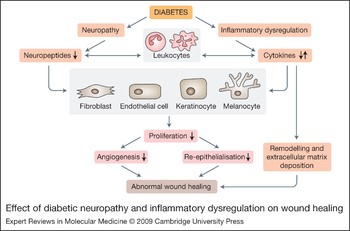
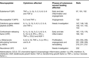
The studies discussed here unmistakably point to the prominent role played by nerves and their mediators in inflammation. Nerves can no longer be viewed simply as transmitters of impulses but need to be recognised as integral components of the inflammatory cascade in various tissues including skin. Cutaneous nerves, both autonomic and sensory, participate in the immune modulation of the skin and can therefore influence wound healing (Fig. 2). They do so by affecting the function of immune and cutaneous cells such as keratinocytes, melanocytes, fibroblasts and dermal microvascular endothelial cells. In the skin, neuropeptides and cytokines function in an endocrine, paracrine and in some cases autocrine fashion to ensure proper wound healing. Although some neuropeptides have received recognition as important players in cutaneous wound healing, a few still need investigation (Table 1).
Effect of diabetic neuropathy and inflammatory dysregulation on wound healing. Diabetes leads to neuropathy and inflammatory dysregulation, which manifests in decreased neuropeptide expression and imbalance in the inflammatory cytokine response. Neuropeptides directly affect leukocytes and monocytes, thereby further contributing to the imbalance in cytokine expression. In addition, neuropeptides and cytokines also directly affect fibroblasts, endothelial cells, keratinocytes and melanocytes, thereby reducing their proliferation and leading to decreased angiogenesis and re-epithelialisation. Decreased angiogenesis and re-epithelialisation, and dysregulation in remodelling and extracellular matrix deposition (affected by disrupted cytokine expression), result in the final outcome of abnormal wound healing.

Phases of cutaneous wound healing affected by neuropeptide–cytokine interaction

Abbreviations: CCL3, CC chemokine ligand 3 (macrophage inflammatory protein 1α); IFN, interferon; IL, interleukin; KGF, keratinocyte growth factor; TGF-β, transforming growth factor β; TNF-α, tumour necrosis factor α.
Diabetes, the endemic disease of the 21st century, and its complications are burdensome on the economy. Neuropathy and peripheral vascular disease result in impaired wound healing. The homeostasis maintained by the nerve–immune function in the skin is disrupted in diabetes. In diabetes some neuropeptides are downregulated (SP, NPY and CGRP) and others upregulated (CRF, α-MSH and NT) with the net effect being that downstream cytokines in the skin are dysregulated. Thus, it is not the gross upregulation or downregulation of cytokines that disrupts wound-healing pathways but the disruption in the balance of cytokines that leads to impaired wound healing.
Current principles of treatment for diabetic wounds include debridement, pressure off-loading, infection control, judicious wound care, tight glycaemic control, and optimisation of cholesterol, nutrition and other lifestyle factors (Refs Reference Dinh and Veves233, Reference Steed234, Reference Pradhan, Andersen, Nabzdyk, LoGerfo and Veves235). Despite this current multifaceted approach, treatment success is limited. Therefore, future therapeutics need to rely on the successful integration of mediators of different pathways involved in wound healing. A combination of deficient neuropeptides and cytokines and inhibitors of upregulated neuropeptides and cytokines could be one such approach. It is extremely important to underscore the complex molecular events that underlie successful wound healing and thus monotherapy is doomed to fail. In the past, growth factors were considered as important potential targets for therapy but to this day the only growth factor approved for treatment of diabetic wounds is PDGF. Combining different growth factors or using growth factors in concert with neuropeptides may be a key strategy in achieving high rates of wound healing in the clinic. Another limiting factor in treating diabetic wounds is the delivery of wound-healing agents. Efflux of these agents and degradation by proteases of protein-based agents in the wound microenvironment is problematic. Delivery of protein-based agents by gene therapy using viral vectors, although promising, poses a serious risk of local and systemic inflammation. Nonviral plasmids could prove to be better vectors. However, in vivo plasmid delivery using chemical transfection reagents is difficult to achieve. Alternatively, physical methods such as electroporation are easy, feasible, safe and efficacious (Refs Reference Denet, Vanbever and Preat236, Reference Vanbever and Preat237, Reference Lee, Chesnoy and Huang238, Reference Lin239). Transdermal delivery of keratinocyte growth factor using these methods in a rat model successfully healed wounds (Ref. Reference Lin239). Thus, identification of therapeutic mediators and therapeutic feasibility are equally important. We have already recognised the importance of the neuropeptide–immune axis mediators in diabetic wound healing and now the future lies in the successful translation of these mediators into treatments for this insidious complication.
Acknowledgements and funding
This work was partly supported by National Institutes of Health Grants R01-HL075678, R01-NS046710 and R01 DK076937 (to A.V.), by the William J. von Liebig Foundation (to F.W.L. and L.P.) and a Howard Hughes Medical Institute Research Training Fellowship for Medical Students (to N.D.A)