Persistent pulmonary hypertension of the newborn (PPHN) is associated with the lack of relaxation of the pulmonary vasculature. This condition requires quick diagnosis and treatment to prevent hypoxia-related morbidities, with diagnostic indicators including right-to-left shunting of blood through the patent foramen ovale (PFO) and patent ductus arteriosus (PDA). Additionally, right ventricular (RV) dilation and septal shifts reflecting elevated right-sided pressures are not uncommon. Reference Puthiyachirakkal and Mhanna1
In this case report, we present a newborn with severe PPHN exacerbated by functional mitral regurgitation (MR), both of which is resolved after veno-arterial extracorporeal membrane oxygenation (VA ECMO) and medical therapy.
Case report
A full-term, 4 kg female was born at an outside hospital via C-section and was hypoxic, with significant pulmonary haemorrhage, requiring intubation. Chest X-rays showed a large heart and hazy lung fields, and an echocardiogram raised concern for mitral valve (MV) disease. A prostaglandin infusion was then started to maintain a PDA. The mother’s infectious disease laboratory results were normal, and she had continuous prenatal care without complications. Prenatal ultrasounds did not reveal any cardiac or pulmonary abnormalities.
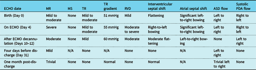
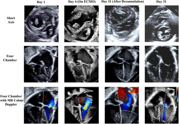
On the day of birth, she was transferred to our centre where an echocardiogram demonstrated a small fenestrated PFO with left-to-right flow and up to a 6 mmHg mean gradient. The left atrium was moderately dilated with rightward atrial septal bowing (Table 1). The mitral valve annulus (MVA) was 10–11 mm (z score −1.43 to −0.79) and moderate MR was noted, likely secondary to anterior leaflet prolapse (Fig 1). There were two, well-spaced papillary muscles with shortened chordae on the posterior leaflet and left ventricular (LV) dimensions were borderline with a diameter of 13.3 mm (z score −3.8) in diastole and 9 mm (z score −2.3) in systole. The aortic valve was hypoplastic with an annular diameter of 5 mm (z score −3.6) and no significant aortic stenosis or LV outflow tract obstruction. The ductus arteriosus remained patent with a systolic right-to-left shunt, demonstrating supra-systemic RV pressures, and the patient had significant pulmonary hypertension (PH) necessitating 100% FiO2 and inhaled nitric oxide. The interventricular septum (IVS) bulged into the LV, consistent with severely elevated pulmonary artery pressures.
Echocardiography

ASD = atrial septal defect; Echo = echocardiogram; MR =mitral regurgitation; MS = mitral stenosis; PDA = patent ductus arteriosus; RVD = right ventricular dilation; TR = tricuspid regurgitation; TR gradient = tricuspid gradient.
Echocardiography over time. All images are in the systolic phase.

The patient’s respiratory failure worsened and she entered cardiovascular shock. Pressor support was initiated but was inadequate to restore appropriate haemodynamics. Consequently, she was cannulated to VA ECMO 2 days after birth. While on VA ECMO support, the patient was maintained on stress dose hydrocortisone. Significant pulmonary rehabilitation was required during her circulatory support.
The patient’s VA ECMO was discontinued after a week, but decannulation did not occur until 2 days later due to recurrent pulmonary haemorrhage. Nine days after decannulation, the patient’s repeat echocardiogram revealed normal pulmonary pressures and an appropriately sized LV with good systolic function. She was extubated 2 days later and was transitioned off nitric oxide to sildenafil, which was discontinued after 10 days when she demonstrated normal right-sided pressures on echocardiogram.
The patient’s discharge echocardiogram, 35 days after birth, showed no evidence of PH and normal RV and LV systolic function with no dilation. Of note, the once severe MR had nearly resolved, displaying only mild insufficiency. She was discharged on clonidine for sedation wean and tachycardia, along with chlorothiazide and fluticasone nebuliser for chronic lung disease.
Discussion
In this case report, we present a newborn with severe PPHN and significant MR. While PPHN and treatment with VA ECMO are not novel, we believe the pathophysiology in this child is unique and has not been previously reported in newborns. Reference Fuloria and Aschner2,Reference Maeder, Weber and Buser3
In this patient, we believe MR was a component of a unique positive feedback loop resulting in worsening PH and cardiac parameters, and not a structural defect; the mitral valve’s return to normal function following the restoration of normal pulmonary and RV pressures would not have been expected with an anatomically defective valve. We believe the PPHN in this case had a common aetiology and resulted in elevated pulmonary and RV pressures. Due to RV dilation and leftward IVS shift, the positioning of the papillary muscles, and therefore the directionality of tension forces placed on the MV leaflets by the chordae, altered MV function and caused MR. This MR led to elevated left atrial (LA) pressures and LA dilation, which caused a rightward atrial septal shift and may have progressed back through the pulmonary veins, potentially worsening PPHN as previously described in the literature. Reference Maeder, Weber and Buser3 This would further increase RV pressures and worsen RV dilation, magnify the septal shift, and create a vicious cycle of worsening PPHN and MR. The rightward atrial septal shift could have also resulted in malpositioning of the MVA, thereby contributing to the development of MR.
Prior studies in adults have demonstrated the capacity for RV hypertrophy and leftward IVS shifts to result in MR by altering LV anatomy and the arrangement of the papillary muscles. This phenomenon was demonstrated by Garcia-Dorado et al. In this study of adults who developed PH, 9.3% of patients with no RV hypertrophy developed MV prolapse, in contrast to 56% of those with severe RV hypertrophy. Reference García-Dorado, García and Bello4 In patients with congenitally corrected transposition of the great arteries and tricuspid regurgitation, pulmonary artery banding has been shown to alter the geometry of the IVS by raising right-sided pressures, thereby restoring tricuspid anatomy and function. Reference Poirier and Mee5,Reference Halpern, Nathan, Yaacov and Marx6 Beyond the role of ventricular anatomy, MR has been shown to commonly result in LA dilation. Park et al, argued that in patients with dilated cardiomyopathy, the primary factor affecting MVA geometry was not LV dilation, but rather LA dilation. Reference Park, Park and Casaclang-Verzosa7 We suspect that abnormal IVS motion due to RV dilation and elevated pulmonary pressures, in conjunction with altered motion and shape of the LA due to elevated LA pressures, was the cause of MR in this patient, explaining the simultaneous resolution of PPHN and MV function.
Acknowledgements
None.
Financial support
This research received no specific grant from any funding agency, commercial, or not-for-profit sectors.
Conflicts of interest
None.
Ethical standards
This research does not involve human or animal experimentation and was exempt from IRB review at our institution.