Case presentation
A 13-year-old boy was undergoing follow-up at our hospital every 2 years, since being diagnosed with mitral regurgitation with mitral valve prolapse in the wake of a systolic murmur at 6 months of age.
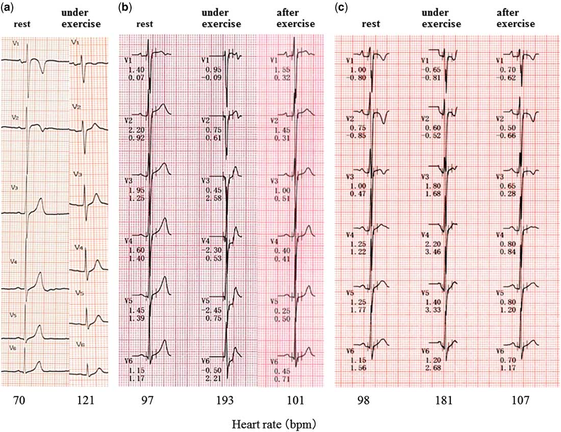
There was no significant ST change in the double master two-step test at 7 years of age. His heart rate at rest was 70 beats per minute, and during exercise his heart rate was 121 beats per minute (Fig 1). There was no ST-T change in the double master test at the age of 10 as well.
He had two instances of transient chest pain, dimmed vision, and syncope, after endurance running at the ages of 11 and 12 years. He was admitted after demonstrating ST depression on exercise electrocardiography at 13 years of age; however, he had no symptoms on admission. Physical examination showed a heart rate of 76 beats per minutes, and a regular rhythm with no heart murmurs. His respiratory rate was 18 per minute, and there was no rale or laboured breathing. Levels of human atrial natriuretic peptide and brain natriuretic peptide were within normal limits (human atrial natriuretic peptide 34.7 pg/ml; brain natriuretic peptide<4.0 pg/ml), and the myocardium deviation enzyme was also within normal limits. Chest X-ray showed a cardiothoracic ratio of 40% and no pulmonary congestion. He had a normal sinus rhythm, and no ST-T change on electrocardiography at rest; however, ST depression at V4–V6 was detected on exercise electrocardiography (Fig 1).
( a ) No significant ST depression in the double master two-step test at 7 years of age. ( b ) Preoperative treadmill electrocardiography (ECG) showing significant ST depression at V4–V6 at 13 years of age. ( c ) After 3 years of coronary artery bypass grafting, no ST depression on treadmill ECG. bpm=Beats per minute.

His heart rate at rest was 97 beats per minute, during exercise it was 193 beats per minute, and after exercise it was 101 beats per minute (Fig 1).
Echocardiography showed high-echoic antero-lateral papillary muscle of the mitral valve, prolapse of the mitral anterior leaflet, mild mitral valve regurgitation, dilatation of the right coronary artery, and the origin of the left main coronary artery was indistinct; however, there was no asynergy of left ventricle performance. There were similar findings for the left coronary artery on cardiac contrast-enhanced CT. Cardiac perfusion MRI showed no early defect and late gadolinium enhancement, but exercise myocardial perfusion scintigraphy showed ischaemia and hypokinesis at the area of the lateral branch in the early phase. No ischaemic symptoms occurred during scintigraphy, and therefore we suspected asymptomatic myocardial ischaemia.
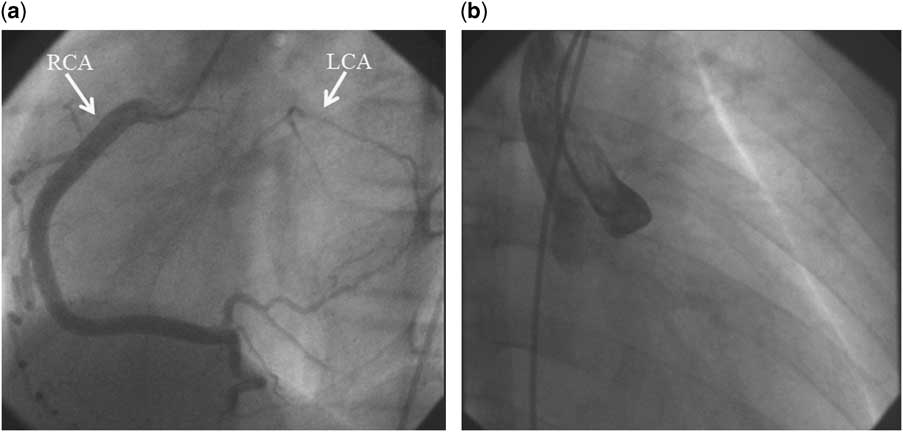
Selective right coronary angiography demonstrated a dilated right coronary artery, and the left coronary artery was filled in a retrogra manner via the right coronary artery, but the left main coronary artery showed no accumulation of contrast fluid. Aortography showed no antegrade left coronary flow (Fig 2); thus, we made a diagnosis of left coronary artery atresia.
We performed coronary artery bypass grafting using the left internal thoracic artery. There were no complications in the perioperative period, and the left intrathoracic artery-to-left anterior descending artery flow was well maintained. We commenced combined aspirin and ticlopidine therapy postoperatively.
His exercise tolerability improved significantly after the operation, and after 3 years of follow-up there has been no ST-T change on exercise electrocardiography (Fig 1). His heart rate at rest was 98 beats per minute, during exercise it was 181 beats per minute, and after exercise it was 107 per minute (Fig 1).
( a ) Selective right coronary angiography showing diffuse dilated right coronary artery (RCA), and left anterior descending coronary artery and left circumflex coronary artery were retrogradely contrasted through collaterals from RCA. ( b ) Left main trunk antegrade flow from the ascending aorta was not contrasted in aortography. LCA=left coronary artery.

Discussion
Left coronary artery atresia is a rare coronary anomaly. Although some children have concomitant anomalies of the bicuspid aortic valve, subaortic stenosis, pulmonary stenosis, and ventricular septal defect,Reference Satran, Dawn and Leesar 1 it is usually an isolated lesion. Reported aetiologies of this anomaly include congenital absence of one coronary anlage, failure of development or displacement of one coronary anlage, failure of canalisation of the proximal segment of the left coronary artery, involvement of the left coronary ostium due to fibrotic change in the aortic media, and coronary obstruction by infection or thrombosis in the early embryonic stage.Reference Koh, Nakagawa and Hamaoka 2 The definitive aetiology, however, is still unknown. Symptoms such as chest pain, dyspnoea, and syncope are frequent, but sudden death has also been reported.Reference Fortuin and Roberts 3 , Reference Goormaghtigh, De Vos and Blanquaert 4 The time of presentation and diagnosis varied; many neonates have increased load with feeding and growth, those under 20 years present after an increase in exercise load, and patients in their fourth decade may present after experiencing hypertension or arteriosclerosis.Reference Eguchi, Nomura and Yotsumoto 5
In our patient, ST depression on exercise electrocardiogram, and a history of chest pain and syncope on exertion during puberty resulted in this diagnosis.
Since the start of follow-up, he had no symptoms for more than 10 years, and ventricular contraction was normal without asynergy until diagnosis. Although exercise electrocardiogram was performed at 7 and 10 years of age, a change in ST-T was not observed. As a result, it took 13 years to make a diagnosis. There is a possibility that resulted from a false-negative result because of insufficient exercise load in childhood.
In addition, mitral valve regurgitation with mitral prolapse was demonstrated at 6 months of age, with these findings suggesting a possibility of myocardial ischaemia. HungReference Hung 6 reported that the main mechanism of ischaemic mitral regurgitation relates to the distortion of the spatial relationships between the mitral valve and the papillary muscles secondary to ventricular re-modelling.
In symptomatic cases exhibiting symptoms such as heart failure, echocardiography with attention to abnormalities in the coronary artery seems to provide sufficient findings to reach the diagnosis. In asymptomatic cases, however, the diagnosis of left coronary atresia by echocardiography alone is difficult. In retrospect, the only abnormal finding in our case in the asymptomatic period was mitral regurgitation with mitral valve prolapse and a high-echoic papillary muscle. When these abnormal mitral findings are confirmed, we should suspect a congenital coronary abnormality. In addition, in cases exhibiting findings as described above, confirming the retrograde flow of the left coronary artery by pulse Doppler is also indicative of an increased possibility of disease.
Although left coronary artery atresia in the literature is limited to case reports, Musiani et alReference Musiani, Cernigliaro and Sansa 7 reported 28 cases (15 children and 13 adult) with left coronary artery atresia in a literature review. In summary, all paediatric cases were symptomatic; only one asymptomatic adult case was found, and in most of the adult cases onset occurred at advanced age; arteriosclerosis of the coronary arteries occurred only in two patients (15% of adult cases); all four cases who did not undergo treatment died; the prognosis was good in all patients who underwent surgical re-vascularisation. Amaral et alReference Amaral, Tanamati and Granzotti 8 reported two cases of successful surgical re-vascularisation in children, including an 8-month-old infant, and suggested that surgical intervention should be performed immediately after diagnosis to avoid the progress of the myocardial lesion. D’Souza et alReference D’Souza, Samuel and Vettukattil 9 reported a case of a neonate in whom coronary artery bypass grafting was successful using the left internal mammary artery and a good patency of the graft was achieved for 1 year after the surgery.
In our case, surgical re-vascularisation was performed immediately after the diagnosis, and ischaemic changes improved after the operation. In retrospect, even if we had obtained a diagnosis of left coronary atresia at 6 months of age in our case, we may have decided against aggressive re-vascularisation because he was asymptomatic, and there were no findings of impairment of ventricular contraction.
Although such reports are limited, successful cases of surgery in neonates and infants with a good clinical course have been reported.
Considering the risk of sudden death and the development of myocardial ischaemia, we believe that aggressive surgical treatment for left coronary atresia improves the prognosis, and even in the infant patient, when diagnosis of left coronary artery atresia is reached, consultation with the surgeon about aggressive surgical intervention is warranted.
Long-term prognosis has not been reported; therefore, long-term postoperative follow-up after surgery is essential.
Conclusion
ST depression and history of chest pain and syncope on exertion resulted in the diagnosis of congenital atresia of the left coronary artery. In case of chest pain or syncope during exercise, left coronary artery atresia should be taken into account in the differential diagnosis. In addition, in case of mitral valve prolapse and regurgitation, even in the absence of symptoms, we need to carefully follow the possibility of congenital coronary atresia. Early and aggressive surgical re-vascularisation should be performed after the diagnosis. Long-term follow-up is essential, because the long-term prognosis is unclear.
Acknowledgements
The authors would like to thank T. Komiya of our hospital cardiovascular surgeon who performed the operation of this patient.
Financial Support
This research received no specific grant from any funding agency or from commercial or not-for-profit sectors.
Conflicts of Interest
None.