Introduction
Gnathostomiasis is a food-borne zoonosis caused by infection with third-stage larvae (L3) of Gnathostoma spp. from second intermediate hosts, such as freshwater fish, shellfish, frogs and poultry (Daengsvang, Reference Daengsvang1981; Eamsobhana et al., Reference Eamsobhana, Wanachiwanawin, Roongruangchai, Song and Yong2016). Human gnathostomiasis is primarily endemic in South-East Asia and Latin America, where people consume raw or undercooked fish and is incidentally detected in travellers returning from endemic areas as well as natives who eat raw imported dishes in non-endemic regions (Herman & Chiodini, Reference Herman, Wall, van-Tulleken, Godfrey-Faussett, Bailey and Chiodini2009; Diaz, Reference Diaz2015). During the past few years, cases have been diagnosed outside endemic areas with increased frequency (Herman et al., Reference Herman, Wall, van-Tulleken, Godfrey-Faussett, Bailey and Chiodini2009; Jarell et al., Reference Jarell, Dans, Elston, Mathison and Ruben2011; Jeremiah et al., Reference Jeremiah, Harangozo and Fuller2011; Diaz, Reference Diaz2015). In China, only 57 cases of human gnathostomiasis were reported from 1919 to 2008 (Chen & Li, Reference Chen, Li, Tang, Xu and Chen2012); however, in recent years, several patients have been detected based on the triad of clinical symptoms, exposure risks and specific antibodies (unpublished data). Although the current endemic status of this disease in China is unclear, the number of patients is increasing.
A definitive diagnosis for gnathostomiasis could be made by isolating Gnathostoma worms from the lesions, but discovery of a worm from the lesions happens rarely, even in cutaneous patients, and is also impractical for visceral infection. Currently, enzyme-linked immunosorbent assays (ELISA) for the measurement of specific antibodies against Gnathostoma spp. and immunoblot analysis for the detection of the Gnathostoma spinigerum-specific 24-kDa band are commonly applied to support clinical diagnosis (Díaz Camacho et al., Reference Díaz Camacho, Willms, de la Cruz Otero, Zazueta Ramos, Bayliss Gaxiola, Castro Velázquez, Osuna Ramírez, Bojórquez Contreras, Torres Montoya and Sánchez Gonzáles2003; Cornaglia et al., Reference Cornaglia, Jean, Bertrand, Aumaître, Roy and Nickel2016; Mulroy et al., Reference Mulroy, Simpson and Frith2016). The L3 crude extract, used as antigen in ELISA, presented poor specificity and sensitivity (Suntharasamai et al., Reference Suntharasamai, Desakorn, Migasena, Bunnag and Harinasuta1985; Dharmkrong-at et al., Reference Dharmkrong-at, Migasena, Suntharasamai, Bunnag, Priwan and Sirisinha1986). Studies on the analysis and identification of antigenic components of G. spinigerum and Gnathostoma binucleatum have been conducted in several laboratories (Wongkham et al., Reference Wongkham, Maleewong, Ieamviteevanich, Intapan and Morakote2000; Campista-León et al., Reference Campista-León, Delgado-Vargas, Landa, Willms, López-Moreno, Mendoza-Hernández, Ríos-Sicairos, Bojórquez-Contreras and Díaz-Camacho2012; Zambrano-Zaragoza et al., Reference Zambrano-Zaragoza, Durán-Avelar Mde, Messina-Robles and Vibanco-Pérez2012; Janwan et al., Reference Janwan, Intapan, Laummaunwai, Rodpai, Wongkham, Insawang, Thanchomnang, Sanpool and Maleewong2015). However, the availability of specific antigens remains a major issue for developing serodiagnostic tests, particularly in non-endemic areas. Immunoblot analysis for the detection of the G. spinigerum-specific 24-kDa band is the most widely used method to confirm a clinical diagnosis with 100% specificity (Tapchaisri et al., Reference Tapchaisri, Nopparatana, Chaicumpa and Setasuban1991; Anantaphruti et al., Reference Anantaphruti, Nuamtanong and Dekumyoy2005; Sawanyawisuth et al., Reference Sawanyawisuth, Intapan, Khotsri, Kanpittaya, Chotmongkol and Maleewong2011). However, the immunoblot method is labour-intensive and time-consuming because it requires sophisticated instruments and well-trained operators. To date, services that include immunoblotting for serological diagnosis of gnathostomiasis are available in several research institutions worldwide.
In the present study, we developed a simple and rapid filtration-based test, dot immune–gold filtration assay (DIGFA), using a partially purified antigen of Gnathostoma third-stage larvae (L3), bright-red colloidal gold as a visual label and porous nitrocellulose membrane as a solid capture matrix, to detect specific IgG in human serum. The DIGFA technique has been utilized successfully for immunodiagnosis of other helminthiases, for example angiostrongyliasis (Gan et al., Reference Gan, Wang, Yang, Zhang and Eamsobhana2007; Eamsobhana et al., Reference Eamsobhana, Gan, Ma, Wang, Wanachiwanawin and Yong2014, Reference Eamsobhana, Prasartvit, Gan and Yong2015). To our knowledge, the present study shows the first application of DIGFA for the serological diagnosis of gnathostomiasis.
Materials and methods
Serum samples
Thirty serum samples from gnathostomiasis patients, including two pathologically confirmed cases and 28 clinically diagnosed patients, were used in the present study. The triad of clinical signs and symptoms, obvious exposure risk and positive reactivity based on G. spinigerum 24-kDa antigen-based immunoblotting were used as diagnostic criteria for clinical gnathostomiasis. In addition, 50 negative control serum samples were collected from healthy donors with no history of exposure to parasites and no indication of helminth infection based on a stool examination at the time of blood collection. To observe cross-reactivity, sera from patients with angiostrongyliasis (n = 25), cysticercosis (n = 25), paragonimiasis (n = 25), schistosomiasis (n = 25) and sparganosis (n = 25) were tested. Patients with schistosomiasis japonica, paragonimiasis westermani and sparganosis mansoni were diagnosed parasitologically. Cases of angiostrongyliasis and cysticercosis were confirmed by a computerized tomography scan combined with specific serological tests (Gan et al., Reference Gan, Wang, Guo, Zhu and Shi2006, Reference Gan, Wang, Yang, Zhang and Eamsobhana2007). Human serum samples were collected in mainland China with the consent of the subjects or their guardian(s) after verbal explanations of the study. All serum samples were stored at –80°C until further use.
Preparation of L3 crude extract
The Gnathostoma L3 crude extract was prepared as described previously (Dharmkrong-at et al., Reference Dharmkrong-at, Migasena, Suntharasamai, Bunnag, Priwan and Sirisinha1986) with minor modifications. Freshwater eel livers were collected from farmers’ markets, gently sheared with forceps in the laboratory and subsequently digested with a 1% pepsin solution (w/v; pH 2.0) at 37°C for 4 h. Living larvae were harvested from the digested liver tissue under a dissecting microscope. Fifty Gnathostoma larvae were collected and rinsed three times with sterile normal saline, re-suspended in 400 μl of pre-cooled phosphate-buffered saline (PBS, 0.01 mol/l, pH 7.2) containing proteinase inhibitors (Thermo Fisher Scientific, Rockford, Illinois, USA) and 5 mm EDTA, and homogenized using a glass tissue-grinder. The homogenate was sonicated at 20 kHz for 5 min in an ice bath and centrifuged at 20,000 g for 15 min at 4°C. The supernatants were pooled as the L3 crude extract. The protein concentration was 3.2 mg/ml, as measured by a Bradford Coomassie Plus Protein Assay Reagent Kit (Pierce, Rockford, Illinois, USA).
Partial purification of L3 antigen
To enrich specific antigen components from the L3 crude extract, a single-step 2,2,2-trichloroacetic acid (TCA)/acetone precipitation for low-molecular weight proteins was performed as reported previously (Lee et al., Reference Lee, Bae, Kim, Díaz-Camacho, Nawa and Kong2010). Briefly, 400 μl (3.2 mg/ml) of L3 crude extract was blended with an equal volume of a TCA/acetone (10%/60%) mixture and incubated at 4°C for 1 h. The mixture was centrifuged at 20,000 g for 20 min and the supernatant was recovered. After dialysis against PBS for 4 h, the solution was concentrated to 100 μl using a CentriPrep (Millipore, Billerica, Massachusetts, USA), as the partially purified L3 antigen, and stored in aliquots at −80°C until further use. All procedures were performed at 4°C. The protein concentration of the purified antigen was 1.3 mg/ml and protein recovery was 10.2% after partial purification.
Immunoblot analysis
An immunoblot analysis to detect gnathostomiasis-specific antigens was performed as described previously (Towbin et al., Reference Towbin, Staehelin and Gordon1979; Laummaunwai et al., Reference Laummaunwai, Sawanyawisuth, Intapan, Chotmongkol, Wongkham and Maleewong2007). Briefly, 1.5 μg of antigen was resolved by 12% sodium dodecyl sulphate–polyacrylamide gel electrophoresis (SDS–PAGE) and transferred electrophoretically on to a nitrocellulose membrane for immunoblotting. After non-specific binding was blocked with 5% non-fat milk in tris-buffered saline containing tween-20 (TBST) overnight at 4°C, the blot was incubated with each serum sample diluted 1:1000 in blocking solution for 1 h at 37°C. After washing with TBST, the blot was incubated with a 1:10,000 dilution of alkaline-phosphatase-conjugated anti-human IgG (Boster, Wuhan, China) in blocking solution at 37°C for 1 h. After washing, the blot was developed using a BCIP/NBT chromogenic substrate kit (Tiangen Biotech, Beijing, China) until blue-purple bands appeared.
Enzyme-linked immunoabsorbent assay
Indirect ELISA was applied to evaluate the antigenicity of the partially purified L3 antigen for gnathostomiasis. Briefly, each well of a 96-well micro-plate was coated with 100 μl (1 μg/ml) of the L3 antigen in 0.05 m carbonate–bicarbonate buffer (pH 9.6) at 4°C overnight and blocked with 5% non-fat milk at 37°C for 1 h. After washing three times, the serum sample, diluted 1:1000, was added and the mixture was incubated at 37°C for 1 h. Following washing, 100 μl of horseradish-peroxidase-conjugated goat anti-human IgG antibody at 1:10,000 dilution (Boster, Wuhan, China) was added and incubated at 37°C for 1 h. Finally, 50 μl of tetramethylbenzidine (TMB) substrate solution was added and incubated at room temperature for 15 min to determine the binding enzymatic activity. The enzyme reaction was stopped with 50 μl of 1 n H2SO4, and the optical density (OD) was measured at a wavelength of 450 nm using a microtitre plate reader (Bio-Rad, Hercules, California, USA). The mean plus 3SD of 50 healthy control serum samples was set as the positive cut-off value.
Development of DIGFA for gnathostomiasis
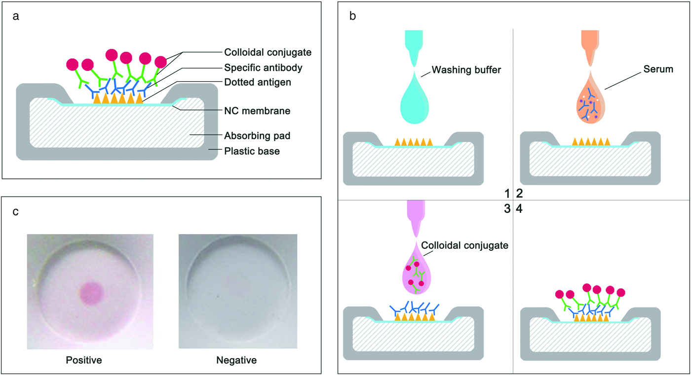
The DIGFA kit comprised a customized device, colloidal gold–protein A conjugate and washing solution. The device was designed as a small plastic housing (40 × 30 × 5 mm) with a test-well on the upper side of the unit. The plastic housing was filled with water-absorbing pads. A piece (10 × 10 mm) of nitrocellulose membrane (NCM) (Φ0.45 μm; Whatman Gmbh, Hahnestr, Germany) was fastened tightly under the test-well (fig. 1a) and 0.5 μl of partially purified L3 antigen (0.5 mg/ml) or L3 crude extract (1.0 mg/ml) was dotted in the centre of the membrane.
A schematic illustration of DIGFA: (a) test device; (b) test procedure; (c) images of the test results.

The colloidal gold–protein A conjugate was prepared according to Horisberger & Clere (Reference Horisberger and Clerc1985). Briefly, 100 ml of 0.01% chloroauric acid solution was boiled, and mixed with 2.8 ml of 1% sodium citrate under constant stirring. When the colour of the solution changed from grey to wine red, boiling was continued for 30 min to obtain mono-disperse gold colloid particles. After the colloidal solution was cooled to room temperature, the pH was adjusted to 6.5 with 1% freshly prepared K2CO3. Subsequently, 1.2 mg of protein A (Nuptec, Hangzhou, China) was added with stirring and incubated at room temperature for 30 min to form a stable colloidal gold–protein A conjugate. Tris-HCl buffer (0.02 mol/l, pH 7.2, containing 1% bovine serum albumin (BSA) and 0.05% Tween-20) was used as a washing solution.
The sample test was performed at room temperature in four steps (fig. 1b). First, one drop (~50 μl) of washing buffer was added to the well of the test device to block non-specific binding of the nitrocellulose membrane; second, 25 μl of the serum sample was added to enable flow through; third, one drop of the colloidal gold–protein A conjugate was added to visualize the specific antigen–antibody reaction; and finally, results were read with the naked eye after washing with Tris-HCl buffer. The appearance of a well-defined pink dot in the centre of the nitrocellulose membrane in the well was determined to be positive; otherwise, the result was determined to be negative (fig. 1c).
Statistical analysis
The ELISA data are reported as the means ± SD. Student's t-test was used to determine the significance of the differences between data groups. A P value of less than 0.05 was considered to indicate statistical significance. The kappa coefficient (κ) was utilized to determine the agreement of two diagnostic tests. Calculations and interpretations of the κ values were performed as described previously (Viera & Garrett, Reference Viera and Garrett2005).
Results
Enrichment of specific antigens from the L3 crude extract using a TCA/acetone precipitation
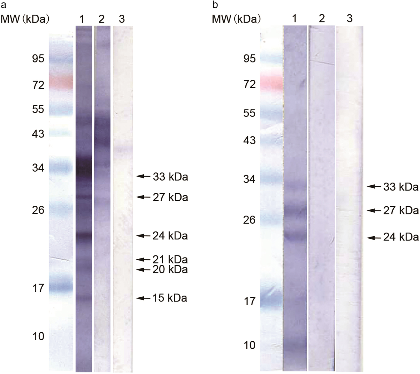
The L3 crude extract was highly complex – more than 15 protein bands were recognized from the pooled serum of patients with gnathostomiasis. Most of the protein bands with molecular weights over 43 kDa reacted strongly with sera from other helminthiasis patients. One protein band (47 kDa) showed a weak reaction with the pooled serum of healthy controls. Six protein bands (15, 20, 21, 24, 27 and 33 kDa) reacted specifically with sera from gnathostomiasis patients (fig. 2a). To isolate the specific antigen components from the L3 crude extract, different concentrations and various combinations of the TCA/acetone mixture were examined to evaluate its effectiveness to precipitate the proteins. A 10% final concentration of TCA in 60% acetone was suitable to precipitate non-specific proteins from L3 crude extracts efficiently. Three specific antigens (24, 27 and 33 kDa), which showed strong reactivity to sera from gnathostomiasis patients but not to sera from other helminthiasis patients or healthy controls, remained in the supernatant fraction (fig. 2b).
Immunoblot analysis of the L3 crude extract (a) and partially purified antigen (b). Lanes: 1, pooled sera from gnathostomiasis patients; 2, pooled sera from other helminthiases; 3, pooled sera from healthy controls. MW, molecular weight; kDa, kilodaltons.

Antigenicity of the partially purified L3 antigen for diagnosis of gnathostomiasis
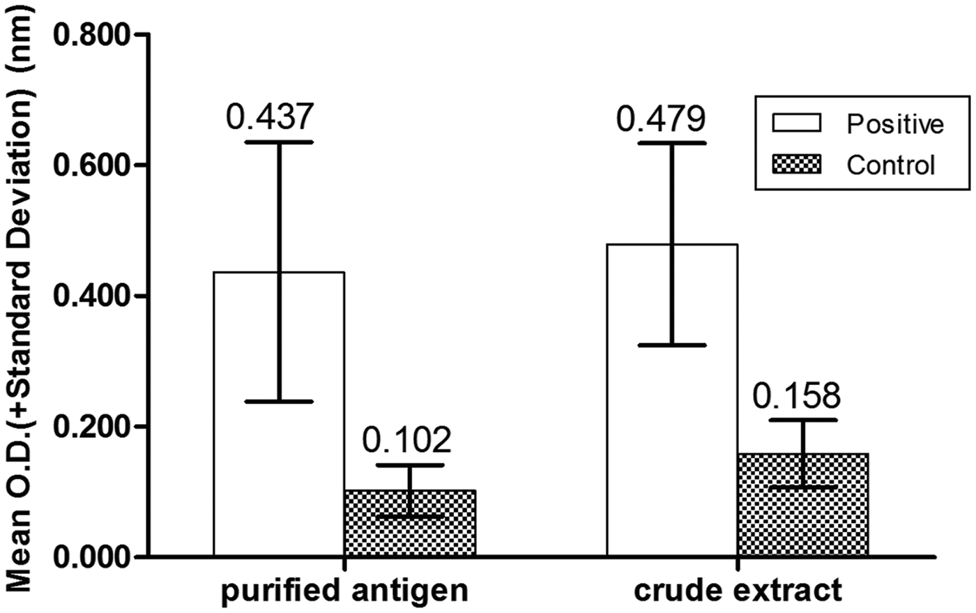
The antigenicity of the partially purified L3 antigen was examined and compared with that of the L3 crude extract using ELISA. Thirty serum samples from gnathostomiasis patients and 50 serum samples from healthy controls were detected by ELISA using the partially purified L3 antigen and L3 crude extract separately. The cut-off value for the partially purified L3 antigen was 0.22 while that for the L3 crude extract was 0.32. All serum samples from gnathostomiasis patients tested positive by ELISA, either using the partially purified L3 antigen or the L3 crude extract. The OD value for the patient group was 0.4367 ± 0.1985, and a significantly higher value than that (0.1017 ± 0.0394) was found for healthy controls, using the partially purified L3 antigen (P < 0.05). The OD value distributions of gnathostomiasis patients based on ELISA with the partially purified L3 antigen were well separated from those of normal healthy controls with the L3 crude extract, indicating the high antigenicity of the partially purified L3 antigen (fig. 3).
Distribution of the optical density (OD) values of serum samples from gnathostomiasis patients and healthy controls determined using ELISA with partially purified antigen and crude extract.

Sensitivity and specificity of DIGFA with the partially purified L3 antigen for gnathostomiasis
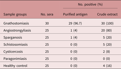
A total of 180 serum samples were tested simultaneously by DIGFA, using the partially purified L3 antigen and the L3 crude extract, separately. The results are summarized in table 1. DIGFA using the partially purified L3 antigen showed good accuracy, with 96.7% sensitivity and 100% specificity. The cross-reactivity with sera from other helminthiasis patients was low, from 0% (schistosomiasis, cysticercosis and paragonimiasis) to 4% (angiostrongyliasis and sparganosis), with an average of 1.6% (2/125). On the other hand, DIGFA using the L3 crude extract presented poor specificity (84%) and high cross-reactivity (25.6%) with sera from other helminthiasis patients, particularly with sera from angiostrongyliasis patients (80%).
Performance of DIGFA using the partially purified L3 antigen and L3 crude extract for the detection of specific antibodies in human serum.

Comparison between DIGFA using the partially purified L3 antigen and immunoblot for the detection of a specific 24-kDa band
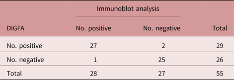
To evaluate the potential of DIGFA for serological diagnosis of gnathostomiasis, a double-blind test was performed on a panel of serum samples (30 from gnathostomiasis patients and 25 normal controls) using both DIGFA and the current gold standard immunoblot analysis. The results are summarized in table 2. Twenty-seven samples tested positive and 25 samples showed negative results in both tests. The concordance of DIGFA and immunoblot tests was 94.5% (52/55). The κ value between DIGFA using the partially purified L3 antigen and immunoblotting for the detection of a specific 24-kDa band was 0.94, indicating an almost perfect agreement.
Comparison between DIGFA using the partially purified L3 antigen and immunoblot analysis for the detection of a specific 24-kDa band.

Discussion
Currently, serological tests are needed for the diagnosis of gnathostomiasis in the clinic and investigations in the field. The L3 crude extract has generally been used as the antigen in sero-immunological assays. ELISA using the L3 crude extract as the antigen produced low sensitivities (59–87%) and specificities (79–96%) (Suntharasamai et al., Reference Suntharasamai, Desakorn, Migasena, Bunnag and Harinasuta1985; Dharmkrong-at et al., Reference Dharmkrong-at, Migasena, Suntharasamai, Bunnag, Priwan and Sirisinha1986; Nopparatana et al., Reference Nopparatana, Tapchaisri, Setasubun, Chaicumpa and Dekumyoy1988; Anantaphruti, Reference Anantaphruti1989). The L3 crude extract was highly complex and contained non-specific components that were able to react with sera from other helminthiasis patients and healthy controls, as reported in previous studies (Nopparatana et al., Reference Nopparatana, Tapchaisri, Setasubun, Chaicumpa and Dekumyoy1988; Tapchaisri et al., Reference Tapchaisri, Nopparatana, Chaicumpa and Setasuban1991). A group of low-molecular-weight proteins (15, 20, 21, 24, 27 and 33 kDa) in the L3 crude extract were found to be specific for gnathostomiasis. Most of the components, which had strong cross-reactivity with sera from other helminthiases, ranged from 43 to 150 kDa (fig. 2a). In the present study, three specific antigens (24, 27 and 33 kDa) were successfully enriched from the L3 crude extract by single-step TCA/acetone precipitation. The results of ELISA indicated that this partially purified L3 antigen had high antigenicity. These findings suggested that a single-step TCA/acetone precipitation would be suitable for separating these specific antigens from the L3 crude extract. TCA precipitation is a common process of sample preparation for proteomics analysis (Peterson, Reference Peterson1983; Jacobs et al., Reference Jacobs, van Rijssen, van der Heijden and Verpoorte2001; Nandakumar et al., Reference Nandakumar, Cheung and Marten2006). The trichloroacetic moiety and acidic property play important roles in protein precipitation (Sivaraman et al., Reference Sivaraman, Kumar, Jayaraman and Yu1997; Rajalingam et al., Reference Rajalingam, Loftis, Xu and Kumar2009). Acetone induces hydrophobic aggregation of proteins by reducing the water-attracting power of charged hydrophilic moieties of protein molecules (Lee et al., Reference Lee, Bae, Kim, Díaz-Camacho, Nawa and Kong2010). Using TCA/acetone precipitation, specific antigens of Taenia solium ranging from 7 to 38 kDa have been enriched from metacestode cysts (Lee et al., Reference Lee, Bae, Kim, Díaz-Camacho, Nawa and Kong2010). This single-step method could easily be performed, with no requirement for special instruments, illustrating a potential protocol in primary laboratories for the preparation of native antigens from helminths. Considering the high protein recovery (10.2%) after purification, 50 worms were sufficient for each antigen batch. Since freshwater eels from endemic regions had high rates and intensities of infection, dozens of eel livers were collected and used to isolate Gnathostoma larvae for each batch. The preparation of a partially purified antigen is more economical and practical than that of recombinant antigens and synthetic peptides, particularly in less-developed endemic areas.
Immunoblotting for the detection of the 24-kDa protein band using the L3 crude extract of G. spinigerum has been most widely used as the gold standard to confirm a clinical diagnosis of gnathostomiasis in Asia and Europe (Herman & Chiodini, Reference Herman, Wall, van-Tulleken, Godfrey-Faussett, Bailey and Chiodini2009; Diaz, Reference Diaz2015). However, the test involves multiple steps and requires 2 days, although it provides the advantages of sensitivity and specificity. In the present study, DIGFA using the partially purified L3 antigen presented almost perfect agreement (κ = 0.94) with immunoblotting. Moreover, the DIGFA test has the advantages of speed, ease of use, low cost and good long-term stability of the reagents (12 months or more). A sample test with DIGFA can be performed within 5 min and costs US$1, which is much less than that for immunoblotting.
In Mexico, where G. binucleatum is the pathogenic nematode for human gnathostomiasis, only 26.3% of patients’ serum samples reacted with the 24-kDa protein band, whereas 65.2% of these samples could recognize the 33-kDa protein (Zambrano-Zaragoza et al., Reference Zambrano-Zaragoza, Durán-Avelar Mde, Messina-Robles and Vibanco-Pérez2012). American patients with gnathostomiasis showed negative results in G. spinigerum-antigen-based immunoblotting, reflecting incomplete cross-reactivity between the 24-kDa antigens of G. spinigerum and G. binucleatum (Neumayr et al., Reference Neumayr, Ollague, Bravo, Gotuzzo, Jimenez, Norton, Doanh, Nawa, Horii, Nickel and Marti2016). These findings suggest that the 24-kDa protein is the immunodominant antigen for G. spinigerum and the 33-kDa protein is the immunodominant antigen for G. binucleatum. Therefore, immunoblot analysis for the detection of a specific 24-kDa antigen might not be appropriate for the diagnosis of G. binucleatum infection. In the present study, the partially purified L3 antigen contained specific 24 and 33-kDa protein components and presented high specificity and sensitivity for the diagnosis of patients with gnathostomiasis in China, where Gnathostoma hispidum, Gnathostoma doloresi and G. spinigerum have been identified as aetiological agents of human gnathostomiasis (Chen & Li, Reference Chen, Li, Tang, Xu and Chen2012). The potential of DIGFA using the partially purified L3 antigen for the diagnosis of infection by the various species – G. binucleatum, G. hispidum and G. doloresi – needs to be investigated.
Simple and rapid serological tests are imperative for the diagnosis of gnathostomiasis and screening of suspected patients, particularly in non-endemic areas where most physicians are not familiar with this disease. There are two forms of rapid tests (lateral-flow and flow-through), with the advantages of low-cost, speed and convenience. A lateral-flow immunochromatographic test (ICT) for gnathostomiasis, with high sensitivity (93.75%) and specificity (97.01%), has recently been developed using a recombinant protein (rGslic18) as the antigen (Janwan et al., Reference Janwan, Intapan, Yamasaki, Rodpai, Laummaunwai, Thanchomnang, Sanpool, Kobayashi, Takayama, Kobayashi and Maleewong2016). In the present study, the DIGFA rapid test, in flow-through format, was developed and evaluated for gnathostomiasis. The filtration-based design completely eliminated the diffusion limitation and improved the overall reaction kinetics tenfold compared to the classic shaking incubation method (Xu & Bao, Reference Xu and Bao2003). In addition, the filtration-based test decreased non-specific binding between proteins, while the reaction kinetics were effectively accelerated and the reaction time of each step was shortened. In the present study, DIGFA using the partially purified L3 antigen had high sensitivity (96.7%) and low cross-reactivity (1.6%), showing promise as a rapid test for the serological diagnosis of gnathostomiasis in clinics or large-scale investigations in the field.
The limitation of the present study is the low number of serum samples from parasitologically definitive gnathostomiasis patients, and of sera from patients with different types of parasitosis. Out of 30 positive control sera, 28 samples were collected from clinically diagnosed gnathostomiasis patients. All patients were confirmed by immunoblotting for the detection of the G. spinigerum-specific 24-kDa band. This G. spinigerum 24-kDa-antigen-based immunoblot showed 100% specificity (Tapchaisri et al., Reference Tapchaisri, Nopparatana, Chaicumpa and Setasuban1991; Anantaphruti et al., Reference Anantaphruti, Nuamtanong and Dekumyoy2005; Laummaunwai et al., Reference Laummaunwai, Sawanyawisuth, Intapan, Chotmongkol, Wongkham and Maleewong2007; Sawanyawisuth et al., Reference Sawanyawisuth, Intapan, Khotsri, Kanpittaya, Chotmongkol and Maleewong2011). Thus, these 28 clinical gnathostomiasis patients represented true infections, and the evaluation of sensitivity is therefore accurate in the present study. Sera from patients with five types of helminthiases (angiostrongyliasis, cysticercosis, paragonimiasis, schistosomiasis and sparganosis), with high cross-reactivity with the L3 crude extract, were selected to evaluate the diagnostic specificity of DIGFA using a partially purified antigen. If more human sera samples from more types of parasitoses are utilized, the total cross-reactivity might decrease. More types and numbers of patient sera need to be used in future studies to determine the accuracy and specificity of the evaluation.
Acknowledgements
The authors would like to thank the Director of Zhejiang Academy of Medical Sciences for permission to conduct the present study. The authors would also like to thank Yin Kang, Qing-An Zhou and Lei Yang for their assistance with the collection of Gnathostoma larvae.
Financial support
This work was supported by a grant from the Medical Science and Technology Project of Zhejiang Province (grant number 2011KY001).
Conflict of interest
None.
Ethical standards
The Ethics Committee of Zhejiang Academy of Medical Sciences approved the present study.