Anomalous drainage of the right superior caval vein to the left atrium is a rare congenital systemic venous anomaly.Reference Park, Smith and Silberstein 1 , Reference Baggett, Skeen, Gantt, Trotter and Birkemeier 2 Patients with this unique condition present with cyanosis and its secondary effects including polycythaemia, clubbing, and brain abscess due to the persistent right-to-left shunting. The choice of therapy is surgical re-implantation of the right superior caval vein to the right atrium.Reference Braudo, Beanlands and Trusler 3 , Reference Alpert, Rao, Moore and Covitz 4 We report successful transcatheter treatment of anomalous venous drainage of the right superior caval vein to the left atrium. Pre-procedural planning was guided by a life-sized three-dimensional printed model.
Case report
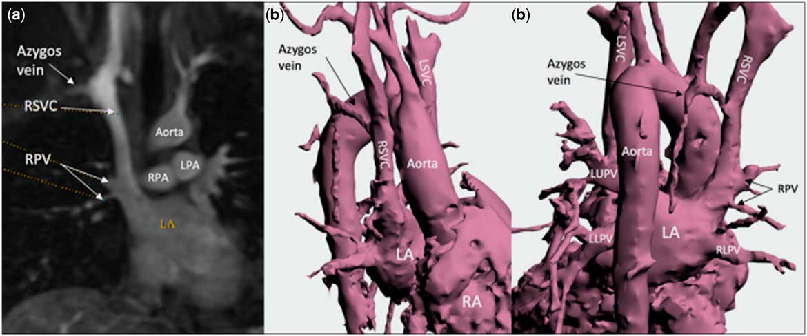
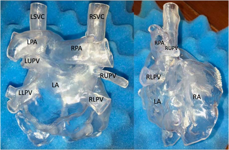
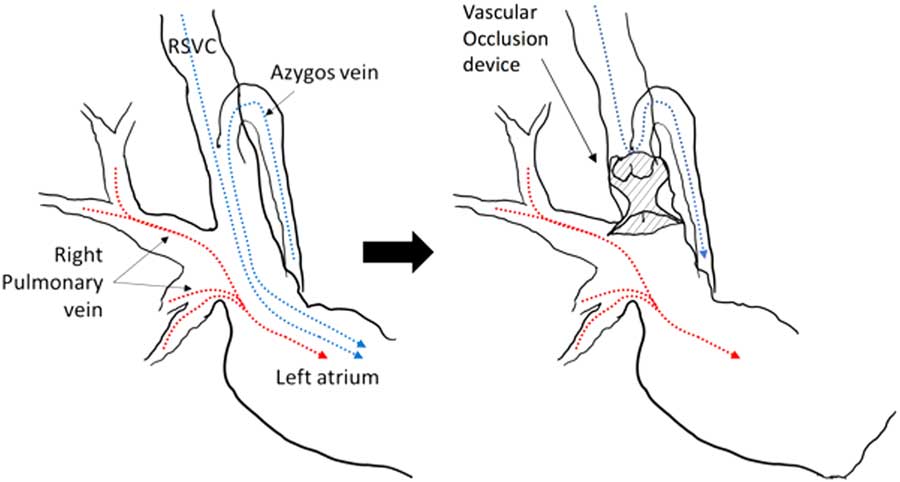
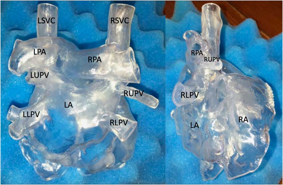
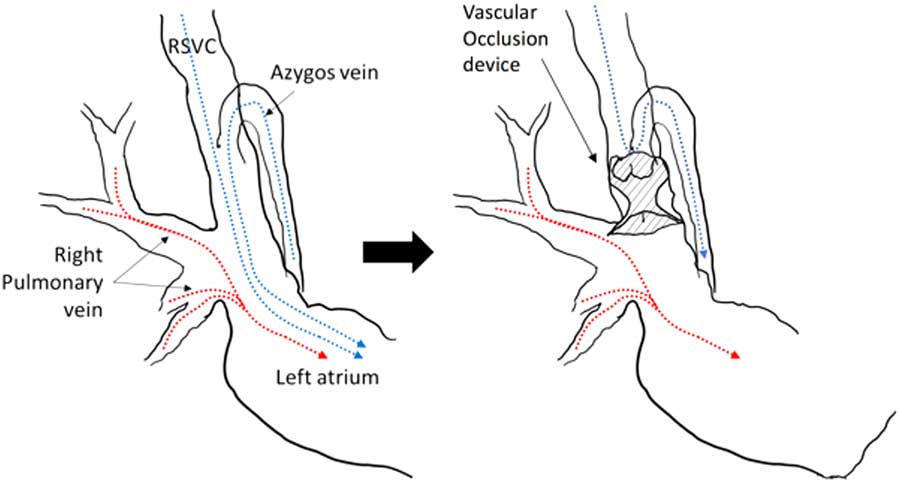
A 32-year-old obese man with a history of brain abscess was referred to our cardiology clinic. He was mildly desaturated at 93% on room air. Other vital signs were unremarkable. He was on aspirin 81 mg once daily. Transthoracic echocardiography raised concern of an anomalous right superior caval vein draining into the roof of the left atrium without an innominate vein, along with a left superior caval vein draining normally into the right atrium through the coronary sinus. Agitated saline injection from a peripheral intravenous line in the right arm showed complete opacification of the left atrium. Cardiac magnetic resonance imaging (Fig 1a) demonstrated that the right upper pulmonary vein and right middle pulmonary vein drained into the right superior caval vein within 25 mm from its entry to the left atrium. Other pulmonary veins drained normally to the left atrium. Transcatheter management of this unique anomaly was undertaken. Before the catheterisation, a life-sized cardiac three-dimensional printed model of the anomalous drainage was created for pre-procedural planning. A three-dimensional digital model (Fig 1b and c) was made based on the three-dimensional data set acquired by magnetic resonance angiography, using the software Mimics® (Materialise, Plymouth, MI, United States of America). The three-dimensional printed model (Fig 2) was printed by sterolithographic technology. The intended procedure was to occlude the right superior caval vein with a vascular occlusion device inferior to the azygous vein but superior to the entries of the right upper and middle pulmonary vein. This would divert the right superior caval vein flow to the inferior caval vein system through the azygos vein in a retrograde manner and allow the right upper pulmonary vein and right middle pulmonary vein flow to drain into the left atrium normally, achieving exclusion of right-to-left shunting (Fig 3).
( a ) Magnetic resonance angiography. Right superior caval vein (RSVC) drains into the left atrium (LA). Right pulmonary vein (RPV) enters the inferior portion of RSVC. ( b and c ) Three-dimensional digital model. LA=left atrium; LPA=left pulmonary artery; LPV=left upper pulmonary vein; LSVC=left superior caval vein; RA=right atrium; RLPV=right lower pulmonary vein; RPA=right pulmonary artery.

Three-dimensional printed model using sterolithography technology. LA=left atrium; LLPV=left lower pulmonary vein; LPA=left pulmonary vein; LSVC=left superior caval vein; LUPV=left upper pulmonary vein; RA=right atrium; RLPV=right lower pulmonary vein; RPA=right pulmonary artery; RSVC=right superior caval vein; RUPV=right upper pulmonary vein.
Schematic figures of intended intervention. Venous return (blue dotted line) from the right superior caval vein (RSVC) drains into the left atrium. After placing a vascular occlusion device inferior to the orifice of azygos vein in the RSVC, venous return is directed to the inferior caval vein system through the azyos vein in a retrograde manner. Pulmonary venous drainage from the right pulmonary vein normally drains into the left atrium.
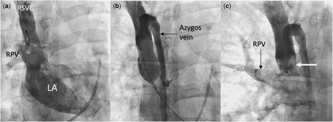
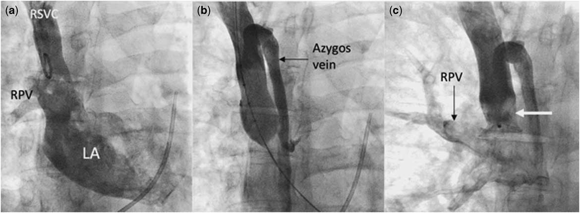
Cardiac catheterisation was performed under general anaesthesia. An 8-Fr sheath was placed in the right internal jugular vein and was exchanged to a 12-Fr sheath subsequently. Another 8-Fr sheath was placed in the right femoral vein. Baseline haemodynamic data showed normal mean pulmonary artery pressure of 14 mmHg. Mean superior caval vein and left atrial pressures were 10 mmHg. His systemic saturation was 93%, compared with RUPV saturation of 99% on room air. By Fick method, Qp:Qs ratio was 0.8:1, consistent with right-to-left shunting. Angiography demonstrated that the right upper pulmonary vein and right middle pulmonary vein anomalously drained into the inferior part of the right superior caval vein (Fig 4a). Nominal diameter of the right superior caval vein was 13.1 mm superiorly, expanding to 18 mm at the entrance of the right pulmonary veins into the inferior portion of the right superior caval vein, and expanding to 23 mm at its entrance into the left atrium. The distance between the takeoff of the azygous vein and the entrance of the right pulmonary veins to the distal right superior caval vein was 13.6 mm in length. Balloon test occlusion of the right superior caval vein was performed using a 25-mm NuMed sizing balloon catheter (PTS®; NuMED Inc., Hopkington, NY, United States of America). Subsequent angiography showed adequate occlusion of the right superior caval vein with azygos vein acting as a pop-off of blood flow from the superior right superior caval vein to the inferior caval vein system (Fig 4b). Balloon diameter measured 13.9 mm. After 10 minutes of test occlusion, the mean pressure of the superior portion of the right superior caval vein minimally increased by 1 mmHg. As test occlusion of the mid-portion of the right superior caval vein was deemed satisfactory, we proceeded with vascular occlusion of the inferior right superior caval vein. Two different devices were attempted, based on the response of RSVC to the balloon test occlusion. Those devices were 18 mm AMPLATZER™ Vascular Plug II (St. Jude Medical, St. Paul, Minnesota, United States of America) and 18 mm mm AMPLATZER™ Muscular Ventricular Septal Defect Occluder (St. Jude Medical). Both devices either occluded the entry of azygos vein or protruded into the orifice of the right upper pulmonary vein. Satisfactory occlusion was obtained using a 16-mm AMPLATZER™ Muscular Ventricular Septal Defect Occluder in the mid-section of the right superior caval vein, just inferior to the takeoff of the azygous vein and superior to the entrance of the right pulmonary veins to the distal right superior caval vein (Fig 4c). The mean pressure of the right superior caval vein was 13 mmHg at the end. The next day, his saturations improved to 97% and transthoracic echocardiography showed satisfactory device position in the right superior caval vein. At 6-month follow-up, he had no symptoms of superior caval vein syndrome nor desaturation.
Cardiac catheterisation, anteroposterior view. ( a ) Baseline angiography shows right superior caval vein (RSVC) drains into the left atrium (LA). Right pulmonary vein (RPV) is shown. ( b ) Balloon test occlusion of RSVC shows satisfactory occlusion and azygos vein, shown by contrast injection in the superior portion of RSVC. ( c ) RSVC angiography, post-device occlusion. A 16-mm Amplatzer muscular ventricular septal defect occluder (white arrow) is positioned between the orifice of RPV and azygos vein.
Discussion
Anomalous drainage of the right superior caval vein to the left atrium is extremely rare, especially given the absence of other congenital heart disease.Reference Park, Smith and Silberstein 1 , Reference Baggett, Skeen, Gantt, Trotter and Birkemeier 2 Anomalous drainage of the left superior caval vein to the left atrium is better described and referred to as Raghib complex.Reference Raghib, Ruttenberg, Anderson, Amplatz, Adams and Edwards 5 , Reference Mazzucco, Bortolotti, Stellin and Gallucci 6 In both these anomalous systemic venous connections, right-to-left shunting results in systemic desaturation and may lead to occult bacterial infection. As in our patient, desaturation can be mild and may be missed for decades.Reference Baggett, Skeen, Gantt, Trotter and Birkemeier 2 , Reference Ezekowitz, Alderson, Bulkley, Dwyer, Watkins and Lappe 7 Because of the untoward consequences of right-to-left shunting (like brain abscess), anatomical correction of this anomaly is generally recommended at the time of diagnosis. Surgical correction is the standard choice of therapy.Reference Braudo, Beanlands and Trusler 3 , Reference Alpert, Rao, Moore and Covitz 4 We report successful transcatheter therapy of this anomaly, which has been rarely reported.Reference Leventhal, Shah, Crean, Osten, Horlick and Benson 8
Normally, blood flow from azygos vein drains into the right superior caval vein. In our case, occlusion of the inferior part of the right superior caval vein forced the venous flow to course from the right superior caval vein to the inferior caval vein system through the azygos vein in a retrograde manner. Because our patient had bilateral superior caval vein, each superior caval vein system had half of the venous return from the upper half of the body. Furthermore, the azygos vein was large enough to act as a pop-off. All of these anatomical factors allowed us to successfully occlude the inferior part of the right superior caval vein without development of superior caval vein syndrome.
The three-dimensional printed model is increasingly used for the care of patients with congenital heart disease.Reference Giannopoulos, Mitsouras, Yoo, Liu, Chatzizisis and Rybicki 9 , Reference Yoo, Thabit, Kim, Ide, Yim, Dragulescu, Seed, Grosse-Wortmann and van Arsdell 10 The 3D printed model allows clinicians to understand complex anatomy, help pre-procedural planning and is greatly helpful in explaining the procedure to the patient. It is important to note that case simulation on a 3D-printed model does not account for vascular elasticity and needs to be interpreted carefully. Although such printing is useful, interventional cardiologists should continue to be meticulous while doing such complex procedures in the catheterisation lab and utilise all available resources to increase procedural success.
Conclusion
We were able to achieve physiologic correction of an anomalous systemic venous drainage of the right superior caval vein to the left atrium by an innovative transcatheter approach without any adverse effects. Pre-procedural planning was augmented by the examination of the three-dimensional printed model.
Acknowledgements
The authors would like to thank the pediatric cardiac catheterization nursing and technical staff for their hard work and support in the catheterization laboratory.
Financial Support
No honorarium, grant, or other form of payment was given to any of the authors to produce the manuscript.
Conflicts of Interest
None.
Ethical Standards
All procedures performed in this study involving human participants was in accordance with the ethical standards of the institutional and/or national research committee and with the 1964 Helsinki declaration and its later amendments or comparable ethical standards.