An 8-year-old patient with previously repaired tetralogy of Fallot underwent relief of proximal pulmonary artery stenoses and orthotopic pulmonary homograft implantation. On completion of surgery, the tip of a Ross Sump sucker (Tricomed®) was missing. On-table fluoroscopy showed a metal object in the right posterior costodiaphragmatic angle. The sucker tip was palpable within the right atrium, but mobile and thought to be difficult to approach surgically.
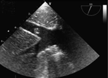
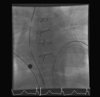
Subsequent intra-operative transesophageal echocardiography (Fig 1), visualised the sucker tip within inferior caval vein. The child was transferred to the catheterisation laboratory, where fluoroscopy (Fig 2) now showed it to be embedded in the pectinate muscles of the right atrium. Given its apparent stability within the atrium, a multi-disciplinary decision was made to leave the instrument part in situ.
Figure 1

Figure 2

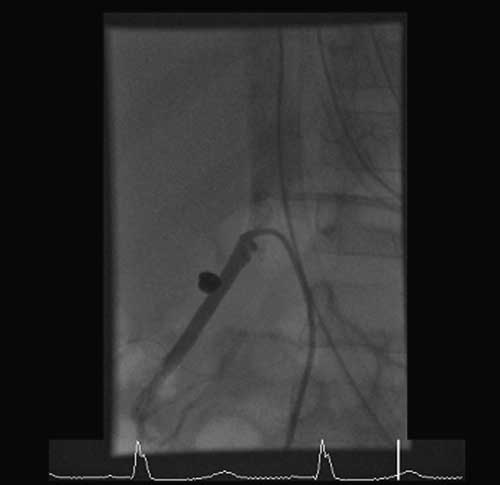
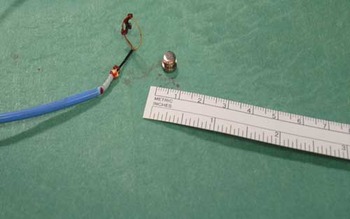
However, further X-rays on the night of operation showed subdiaphragmatic migration, and the patient was returned to the catheterisation laboratory. The flat sucker tip was retrieved from the right renal vein (Fig 3) using a 6 French Amplatzer® gooseneck snare (Fig 4) to manoeuvre it into the right femoral vein, from where it was extracted surgically. The patients’ recovery was otherwise uneventful.
Figure 3
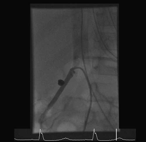
Figure 4

Device embolisation, a well-recognised problem in interventional cardiology, occurs infrequently in the operating room. Decision-making may be complex, taking into consideration the post-operative condition of the patient, possible pathways of embolisation, the object itself, and resources available for its retrieval. As shown in this patient, multi-disciplinary collaboration is essential, and a so-called “hybrid” combination of a transcatheter and surgical techniques may prove optimal.