Case report
A 19-year-old female patient was followed up for history of partial anomalous pulmonary venous return. As a newborn, a chest x-ray conducted for respiratory distress showed cardiomegaly with increased pulmonary vascular markings and an opacified density at the right paravertebral area. On examination, she had a significantly split S2; an electrocardiogram showed mild right ventricular hypertrophy with right-axis deviation, and an echocardiogram demonstrated a secundum atrial septal defect, a small persistent ductus arteriosus, and partial anomalous pulmonary venous return to the inferior vena cava.
Abdominal ultrasound revealed a solid mass with the same density as the hepatic tissue, located posterior and lateral to the inferior vena cava; it was contiguous to the liver without compressing it and produced focal displacement of the lung. Chest computed tomography showed a smaller right lung and confirmed the contiguous position of the mass to the liver. The findings were considered to be pseudo-sequestration owing to the focal herniation of the liver through thinning or focal defect in the diaphragm.
Cardiac catheterisation performed at 2 months of age showed a small secundum atrial septal defect, with partial anomalous venous return to the inferior vena cava with obstruction at the right lower pulmonary vein orifice size and calculated Qp/Qs of 1.3:1. After multidisciplinary discussions, it was decided that there was no indication for surgical correction of the anomalies. Subsequent echocardiograms revealed spontaneous closure of the atrial septal defect.
She had other anomalies including left esotropia, a large haemangioma of the dorsal aspect of her right foot and ankle, mild developmental delay, a didelphic uterus, chronic dysmenorrheoa, and left pelvic splenule. As a teenager she had neurocardiogenic syncope, which is now resolved, and she has been asymptomatic over the years with no increase in the frequency of respiratory infections or liver dysfunction.
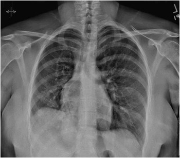
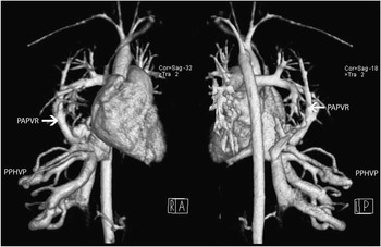
On her last visit, a chest radiograph (Fig 1) showed normal heart size and contour with dextroposition. The right costophrenic angle was obliterated, and a band-like density was noted at the posterior medial aspect of the right lung, most likely related to the anomalous vein draining into the inferior vena cava (Scimitar). Cardiac magnetic resonance imaging (MRI) (Fig 2) revealed the previously described partial anomalous pulmonary venous return and Scimitar syndrome, but further characterised her hepatic venous anomalies to be consistent with persistent primitive hepatic venous plexus. As the patient continued to be asymptomatic and there was no evidence of any haemodynamic consequences of her lesions, expectant management was recommended.
Chest radiograph shows normal heart size and contour with somewhat rightward deviation, probably related to the relatively smaller right lung. The right costophrenic angle was obliterated and a band-like density was noted at the posterior medial aspect of the right lung, most likely related to the anomalous vein draining into inferior vena cava (IVC) (Scimitar).

Cardiac magnetic resonance imaging (MRI) revealed the previously described partial anomalous pulmonary venous return (PAPVR) and Scimitar syndrome, but further characterised her hepatic venous anomalies to be consistent with persistent primitive hepatic venous plexus (PPHVP).

Discussion
The aetiology of persistent primitive hepatic venous plexus is largely unknown. Embryologically, the intrahepatic portion of the inferior vena cava is derived from the proximal portion of the right vitelline vein and the hepatic sinusoids. It is thought that the hepatic venous plexus may represent the precursor of the hepatic veins. Some suggest that the persistence of the foetal intrahepatic plexus leads to an abnormality in the development of the hepatic veins and infrahepatic portions of the inferior vena cava.Reference Blasco 1 – Reference MacDonald 3
Others theorise that the lower boundary of the hepatic segment of the inferior vena cava is the junction that unites the ductus venosus and the inferior vena cava; therefore, an interruption or underdevelopment of this structure may cause maldevelopment of the ductus venosus, which might result in the persistence of the hepatic, renal, and portal vein connections.Reference MacDonald 3
The embryological changes of Scimitar syndrome are also not well understood.Reference Hall, Hislop and Haworth 4 Pulmonary venous drainage is the product of an initial vasculogenesis from the splanchnopleural mesoderm followed by angiogenesis during the development of the intraacinar region. This process is mediated by the interaction of vascular endothelial growth factor and precursors such as Tenascin, and the expression of the sonic hedgehog morphogen. During the development of the lung bud, its primary blood supply changes from a plexus derived from the post-branchial descending aorta to the portion of the sixth aortic arch that becomes the pulmonary artery. Any insult during this period that culminates by the seventh week of gestation can cause persistence of systemic arterial supply to the right lung from the abdominal aorta, and the underdevelopment of the right pulmonary artery and right lung.Reference Hall, Hislop and Haworth 4
Persistent primitive hepatic venous plexus and Scimitar syndrome may remain undetected because of lack of symptoms; therefore, their true incidence is unknown.Reference Agnoletti, Bonnet and De Blic 2 Their association is uncommon and has been reported only a few times in the literature.Reference Blasco 1 – Reference MacDonald 3 , Reference Madan and Moore 5 Both lesions can present in isolation, but most commonly they are associated with other anomalies.Reference Blasco 1 – Reference MacDonald 3 , Reference Madan and Moore 5 – Reference Jolly, Kumar and Arora 9
The presentation is variable and depends on the haemodynamic significance of each lesion. In Scimitar syndrome, the patient may present with signs and symptoms of severe pulmonary hypertension, associated cardiac malformations, and large systemic collateral arteries feeding the right lung. Persistent primitive hepatic venous plexus is usually unsuspected as in most cases it is haemodynamically insignificant. It has occasionally been diagnosed at the time of femoral access during cardiac catheterisation, and in some cases it has also been found incidentally during imaging for other abnormalities.Reference Blasco 1 – Reference MacDonald 3 , Reference Madan and Moore 5
Review of the literature revealed five previously published cases of Scimitar syndrome associated with persistent primitive hepatic venous plexus.Reference Blasco 1 – Reference MacDonald 3 , Reference Madan and Moore 5 All cases were diagnosed by angiography. To the best of our knowledge, this is the first report of this rare combination of lesions imaged by cardiac MRI with three-dimensional reconstruction. A majority of the previously published case reports are female with a wide range of age at diagnosis (18 months–16 years).
Making the appropriate diagnosis can be important in patients with complex cardiac anatomy, who require palliation. The presence of persistent primitive hepatic venous plexus can also interfere with the management of other cardiovascular lesions – diagnostic catheterisations, percutaneous device placement, and cannulation during cardiac surgeries.Reference Agnoletti, Bonnet and De Blic 2 – Reference Hall, Hislop and Haworth 4 , Reference Gajinov 7
Complete diagnosis is achieved by a combination of several imaging techniques. Angiography had been the preferred modality to delineate persistent primitive hepatic venous plexus and Scimitar syndrome; however, more recent advances in cardiac computed tomography and magnetic resonance, especially with 3D reconstruction, allow a non-invasive diagnosis with similar or even better definition of the anatomy.Reference Agnoletti, Bonnet and De Blic 2 , Reference Gajinov 7 , Reference Bret-Zurita 8
There is no specific management of these entities. Usually associated lesions take precedence, and rerouting of venous return is performed when necessary. Coil embolisation of the venous fistula before or early after Kawashima operation has been described in patients with left atrial isomerism and complex congenital heart disease.Reference Gajinov 7
Financial Support
This research received no specific grant from any funding agency, commercial, or not-for-profit sectors.
Conflicts of Interest
None.