Introduction
During the latter part of the 1950s, it was discovered that selenium (Se) played an important physiological role in animal nutrition and that suboptimal concentrations often resulted in the appearance of myopathies (Schubert et al., Reference Schubert, Muth, Oldfield and Remmert1961).
Diets for ruminants are typically of plant origin, and as plant Se concentrations can be extremely variable, dietary Se supplements are generally required for ruminant diets. Selenium supplements are in two forms: inorganic mineral salts, such as sodium selenite (SS; Na2SeO3) or selenate (Na2SeO4), or organic forms, such as Se-enriched yeast (SY), in which selenomethionine (SeMet) is the predominant form of Se (Korhola et al., Reference Korhola, Vainio and Edelmann1986). However, the use of SY has only recently been authorised within the European Union (EU) after a series of tolerance studies established that animal health was not adversely affected by the administration of at least 10 times the maximum permitted concentration, a level thought to represent an accidental over dosage (Commission Directive 87/153/EC amended by 2001/79/EC). However, at the completion of such studies there are no such requirements to investigate the persistency of Se within the blood, visceral, and in particular edible tissues, of animals that could be intended for human consumption.
Studies with both dairy cows and beef cattle have shown that SY is more efficiently absorbed and assimilated than inorganic Se, as SeMet can be taken up via methionine transporter mechanisms within the gastro-intestinal tract and non-specifically incorporated into body proteins in place of methionine (Weiss, Reference Weiss2003), resulting in elevated concentrations of Se in both milk (Juniper et al., Reference Juniper, Phipps, Jones and Bertin2006) and body tissues (Qin et al., Reference Qin, Gao and Huang2007). Surai (Reference Surai2006) reported that SeMet is retained in tissue proteins to a greater extent than selenocysteine (SeCys) or Se derived from inorganic forms. A number of studies have detailed the accumulation of Se within the tissues of animals exposed to supranutritional concentrations of organic Se (Lawler et al., Reference Lawler, Taylor, Finley and Caton2004; Taylor, Reference Taylor2005) but very little information is available on either the persistency of Se or the distribution of selenised amino acids within the tissues of animals following the withdrawal of diets containing high doses of organic Se. This paper aims to redress this shortfall in information and reports on the concentration of total Se and the proportion of total Se comprised as either SeMet or SeCys in post mortem tissues of lambs following the withdrawal of high-dose SY (HSY) diets.
Material and methods
Animal, experimental design and diets
The work was conducted under the authority of the UK Animals (Scientific procedures) Act 1986 (Home office, 1986) and procedures were undertaken by staff holding appropriate licences authorities under the Act. All lambs were housed on straw bedding with sufficient space as prescribed by Home Office guidelines. Fresh potable water was available at all times.
Thirty Texel × Suffolk lambs, that had either been previously exposed to a diet supplemented with high dose of selenised yeast (Sel-Plex® (Saccharomyces cerevisiae CNCM, Collection Nationale de Culture de Micro-organism, I-3060), Alltech, Nicholasville, KY, USA) (HSY; 6.30 ± 0.18 mg Se per kg dry matter (DM)) or an unsupplemented control (C; 0.13 ± 0.01 mg Se per kg DM) during a 91-day period, were enrolled into the study. Lambs, with a mean age of 97 days and body weight of 43.8 ± 1.40 kg, were offered a complete pelleted diet that contained no additional Se over a period of 42 days. The complete pelleted ration (Dairy Direct, Bury St Edmunds, UK) contained 360, 340, 165, 90, 25 and 20 g/kg DM of ground barley, wheat bran, ground wheat, soya-bean meal, molasses and vitamin and mineral supplement, respectively, was offered fresh daily ad libitum (refusal maintained at 100 g/kg DM intake) following the removal of the previous day’s refusal.
Sampling procedures and measurements
Feed analyses
Representative samples of unsupplemented feed were taken weekly and frozen (−20°C). At the end of the study, feed samples were bulked on a weekly basis, sub-sampled and then analysed for nutritional composition and total Se content.
Oven DM concentration was determined by drying samples in a forced draught oven at 100°C for 24 h. Ash was obtained by incineration in a muffle furnace at 550°C for 16 h. Neutral-detergent fibre (NDF) concentration was determined by an ANKOM-Fibre analyser 200 (ANKOM-Technology, Fairpoint, NY, USA) by procedures described by Ministry of Agriculture Fisheries and Food (MAFF; 1993) using SDS and α-amylase solutions and corrected for residual ash (MAFF, 1982). Starch was determined by polarimetry (MAFF, 1982) with random samples analysed using the enzyme technique (MacRae and Armstrong, Reference MacRae and Armstrong1968) to verify the calibration of the polarimeter. Nitrogen concentration was measured by the Kjeldahl technique and water-soluble carbohydrate (WSC) concentration was measured spectrophotometrically. Oil-B was determined and metabolisable energy (ME) was estimated, using the E3 method, by MAFF (1993).
Blood and tissue sampling
At enrolment (PT0), and 21 and 42 days later (PT21 and PT42, respectively), five lambs from each treatment group were blood sampled and euthanased and a range of tissues sampled. Blood samples were taken via jugular venepuncture using the Vacutainer system (BD Diagnostics, Oxford, UK) using a 21-Gauge needle into a 5-ml lithium heparin-pretreated tube, for subsequent determination of total Se and Se species. Following euthanasia, samples of heart, liver, kidney and skeletal muscle (longissimus dorsi and psoas major) were retained to determine total Se and Se species.
Selenium analyses
Total Se for pelleted diets, blood samples and tissues (heart, liver, kidney and skeletal muscle tissue) was determined by mineralising 1 g of each sample in 4-ml HNO3 and 2-ml H2O2 within a closed-vessel heating block system. The solution was further diluted with water and Se was subsequently determined using inductively coupled plasma mass spectrometry (ICP-MS) (Elan 6100 ICPMS; Perkin Elmer, MA, USA).
Selenium speciation was determined using the method described by Palacios et al. (Reference Palacios, Encinar, Bertin and Lobinski2005). Briefly, samples were initially incubated for 5 h with dl-dithiothreitol and iodoacetamide to reduce and alkylate SeCys. Samples were then spiked with SeMet77 (SEMET Ltd, Rypin, Poland) and subsequently incubated for 24 h at 37°C with a mixture of protease and lipase maintained at a pH 7.5. Following incubation, the mixture was centrifuged and the supernatant separated and purified by cell exclusion liquid chromatography. Aliquots of the supernatant were analysed by reversed-phase HPLC-ICPMS using an ICP MS equipped with a collision cell (Elan 6100 ICPMS).
Statistical analysis
Statistically significant differences between the two treatment groups, C and HSY, for whole-blood Se content and post mortem tissue total Se content were determined by ANOVA using the GLM of SAS (v. 8.2; SAS Institute, Inc., Cary, NC, USA). Sources of variation within the model included treatment groups (1 d.f.) and time of sampling (1 d.f.). Statistical tests were undertaken for main effects and treatment × time interactions. Results are presented as least square means with the s.e. and level of significance.
Results and discussion
Feed analyses
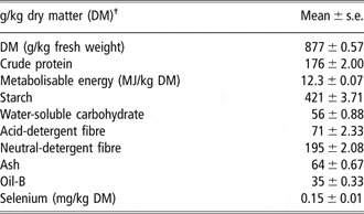
The nutritional composition of the pelleted diet is shown in Table 1. Mean values for DM, crude protein, starch, WSC, ADF, NDF, ash, oil-B and ME were 877 g/kg fresh weight, 176, 421, 56, 71, 195, 64, 35 g/kg DM and 12.3 MJ/kg DM, respectively. The mean total Se content of the diet was 0.15 ± 0.01 mg/kg DM.
Mean (± s.e.) laboratory-determined nutrient composition of experimental diets

†Unless otherwise stated.
Selenium content of blood and tissues
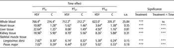
Total Se concentrations of whole blood, heart, liver, kidney and skeletal muscle (longissimus dorsi and psoas major) at PT0, PT21 and PT42 are shown in Table 2.
Mean (±s.e.) for total selenium concentrations in blood (ng/ml) and in heart, liver, kidney and muscle (psoas major and longissimus dorsi) (mg/kg dry matter ) of lambs that had been exposed to a diet supplemented with high dose of selenised yeast (HSY) or unsupplemented (C) after 0 (PT0), 21 (PT21) and 42 (PT42) days of withdrawal

a,b,c,dMean values within a row with different superscripts are significantly different (P < 0.05). Significance levels.
***P < 0.001; ns = P > 0.05.
Selenium concentration in whole blood
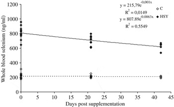
There were significant differences (P < 0.001) in whole-blood total Se concentration between treatment groups. At enrolment, HSY lambs had higher (P < 0.001) whole-blood Se concentrations than C (766.4 v. 216.4 ng Se per ml), as would be expected from animals that had been previously exposed to high doses of Se. These values were similar to those found in pregnant ewes (Davis et al., Reference Davis, McDowell, Wilkinson, Buergelt, Van Alstyne, Weldon and Marshall2006) and wether lambs (Cristaldi et al., Reference Cristaldi, McDowell, Buergelt, Davis, Wilkinson and Martin2005) that had received comparable Se doses in the form of SS. Similarly, whole-blood Se concentrations were still significantly higher (P < 0.001) in HSY animals at PT21 and PT42 when compared with C, although HSY whole-blood total Se values were seen to decline between successive time points (P < 0.001), whereas those of C remained relatively constant.
Trend line analysis of whole-blood total Se content over time is shown in Figure 1. Extrapolation of the resultant curve for whole-blood total Se concentration at PT0, PT21 and PT42 indicated that it would require approximately 210 days for whole-blood Se concentrations in HSY lambs to achieve similar concentrations to those of C lambs.
Trend line analysis of the relation between whole-blood total selenium content and days of sampling of lambs that had been exposed to a diet supplemented with high dose of selenised yeast (HSY) or unsupplemented (C).

Selenium concentration in tissues
Total Se concentrations in heart, liver, kidney and skeletal muscle (longissimus dorsi and psoas major) were significantly (P < 0.001) higher in HSY animals, irrespective of time, when compared with C animals. Tissue total Se concentrations of C animals did not differ appreciably between successive time points, whereas those of HSY animals declined, the rate of decrease varying between differing tissue types.
Among HSY animals, the concentration of total Se at PT0 was highest in the liver (22.64 mg/kg DM), which declined significantly to approximately 35% of PT0 values by PT21 and 20% by PT42). Similar Se concentrations to those of HSY animals at PT0 have been recorded in the liver of pregnant ewes (Davis et al., Reference Davis, McDowell, Wilkinson, Buergelt, Van Alstyne, Weldon and Marshall2006) and wether lambs (Cristaldi et al., Reference Cristaldi, McDowell, Buergelt, Davis, Wilkinson and Martin2005) that had received comparable doses as SS. The total Se content of kidney in HSY animals at PT0 was 18.96 mg/kg DM; this value was higher than those reported for pregnant ewes (Davis et al., Reference Davis, McDowell, Wilkinson, Buergelt, Van Alstyne, Weldon and Marshall2006) and wether lambs (Cristaldi et al., Reference Cristaldi, McDowell, Buergelt, Davis, Wilkinson and Martin2005), indicating that Se source may influence Se retention between different tissues. The concentration of total Se in kidney declined over time to 46% of PT0 values by PT21 and 43% by PT42. Total Se concentrations in both cardiac and skeletal muscle of HSY animals at PT0 were 10.8 and 7.42 mg Se per kg DM, respectively. At PT21 and PT42, there was a marked reduction in total Se within cardiac tissue (50% and 34% of PT0 values, respectively). However, skeletal muscle did not show the same rate of Se clearance, with Se values at PT21 being approximately 90% of PT0 and 70% at PT42. These results indicate that Se clearance rates were greatest in glandular visceral tissues, namely the liver and kidney.
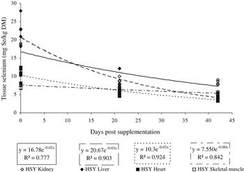
Trend line analysis of the relation between whole-blood and tissue total Se is shown in Figure 2. Extrapolation of the resultant curve for whole-blood Se concentration and the concentration of Se within individual tissue types indicated that the Se content of kidney, liver and heart of HSY animals would decline to those of C animals after 55, 70 and 80 days post supplementation, respectively. However, in contrast regression trend line analysis indicated that skeletal muscle tissue would require considerably longer, with an estimate of approximately 380 days, following the withdrawal of HSY supplementation, to achieve similar Se concentrations to those of C group animals, with average Se clearance rates of 307, 238, 120 and 20 μg Se per day for liver, kidney, heart and skeletal muscle, respectively.
Changes in tissue selenium concentrations in the 42 days period following the withdrawal of long-term high-dose administration of selenised yeast (HSY).

Selenised amino acids contents of whole blood and tissues
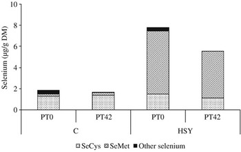
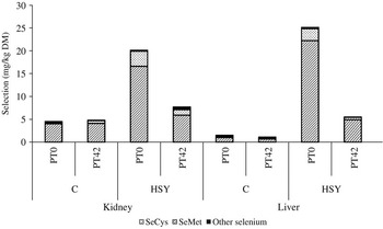
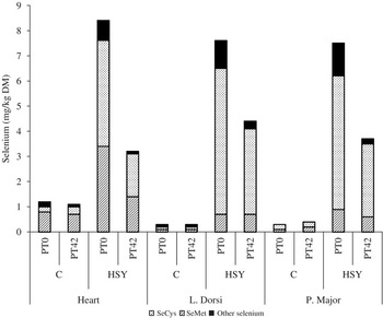
Total Se and the proportion of total Se comprised as either SeMet, SeCys or as other unidentified Se fractions, for whole blood, visceral and muscle tissues are shown in Figures 3, 4 and 5, respectively.
Whole-blood total selenium and proportion of total selenium as selenomethionine (SeMet), selenocysteine (SeCys) and other selenium species of lambs at 0 (PT0) and 42 days (PT42) days after exposure to a diet supplemented with a high dose of selenised yeast (HSY) or an unsupplemented control (C).

Total selenium and proportion of total selenium comprised as selenomethionine (SeMet), selenocysteine (SeCys) and other selenium species in the kidney and liver of lambs that had been exposed to a diet supplemented with high dose of selenised yeast (HSY) or unsupplemented (C) after 0 (PT0) and 42 (PT42) days of withdrawal.

Total selenium and proportion of total selenium comprised as selenomethionine (SeMet), selenocysteine (SeCys) and other selenium species in the muscles of lambs that had been exposed to a diet supplemented with high dose of selenised yeast (HSY) or unsupplemented (C) after 0 (PT0) and 42 (PT42) days of withdrawal.

Selenised amino acids content of whole blood
Speciation of Se fractions showed that much of the large difference in total blood Se concentrations in HSY animals was due to the greater proportion of total Se comprised as SeMet, probably attributable to a combination of the high concentration of SeMet in the SY product (SeMet comprises 54 to 74% of total Se; Rayman, Reference Rayman2004) and the uptake of SeMet from the gastro-intestinal tract via Methionine transporter mechanisms (Weiss, Reference Weiss2003) during the period of supplementation. Furthermore, decreases seen in whole-blood total Se between successive time points was primarily the result of decreases in the proportion of total Se comprised as SeMet, because the proportion of total Se comprised as SeCys was similar between both treatment groups and sequential time points.
Selenised amino acids contents of tissues
The proportion of total Se comprised as either SeMet or SeCys in the tissues of HSY animals was markedly different, irrespective of time point, from those of controls.
Among C animals, the main form of Se in the kidney, liver and heart was SeCys, which comprised 90%, 60% and 60% of total Se, respectively, while in skeletal muscle tissue, there was little difference in the proportions of SeMet and SeCys, which formed approximately 35% of total Se. The differences between cardiac and skeletal muscles in the proportion of total Se comprised as SeCys reflects the relative activity of each muscle type. Active tissues, such as cardiac muscle, are much more likely to be subjected to oxidative damage and the higher SeCys content of cardiac tissue could be indicative of greater antioxidant-specific tissue selenoenzyme activity.
In the tissues of HSY animals, the main selenised amino acid in kidney and liver was SeCys, whereas cardiac and skeletal muscle comprised mainly SeMet. At PT42, SeCys still remained the predominant selenised amino acid within kidney and liver, although much of the reduction in total Se contents within these tissues were attributable to reductions in the proportion of total Se comprised as SeCys. Similarly, SeMet was the predominant Se species within heart and skeletal muscle at PT42; however, the reduction in total Se in cardiac tissue was the result of similar decreases in the proportion of total Se comprised as both SeMet and SeCys, whereas reductions in the total Se content of skeletal muscle were largely attributable to changes in the proportion of total Se comprised as SeMet.
It would therefore appear that the rates of Se clearance observed are dependent on the Se species present, as clearance rates are faster when SeCys is the predominant form of selenised amino acid, and that Se species are dependent on tissue type. Glandular visceral tissues, such as kidney and liver, are both metabolically active tissues and exhibited greatest Se clearance rates. In contrast, heart and skeletal muscle are much less metabolically active and have much lower Se clearance rates. The abundance of SeCys in more metabolically active tissues is maybe the result of the conversion of SeMet to SeCys via the actions of selenocystathionine, as a large proportion of dietary Se was SeMet in origin. This pool of endogenous SeCys could then be available for conversion to selenide and either the subsequent incorporation into selenoenzymes via UGA codon or excretion via methylation (Aspila, Reference Aspila1988; Suzuki and Ogra, Reference Suzuki and Ogra2002). However, neither tissue selenoenzyme activity nor the appearance of methylated Se species within urine or exhaled breath was determined during the course of this study and, as such, it is not possible to state the ultimate fate of the high concentrations of endogenous Se.
Conclusions
Selenium clearance rates in the tissues of animals that had received long-term exposure to high dietary concentrations of SY appear to be dependent on Se species. Tissues that are more metabolically active have a larger proportion of total Se comprised as SeCys and faster Se clearance rates when compared with tissues with a lower metabolic rate in which SeMet is the predominant form of Se. Regression analysis indicated that it would require a period well in excess of 300 days for the Se contents of muscle tissue in HSY lambs to achieve those of C animals, a period much longer than the usual finishing time for commercial lambs. Consequently, Se intake in humans consuming such lambs could most probably exceed recommended levels of Se intake.
Acknowledgements
This research was funded by Alltech EU Regulatory Affairs Department (trial ref. SEL/TOL/LAM/01/0205/UK). The authors would also like to give special thanks to Prof. Lobinski (UT2A laboratories) for performing selenium analyses.