Aortico–left ventricular tunnel is a rare cardiac anomaly, 0.001–0.1% of the CHD, presenting an extracardiac channel that connects the ascending aorta to the left ventricle. Reference McKay1 The timing of surgery varies, however, surgery early in the birth is rare. An aortico–left ventricular tunnel is classified as type IV by Hovaguimian and colleagues. Type IV is poor prognosis because of heart failure due to residual aortic regurgitation. Reference Hovaguimian, Cobanoglu and Starr2 We report a case of a large type IV aortico–left ventricular tunnel successfully repaired at 1 day after birth.
Case report
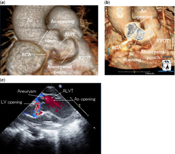
A 32-year-old pregnant woman at 30 weeks of gestation was referred to us with a fetal diagnosis of ALVT. Fetal echocardiography revealed the left ventricular dilatation and severe regurgitation via the ALVT. The cardiothoracic area ratio was 50%. The fetus survived, and the baby boy was delivered at 36 weeks of gestation by caesarean section with a birth weight of 2796 g. The patient was intubated immediately. Transthoracic echocardiography and enhanced CT confirmed the diagnosis. The ALVT formed an aneurysmal pouch on the right ventricular outflow (Fig 1a). The aortic opening is 10 mm and the LV opening is 6.8 mm. The orifice of right coronary artery was adjacent to the ALVT (Fig 1a,b). The regurgitant flow entered the ALVT towards the cardiac apex, turned around in the blind pouch, and then entered into the LV via the LV opening at the LV outflow (Fig 1c). Despite the large ALVT, the patient’s condition was stabilised by respiratory and inotropic support, therefore, the surgery was performed at 1 day (25 hours) after birth.
Preoperative three-dimensional CT and preoperative transthoracic echocardiogram. (a) The aortico–left ventricular tunnel formed an aneurysmal pouch on the right ventricular outflow tract. (b) The ventricular opening was located just underneath the right coronary cusp of the aortic valve. (c) Parasternal long-axis view of the severe aortic regurgitation via the aortico–left ventricular tunnel. ALVT = aortico–left ventricular tunnel; Ao = ascending aorta; LV = left ventricle; RCA = right coronary artery; RVOT = right ventricular outflow tract; STJ = sinotubular junction.

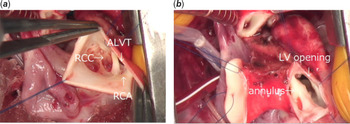
The heart was exposed through a median sternotomy. A patent ductus arteriosus was ligated. The aneurysmal ALVT was identified over the RV outflow tract. Cardiopulmonary bypass was instituted, and cardiac arrest was achieved using antegrade cardioplegia infusion with compression of the ALVT. The ascending aorta was transected, and the aortic valve was examined. There was a large aortic opening at the sinotubular junction of the right coronary sinus, 10 mm in diameter. The orifice of right coronary artery was adjacent to the aortic opening (Fig 2a). The ventricular opening, 10 × 5 mm in size, was located just underneath the right coronary cusp of the aortic valve. The right coronary cusp of the aortic valve was free floating and barely supported by a thin fibrous bar at the annulus. The ALVT extended beyond the LV opening towards the right ventricular apex to form a blind pouch, some 20 mm in depth. The blind pouch was entered longitudinally, and the LV opening was well in sight (Fig 2b). The aortic and left ventricular openings of the ALVT were closed with separate glutaraldehyde-treated pericardial patches using 7-0 polypropylene running sutures, via aortotomy suturing to the thin annular bar and the aortic wall, and via the pouch incision suturing to the thin annular bar and the summit of the ventricular septum, respectively. Obstruction of the right coronary orifice was carefully avoided. Antegrade cardioplegia was infused after the aorta was closed, and no leak into the ALVT was confirmed. Finally, the aneurysmal pouch of ALVT was plicated longitudinally to support the right coronary cusp of the aortic valve. The aortic clamp times were 96 minutes. The left ventricular function was good with catecholamine supports, weaning from cardiopulmonary bypass was smooth and the sternum was closed. Post-operative echocardiography at discharge demonstrated no leak into the ALVT and trivial aortic regurgitation, and the patient was discharged 1 month after the operation.
Surgical anatomy of the aortico–left ventricular tunnel. (a) The orifice of right coronary artery was adjacent to the aortic opening. (b) The ventricular opening was located just underneath the right coronary cusp of the aortic valve. ALVT = aortico–left ventricular tunnel; LV= left ventricle; RCA = right coronary artery; RCC = right coronary cusp.

Discussion
It has been reported that most of the patients with ALVT lead to heart failure within 1 year of life, and left ventricular function does not normalise without treatment within half a year Reference McKay1 . The reported maximum diameter of the ALVT in the cases of surgery in the neonatal period was 7 mm in the literature. Reference Nakamura, Kagami and Takeshi3 The larger the ALVT, the more unlikely it to survive. The diameter of ALVT was 10 mm in the present case, a far larger than the aortic valve diameter. The reason for the patient’s preoperative survival seemed the blind pouch formation in the ALVT that prevented a direct blood flow from the aorta to the LV.
ALVT is classified type I (A simple tunnel with a slitlike), type II (A tunnel with extracardiac aortic wall aneurysm), type III (A tunnel with intracardiac aneurysm of septal portion) and type IV (Type II and III combined). For type III and IV, there is a risk of right ventricular outflow stenosis due to enlargement of the aneurysm when only the aortic opening is closed. Reference Hovaguimian, Cobanoglu and Starr2 Concern exists that direct suture closure of the aortic opening may distort the aortic valve cusp and does not provide adequate support for the valve. Thus, patients may be more prone to long-term development of aortic regurgitation. The aortic regurgitation is a major long-term complication after surgery, requiring valve replacement in as many as 50% of reported cases. Reference Serino, Andrade, Ross, de Leval and Somerville4 Therefore, the two-patch repair is recommended, however, in the case of neonatal surgery, there were many reports of one patch repair because of surgical difficulties. In the present case, incision of the large ALVT allowed a good vision of the left ventricular opening and the aortic valve, and a successful two-patch repair. The thin right coronary cusp annulus was supported by the patches at the aortic and left ventricular openings, and is reinforced by longitudinal plication of the aneurysmal pouch of ALVT. Following up 1 year after the operation, echocardiography and angiography demonstrated only trivial aortic regurgitation.
Acknowledgements
None.
Conflict of interests
None.
Ethical standards
This article does not contain any studies with human participants or animals performed by any of the authors.