I. INTRODUCTION
Magnetoelectrics semiconductors exhibiting both ferromagnetic and ferroelectric properties, which have been investigated for use as optical modulators, memories, computing using electron spins, are experiencing a renewal of interest (Roulland et al., Reference Roulland, Lefevre, Thomasson and Viart2013). Historically, wide bandgap semiconductor materials, such as SiC (Song et al., Reference Song, Bao, Li, Lei, Peng, Jian, Liu, Wang, Wang and Chen2009; Liu et al., Reference Liu, Wang, Wang, Yang, Chen, Qin, Song, Wang and Chen2011), In2O3 (Kotsikau and Ivanovskaya, Reference Kotsikau and Ivanovskaya2015), ZnO (Singh et al., Reference Singh, Dey, Roy and Mandal2014), AlN (Li et al., Reference Li, Bao, Song, Wang and Chen2008), have been widely investigated for their structural, electric, and magnetic properties. Recently, owing to a wide bandgap of ~4.9 eV and chemical and thermal stabilities, Ga2O3 is considered as one of the ideal candidates to fabricate magnetoelectric devices (Guo et al., Reference Guo, Wu, An, Li, Wang, Chu, Guo, Zhi, Lei, Li and Tang2015a). Our group has fabricated the Ga2O3/(Ga1−xFex)2O3 multilayer thin films and investigated their magnetic properties (Guo et al., 2016). Remeika demonstrated that GFO exhibited a single phase with Fe content lower than 1.4 for the first time (Remeika, Reference Remeika1960). It also has been reported that the GFO presents both magnetic and electric properties in a single phase, which makes it suitable for a wide range of applications in electronic devices (Frankel et al., Reference Frankel, Blum and Foner1965; Atanelov and Mohn, Reference Atanelov and Mohn2015). However, up to now, there are few journal publications related to the structural, optical, and magnetic properties of GFO with an emphasis on correlation of iron content with properties.
In this work, we fabricated Ga2−xFexO3 (GFO) compounds for x = 0.7–1.3 using a classic solid-state method, and systematically investigated their structural, optical, and magnetic properties. The space group was confirmed and the lattice parameters have been refined using FULLPROF profile fitting of the X-ray powder diffraction data.
II. MATERIALS AND METHODS
All GFO bulk samples (Bulks here after) were prepared using a classical solid-state route using high-purity commercial powders: α-Fe2O3 (99%) and β-Ga2O3 (99%). Initially, powders of α-Fe2O3 and β-Ga2O3 were mixed in a stoichiometric ratio to obtain the composition of GFO. The mixed powder was manually ground for 30 min to prepare a homogeneous mixture using agate mortar and pestle. Then, the powder was sintered in the air at 1350 °C for 7 h. The heated sample was converted into a powder form. Next, the powder mixture was sintered in the air at 1350 °C for 7 h again and then well ground. The obtained powders were pressed into a pellet of 10 mm in diameter under uniaxial pressure of 8 MPa for 10 min.
The element composition of the samples was measured by energy-dispersive spectroscopy (EDS), and the nanostructures were investigated using scanning electron microscopy (SEM). Room temperature X-ray diffraction (XRD) spectra of the samples were taken using CuKα radiation (λ = 1.540560 Å) in the 2θ range of 15–85°. The valences of Fe ions and element distribution were analyzed by X-ray photoelectron spectroscopy (XPS). The diffuse reflectance was measured using a Hitachi U-3900 ultraviolet–visible (UV–vis) spectrophotometer equipped with an integrating sphere. The magnetic measurements were carried out using a vibrating sample magnetometer attached to the physical property measurement system (PPMS).
III. RESULTS AND DISCUSSION
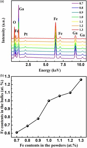
Figure 1(a) presents the EDS spectra of GFO bulks with different Fe content x from 0.7 to 1.3. From Figure 1(a), elements of Ga, Fe, O, and C are observed; the appearance of Pt is because of the spray process required for testing and no other elements are detected. It is obvious that the intensity ratio of Fe/Ga increases up to the Fe content x of GFO bulks. The Fe content x in GFO bulks obtained from the EDS spectra is shown in Figure 1(b) as a function of Fe content x in the GFO bulks. From Figure 1(b), the Fe content x in the bulks increases almost linearly with the composition of Fe content x in the GFO powders. The results suggest that the Fe content in the bulks can be controlled by adjusting the Fe content in the powders, opening the possibility of designing GaFeO3-based devices.
(Color online) Results of EDX measurement of Ga2−xFexO3 bulks. (a) EDS spectra of Ga2−xFexO3 bulks with different Fe content with x = 0.7, 0.8, 0.9, 1.0, 1.1, 1.2, and 1.3; (b) Fe content x in GFO bulks as a function of Fe content in the powders.

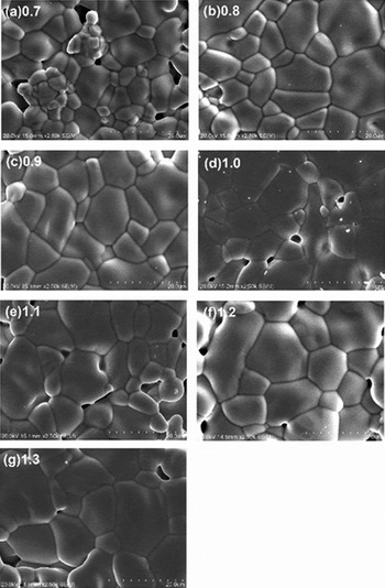
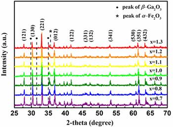
In order to further insight into the microstructure properties of the GFO bulks, we measured surface morphology by SEM. Figures 2(a)–2(g) show the typical SEM images of the GFO samples with x = 0.7, 0.8, 0.9, 1.0, 1.1, 1.2, and 1.3, respectively. The images reveal that the nanoparticles are arranged in a uniform manner having clear boundaries, well-defined, and distinct shapes. The grain size becomes larger with the increase of x. Figure 3 shows the XRD patterns of GFO with x range from 0.7 to 1.3. For x = 1.0, peaks appear at 27.84°, 30.27°, 33.04°, 36.61°, 41.68°, 46.08°, 47.18°, 53.15°, 60.55°, 61.39°, and 63.46°, which can be assigned as (121), (130), (221), (012), (122), (331), (132), (341), (530), (351), and (432) of spinel GaFeO3 (ICDD-PDF # 01-076-1005). The peaks of GFO shift to smaller 2θ with the increase of x, it is because that the larger ion radius of Fe3+ (~0.67 Å) compared with that of Ga3+ (~0.62 Å). The incorporation of Fe into Ga2O3 in a form of Fe3+ substituting for Ga3+ leads to the expansion of lattice constants of Ga2O3. A few additional peaks were noted and (*, ·) marked in the spectra. As shown in Figure 3, when Fe content x is lower than 0.8, three different diffraction peaks located at 29.9°, 31.6°, and 35.0°, which can be assigned as the (400), (002), and (111) planes of monoclinic Ga2O3, respectively. As x increases to be 1.3, a peak related to Fe2O3 begins to appear at 35.7°, indicating that the phase separation appeared here.
SEM images of the Ga2−xFexO3 bulk with x = 0.7, 0.8, 0.9, 1.0, 1.1, 1.2, and 1.3. (a) x = 0.7, (b) x = 0.8, (c) x = 0.9, (d) x = 1.0, (e) x = 1.1, (f) x = 1.2, (g) x = 1.3.

(Color online) X-ray diffraction pattern of Ga2−xFexO3 bulk with x = 0.7, 0.8, 0.9, 1.0, 1.1, 1.2, and 1.3.

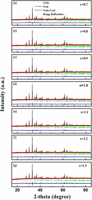
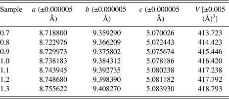
FULLPROF profile fitting was carried out to analyze the structure data. Cell parameters [lattice constants: a, b, c, and cell volume (V)] of the samples have been calculated from profile fitting of XRD patterns using FULLPROF program and the values are shown in Table I. We find that the lattice parameters and cell volume of the samples increase with the increase of Fe content x, indicating the successful doping of Fe ions. The corresponding generated patterns along with the observed data are shown in Figure 4. From the profile fitting, it is observed that GFO belongs to the orthorhombic structure with the space group Pc21n. The lattice parameters extracted from the profile fitting, namely, a = 8.7 ± 0.1 Å, b = 9.4 ± 0.1 Å, and c = 5.1 ± 0.1 Å, are found to be the same for all the samples within the experimental errors (Atanelov and Mohn, Reference Atanelov and Mohn2015).
(Color online) Results of profile fitting of Ga2−xFexO3 with x = 0.7, 0.8, 0.9, 1.0, 1.1, 1.2, and 1.3 using the FULLPROF program. (a) x = 0.7, (b) x = 0.8, (c) x = 0.9, (d) x = 1.0, (e) x = 1.1, (f) x = 1.2, (g) x = 1.3. The circles are the observed data (Y obs), the solid line is 95 the calculated pattern (Y cal), the bottom line is the difference between the observed and calculated patterns (Y obs−Y cal), and the vertical bars are the Bragg reflections.

Cell parameters (a, b, c, V) of different samples calculated from profile fitting of XRD patterns using FULLPROF program.

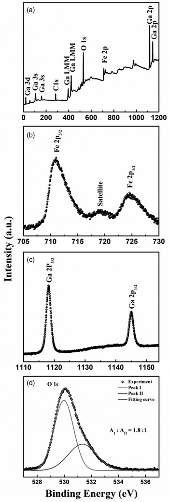
In order to analyze the valences of ions and element distribution in the bulks, we chose GFO with x = 1.0 as a typical sample. XPS spectra of GaFeO3 are shown in Figures 5(a)–5(d). From Figure 5(a), elements of Ga, Fe, O, and C are observed, and no other elements are detected from the wide scan spectra. The C–C/C–H, C 1s peak is located at the binding energy of 284.6 eV as the standard reference. The Fe 2p spectra obtained from the reference sample is shown in Figure 5(b). The binding energies of Fe 2p 3/2 and Fe 2p 1/2 obtained from the present study are 711.05 and 724.74 eV, respectively, which have been reported by many researchers (Abrahams et al., Reference Abrahams, Reddy and Bernstein1965). Of the two peaks, Fe 2p 3/2 is narrower and stronger than Fe 2p 1/2 and the area of Fe 2p 3/2 peak is greater than that of Fe 2p 1/2 because in spin–orbit (j–j) coupling (Muhler et al., Reference Muhler, Schuetze, Wesemann, Rayment, Dent and Schloegl1990). The satellite peak of Fe 2p 3/2 is located ~8 eV higher than the main Fe 2p 3/2 peak, which is clearly distinguishable and does not overlap either the Fe 2p 3/2 or Fe 2p 1/2 peak. In Figure 5(c), it is clear that two symmetrical peaks of Ga 2p 3/2 and Ga2p 1/2 are located at 1118.1 and 1145.0 eV, respectively. The separation energy between these two peaks is about 27 eV, which is in good agreement with the reported values (Xiao et al., Reference Xiao, Ma, Xue, Zhuang, Ma and Zong2007; Yamashita and Hayes, Reference Yamashita and Hayes2008). As is shown in Figure 5(d), the O 1s core-level peaks can be divided into two components: I and II, located at 529.98 and 531.34 eV, respectively. The peak I is assigned to lattice oxygen ions in the GFO bulks, and the peak II is assigned to the oxygen ions in the oxygen vacancies region as well as C/O or OH− adsorbed species on the surface (Scott et al., Reference Scott, Ross, Araujo, Scott and Huffman1996; Xu et al., Reference Xu, Liu, Sun, Liu, Han and Wang2008; Guo et al., Reference Guo, Wu, Li, Wang, Lei, Li and Tang2015b, Reference Guo, Wu, Zhang, Yang, Hu, Lei, Li, Li and Tangc). The peak ratio of I/II is about 1.8/1, implying the existence of abundant oxygen vacancies in the GFO bulks.
XPS spectra of the GaFeO3: (a) wide scan, (b) Fe 2p, (c) Ga 2p, (d) O 1s.

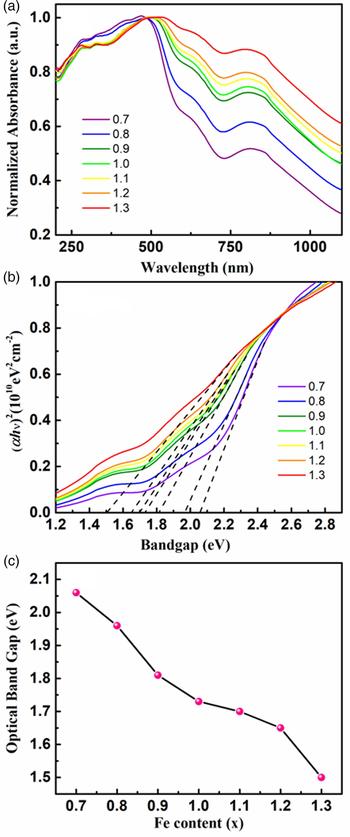
Figure 6(a) shows the optical absorbance spectra for GFO with x from 0.7 to 1.3 as a function of wavelength in the range of 200–1200 nm. It is clear that GFO bulks show sharp absorption edges at 602 nm, which shift toward longer wavelength with the increasing Fe content x. It is well known that for the bulks with direct bandgap, the absorption follows a power law of the form: (αhν)2 = A(hν−E g), where α is the absorption coefficient, hν is the energy of the incident photo, A is the absorption edge width parameter, and E g is the bandgap energy. The plot of (αhν)2 as a function of the bandgap is shown in Figure 6(b). As can be seen in Figure 6(c), we can find that the bandgaps decrease almost linearly with the increase of Fe content. The fact suggests that the bandgap of GFO can be controlled by adjusting the Fe content in the bulks, which is of great importance for future application. The bandgaps for GFO with x = 0.7–1.3 are obtained within 2.06–1.51 eV, which is consistent with the previous works (Kalashnikova et al., Reference Kalashnikova, Pisarev, Bezmaternykh, Temerov, Kirilyuk and Rasing2005; Thomasson et al., Reference Thomasson, Ibrahim, Lefevre, Autissie, Roullan and Mény2013). The decrease of the bandgap is ascribed to new unoccupied electron states in the gap below the conduction band edge because of the incorporation of Fe ions on the substitutional sites of Ga2O3 (Guo et al., Reference Guo, An, Cui, Zhi, Zhao, Lei, Li, Li, Wu and Tang2016).
(Color online) (a) UV–vis absorption spectra of Ga2−xFexO3 with x = 0.7, 0.8, 0.9, 1.0, 1.1, 1.2, and 1.3; (b) the plot of (αhν)2 vs. photon energy (hν) to calculate E g; (c) bandgap along with the Fe content.

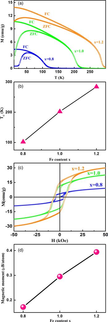
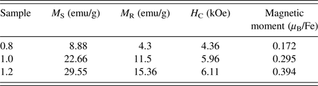
Thermal variation of the magnetization (M–T) data measured in 500 Oe in the range of 300–5 K applied magnetic field GFO is shown in Figure 7(a). The zero field cooled (ZFC) magnetization data are obtained by cooling the sample in the absence of field from 300 K down to 5 K and the magnetization is measured during the increase of temperature. In a similar way, the field cooled (FC) magnetization data of the sample are measured in the presence of magnetic field to 5 K and then the corresponding data are recorded in warming. The M–T data show very interesting features such as bifurcation between the FC–ZFC data and cusp in the ZFC data. Another noteworthy point to be observed in the M–T data as shown in Figure 7(a) is the non-monotonous behavior of ZFC curve as a function of temperature, especially in the low-temperature range. These phenomena were also found in other polycrystalline, which shows that these features are indeed because of the anisotropic nature of GFO and not because of any spin-freezing/spin-blocking phenomenon (Arima et al., Reference Arima, Higashiyama, Kaneko, He, Goto and Miyasaka2004; Wu et al., Reference Wu, Bai, Hu, Guo, Sun, Ji, Lei, Li, Li, Hao and Tang2015). The Curie temperature (T C), namely the magnetic transition temperature, is obtained as the differential maximum of FC curves. The T C value vs. Fe content is shown in Figure 7(b). As can be seen, T C appears to be 102, 202, and 284 K for x = 0.8, 1.0, and 1.2, respectively. The value of T C shifts to the higher temperatures with the increase of Fe content, which is consistent with the previous works (Han et al., Reference Han, Ozaki and Yu2007; Sharma et al., Reference Sharma, Raghavendra, Gupta, Banerjee and Awasthi2013). Figure 7(c) shows the magnetization vs. magnetic field (M–H) curves of GFO bulks at 4 K. Different magnetic quantities, saturation magnetization (M S), remanent magnetization (M R), coercive field (H C), are obtained from the displayed loops. Along with the relevant quantities extracted from different loops, all these data are shown in Table II. As can be seen from Table II, coercive field and saturation magnetization both get improved monotonically with the increase of x, revealing apparent enhancement of ferromagnetism with the increase of Fe content. Magnetization increases with the increase of field slowly, which may attribute to the mixture of ferromagnetism and antiferromagnetism (Levine et al., Reference Levine, Nowlin and Jones1968; Trassin et al., Reference Trassin, Viart, Versini, Loison, Vola and Schmerber2007). Meanwhile, the magnetic moment of each sample is calculated and shown in Figure 7(d). The magnetic moments obtained to be 0.172, 0.295, and 0.394 µB atom−1 for x = 0.8, 1.0, and 1.2, respectively. As can be seen, the magnetic moment increases with the increase of x. To explain those results, the changes of interaction force between Ga and Fe or Fe and Fe atoms depending on the Fe concentration were expected to play an important role (Arima et al., Reference Arima, Higashiyama, Kaneko, He, Goto and Miyasaka2004). None of the samples is saturated even with the application of a magnetic field of 50 kOe, indicating the high magnetic anisotropy value in these samples.
(Color online) Results of PPMS measurement of Ga2−xFexO3 bulks with various Fe content of x = 0.8, 1.0, and 1.2. (a) Dependency of magnetization on temperature measured in the FC and ZFC conditions, (b) the Curie temperatures (T C) vs. various Fe content, (c) the magnetization vs. magnetic field (M–H) curves at 4 K, (d) the magnetic moment vs. various Fe content.

Different magnetic parameters and magnetic moment of Ga2−xFexO3 nanoparticles obtained from the M–H loops.

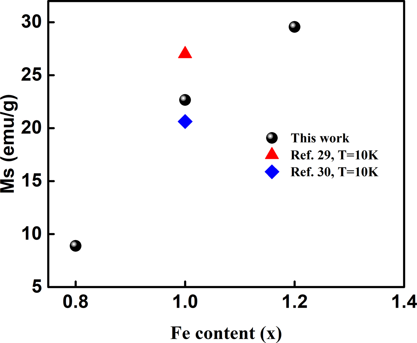
In this work, M S of GFO is 8.88, 22.66, and 29.55 emu g−1 when x = 0.8, 1.0, and 1.2 in 4 K, respectively. Figure 8 summarizes the M S vs. Fe content of GFO in this work and other reported work. Compared with other results (Naik and Mahendiran, Reference Naik and Mahendiran2011; Han et al., Reference Han, Chen and Lee2013), the M S of our work is relatively large. The origin of magnetic properties depends on many parameters, such as the concentration and distribution of the TM ions, type and concentration of defects, n-type doping, p-type doping, the morphology of the host lattice, etc.; whereas many groups have reported that the defects, especially oxygen vacancies, play a dominant role in inducing FM (Kaneko et al., Reference Kaneko, Kakeya, Komori and Fujita2013; Mukhopadhyay et al., Reference Mukhopadhyay, Mahapatra and Chakrabarti2014). Considering the XPS results of O 1s core level in Figure 5(d), the peak ratio of I/II is about 1.8/1, implying the existence of abundant oxygen vacancies in the GFO bulks. It is inferred that the appearance of FM for all films is related not only to the role of Fe dopant but also to the oxygen vacancies.
(Color online) Comparison of the M S of Ga2−xFexO3 in this work with other reported results.

IV. CONCLUSIONS
In conclusion, we fabricated GFO bulks with x from 0.7 to 1.3 using the classic solid-state route, and systematic investigation of the structural, optical, and magnetic properties was carried out. EDS results reveal that the Fe content in the bulks increases almost linearly with the composition of x in GFO. The results suggest that the Fe content in the bulks can be controlled by adjusting Fe2O3 in the mixed powders. XRD spectra and FULLPROF profile fitting indicated that GFO bulks belong to the orthorhombic structure with the space group Pc21n. Phase separation appeared at the Fe content of x = 1.3, which is of vital importance for the application of ferromagnetic semiconductors. The characterization by XPS demonstrated that GFO bulks were composed of Ga, Fe, O elements. UV–vis absorbance spectra confirmed the incorporation of Fe into Ga2O3 and revealed its effects on optical properties. Magnetic property measurements suggested that GFO is ferromagnetic, and the magnetic properties have been enhanced compared with other reported works.
SUPPLEMENTARY MATERIAL
The supplementary material for this article can be found at https://doi.org/10.1017/S0885715618000374
ACKNOWLEDGEMENTS
This work was supported by the National Natural Science Foundation of China (Grant Nos. 11404029, 51572033, 61774019, 51172208), and the Fund of State Key Laboratory of Information Photonics and Optical Communications (BUPT).