


Highlights
-
Patients with CRAO present within hours of vision loss but have not been treated with thrombolysis in our center.
-
Delay in ophthalmological consultation and lack of defined CRAO treatment pathway may be barriers to care.
-
Patients with CRAO frequently have high-risk underlying pathology and uncommonly experience functional visual recovery.
Introduction
Central retinal artery occlusion (CRAO) is an ischemic stroke syndrome characterized by acute, painless and generally profound vision loss with low rates of functional recovery. Reference Mac Grory, Schrag and Biousse1,Reference Quinn2 Like cerebral stroke, CRAO is a medical emergency given its associations with life- and vision-threatening conditions and the risk of additional ischemic events. Reference Scoles, McGeehan and VanderBeek3,Reference Wai, Knapp and Ludwig4 In recent years, multidisciplinary guidelines from American organizations have proposed optimal care for patients presenting with acute vision loss from CRAO. Reference Mac Grory, Schrag and Biousse1,Reference Grzybowski and Kanclerz5 While the 2022 update to Canadian Stroke Best Practice Recommendations (CSBPR) does include limited reference to acute retinal ischemia, Reference Lindsay, Mountain, Gubitz, Dowlatshahi, Casaubon and Smith6 specific treatment recommendations for CRAO are not provided, and there are no comprehensive Canadian guidelines for management of this condition.
Recently, observational studies from American and European centers have sought to characterize patterns in patient presentation and management practices for acute CRAO. Reference Youn, Lavin and Patrylo7–Reference Shah, Gilbert and Melles12 Such studies are an important first step in identifying opportunities to improve care for patients with CRAO; however, Canadian data are limited to a single series of 27 patients. Reference Smith, Benson, Tennant and Jivraj13 We carried out a descriptive study of patients presenting with CRAO to one of Canada’s largest academic health sciences centers. Our study objective was to provide Canadian data on patterns in patient presentation, management and outcomes for acute CRAO.
Methods
Overview
We carried out a retrospective descriptive analysis of consecutive patients with CRAO at our institution to characterize patterns in patient presentation, urgent investigations and interventions and secondary prevention referrals. Data used for the study were generated as part of a quality improvement initiative, and the requirement for full review was waived by the institutional research ethics board.
Study setting
The Ottawa Hospital (TOH) is a multi-campus academic hospital system in Ottawa, Canada, which provides tertiary care medical services to a population of over 1.4 million. TOH is one of the largest academic health sciences center in Canada. TOH also houses the sole provincially designated regional stroke center for Ottawa and the surrounding region.
Patient identification
Patients were identified through a search of the institutional electronic health record (EHR) Epic (Epic Systems Corporation, Madison, WI). A “Find Patients” report was created to identify all patients with a diagnosis of “central retinal artery occlusion” or “retinal artery occlusion” and an encounter during the study window. These diagnoses are selected during clinical encounters from a pre-specified list. Diagnoses can be added to the chart at any time, but the clinician is prompted to select an encounter diagnosis when closing any encounter. We then reviewed all identified charts for inclusion/exclusion criteria. Inclusion criteria were as follows: diagnosis of CRAO verified on review of ophthalmological examination, fundus photographs, optical coherence tomography data or referral letters from external ophthalmologists or optometrists; age 18 or above at time of CRAO; and presentation to our institution between June 1, 2019, and May 31, 2023. Exclusion criteria were isolated branch retinal artery occlusion or cilioretinal artery occlusion and CRAO that occurred prior to the current presentation (i.e., historical diagnosis of CRAO).
Study outcomes
Patient-level data were extracted from the EHR including demographics (sex, age at CRAO), previously diagnosed stroke risk factors and ocular physical examination findings (visual acuity [VA], fundus examination).
To assess patient presentation pathways, we examined the site (provider) of initial presentation and time from vision loss/last known well to initial presentation. We examined whether an urgent consultation with ophthalmology occurred, as well as the time from presentation at our institution to that consultation (for patients presenting to a non-ophthalmology service).
We examined whether patients received brain and carotid imaging and the imaging modality. As more urgent workup may be indicated for patients presenting with very recent vision loss, we examined door-to-imaging time for patients presenting to our institution within 48 hours of vision loss/last known well (without pre-arrival imaging). As giant cell arteritis (GCA) is a high-risk etiology of CRAO (given the risk of contralateral eye involvement), we assessed whether a focused history/physical examination for GCA was documented in the chart and whether serological investigations for GCA were ordered including erythrocyte sedimentation rate (ESR), C-reactive protein (CRP) and complete blood count. We examined rates of temporal artery biopsy (TAB) for suspected GCA.
Urgent intervention provided to patients with recent CRAO was also evaluated, including putative vision-saving therapy: thrombolysis, conservative therapies for CRAO and glucocorticoid for suspected GCA. We evaluated the prescription of antiplatelet therapy (APT).
We examined whether secondary prevention referrals were made, including acute referral for carotid revascularization and outpatient referrals for secondary stroke prevention and retinal neovascularization surveillance, including to which types of providers these referrals were sent. We examined results of vascular and cardiac workup, as well as ocular and neurovascular outcomes in patients with adequate follow-up documented in the EHR.
Descriptive statistics were calculated with Microsoft Excel (Microsoft, Redmond, WA).
Results
Patient characteristics
A total of 76 patients with CRAO were identified (Table 1). The median (interquartile range [IQR]) age was 68.1 (61.4–81.8) years, and 46 (60.5%) were male. Previously diagnosed stroke risk factors were common, including hypertension in 57 (75.0%), dyslipidemia in 43 (56.6%), diabetes in 26 (34.2%) and smoking history in 43 (56.6%).
Characteristics of patients with central retinal artery occlusion

a All values are number (percentage), n = 76; unless otherwise indicated.
b Risk factors known at the time of presentation with vision loss.
c Complete height and weight data to calculate BMI were available for 55 patients.
IQR = interquartile range; TIA = transient ischemic attack; BMI = body mass index.
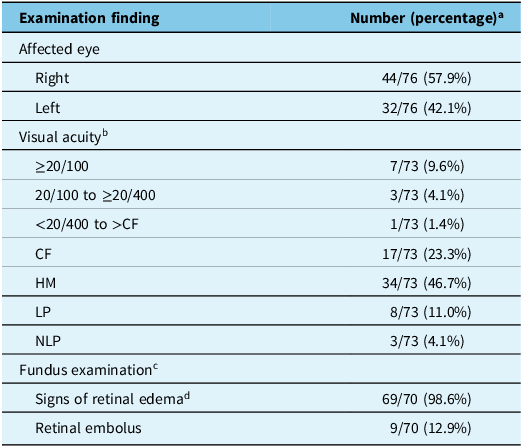
The median presenting VA in affected eyes was hand motions only; 65 (89.0%) of 73 patients presented with VA worse than 20/400 (Table 2). Signs of retinal edema were present in 69 (98.6%) of 70 patients with a complete fundus examination; in 1 patient whose fundus was examined the first 3 weeks after vision loss, no clinically overt retinal edema was seen. A retinal embolus was seen in 9 (12.9%) of 70. Thirteen patients (17.1%) reported transient ipsilateral visual disturbance in the days preceding vision loss from CRAO, including negative visual phenomenon in 9 and positive visual phenomenon in 4.
Presenting examination findings of eyes with central retinal artery occlusion

a All values are number (percentage), n = 76 unless otherwise indicated.
b Visual acuity data available were for 73 patients at presentation.
Previously diagnosed risk factors.
c A complete baseline fundus examination was documented in 70 patients.
d Signs of retinal edema included cherry red spot and retinal whitening.
CF = counting fingers only; HM = hand motions only; LP = light perception only; NLP = no light perception.
Patient presentation and referral pathways
The initial site of presentation (Figure 1a) was an ED in 47 (61.8%) of 76 patients, optometry clinic in 11 (14.5%), ophthalmology clinic in 10 (13.2%) and primary care clinic in 5 (6.6%). Two (2.6%) patients experienced vision loss while admitted at external hospitals, and for one patient, the site of initial presentation could not be determined.
Initial presentation after vision loss from central retinal artery occlusion. (a) Site (provider) of presentation. (b) Time from vision loss/last known well to presentation. ED = emergency department; optom = optometry practice; ophth = ophthalmology practice; GP = general practitioner practice; inpt = vision loss experienced as an inpatient; ? = unknown.

The distribution of times from vision loss/last known well to initial presentation was assessed (Figure 1b): the median (IQR) was 15.0 (3.5–48.0) hours. Twenty-two (28.9%) presented within 4.5 hours, 50 (65.8%) within 24 hours and 61 (80.3%) within 48 hours. Eighteen (25.0%) of 76 patients first noticed vision loss upon waking.
Thirty (39.5%) patients presented directly to our institution, 36 (47.4%) were assessed by 1 external provider, 9 (11.8%) were assessed by 2 external providers and 1 (1.3%) was assessed by 3 external providers prior to arrival at our institution. Arrival at our institution occurred at a median (IQR) of 23.5 (13.1–49.4) hours after vision loss/last known well. Twelve (15.8%) patients arrived within 4.5 hours, 42 (55.3%) within 24 hours and 57 (75.0%) within 48 hours.
Urgent ophthalmological consultations
Urgent ophthalmological consultation (before or after arrival at our institution) to diagnose/confirm CRAO occurred for 73 (96.1%) patients. Another two patients were referred directly to the ED by optometrists who had diagnosed CRAO and did not have urgent ophthalmological consultation. The remaining patient experienced right monocular vision loss shortly after developing left-sided motor weakness; a right-sided hemispheric stroke was diagnosed, but referral to ophthalmology did not occur until 3 weeks later at which time right subacute CRAO was found to be present.
For the 31 patients presenting to non-ophthalmology services at our institution who were subsequently referred to ophthalmology, the median (IQR) door-to-ophthalmology time was 12.0 (4.6–22.6) hours; a door-to-ophthalmology time of greater than 24 hours occurred for 6 (19.4%) of those patients.
Investigations
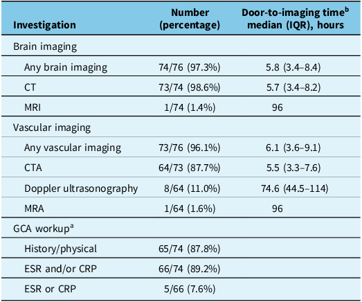
Acute brain imaging (Table 3) was obtained for 74 (97.3%) patients. The modality was CT in all except one patient who underwent MRI. Vascular imaging (Table 3) was obtained for 73 (96.1%) patients. The modality was computed tomography angiography (CTA) in 64 (87.7%), Doppler ultrasonography in 8 (11.0%) and magnetic resonance angiography (MRA) in 1 (1.6%). Two patients underwent neither brain nor vascular imaging, and one patient underwent CT head only; all three presented with high suspicion for GCA, which was later confirmed with TAB. Door-to-brain imaging times for patients presenting within 48 hours of vision loss/last known well are summarized in Table 3.
Urgent investigations at presentation for patients with central retinal artery occlusion

a GCA workup assessed in the 74 patients aged 50 and above.
b For patients presenting to our institution within 48 hours of vision loss/last known well.
IQR = interquartile range; GCA = giant cell arteritis; ESR = erythrocyte sedimentation rate; CRP = C-reactive protein.
We considered clinical and serological screening for GCA in the 74 patients who were aged 50 or above. Reference Flaxel, Adelman and Bailey14 A targeted history and/or physical examination for GCA was documented for 65 (87.8%) of 74 (Table 3). ESR and/or CRP were obtained for 66 (89.2%) patients; however, either ESR or CRP alone was ordered in 5 (7.6%) of 66. TAB was performed for 19 (25.7%) patients, and GCA was ultimately diagnosed in 6 (8.1%) of 74 patients over age 50.
Results of vascular and cardiac studies
Among the 70 patients with non-arteritic CRAO, 18 (25.7%) had ipsilateral carotid stenosis over 50%, including 1 patient who had complete stenosis and 1 patient who had associated free floating thrombus. One patient was found to have a lymphomatous mass encasing the ipsilateral common carotid artery.Footnote *
Completed cardiac investigations, including echocardiogram and/or Holter, as deemed appropriate by the stroke provider, were available in the EHR for 57 patients with non-arteritic CRAO (the remaining 13 patients with non-arteritic CRAO were investigated externally or lost to follow-up). Six of 57 (10.5%) were found to have a presumed cardioembolic source, including atrial fibrillation in 5 (8.8%) and left atrial appendage thrombus in 1 (1.8%).
Interventions
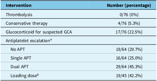
No patient with CRAO was treated with thrombolysis (Table 4). Four (5.3%) patients received intraocular pressure lowering therapy or ocular massage acutely (one patient received topical intraocular pressure lowering therapy, two received ocular massage alone, one received ocular massage with anterior chamber paracentesis). Empiric glucocorticoid therapy for suspected GCA was initiated for 17 (23.0%) of 74 patients over age 50, including intravenous methylprednisolone for 11 patients and oral prednisone for 6. All six patients in whom GCA was later confirmed had received empiric treatment following presentation.
Interventions for patients with central retinal artery occlusion

a Sixty-four patients were eligible for antiplatelet escalation as they did not have contraindications such as active treatment with anticoagulation or dual APT.
b Number of patients receiving a loading dose of aspirin and/or clopidogrel, as a proportion of total patients who received antiplatelet escalation.
GCA = giant cell arteritis; APT = antiplatelet therapy.
Sixty-four patients were eligible for the initiation of APT after presenting with CRAO as they did not have contraindications such as current use of anticoagulation or dual antiplatelet therapy (DAPT). Of those, treatment was with single APT for 16 (25.0%) and DAPT for 29 (45.3%). A loading dose of acetylsalicylic acid (ASA) and/or clopidogrel was administered to 19 (42.2%) of 45 patients initiating APT therapy.
Among the 18 patients with symptomatic carotid stenosis, urgent carotid revascularization was offered to 16 or 22.9% of all patients with non-arteritic CRAO. Nine of 16 patients underwent carotid endarterectomy, 5 underwent carotid stenting and 2 patients declined procedural intervention. One patient with 80% occlusion of the internal carotid artery (ICA) on initial CTA had free-floating thrombus that was managed with heparin; repeat CTA showed 30% defect in the ICA, and revascularization was deferred. One patient had complete occlusion of the ICA and as such was not a candidate for revascularization.
Secondary prevention referrals
Of 70 patients with non-arteritic CRAO, 48 (63.2%) were subsequently referred to a stroke prevention clinic, 13 (17.1%) underwent secondary stroke prevention investigations during inpatient admission and 3 (3.9%) were referred to family medicine for stroke prevention. A referral for secondary stroke prevention was not sent for six patients (7.9%). All patients with arteritic CRAO were referred to neuro-ophthalmology.
Referral for ocular follow-up was made to retina subspecialists for 31 (40.8%) of 76 patients, general ophthalmologists for 20 (26.3%) and neuro-ophthalmologists for 9 (11.8%). No referral for ocular follow-up was made to an optometrist. Sixteen (21.1%) patients were not referred for ocular follow-up.
Ophthalmic and neurovascular outcomes
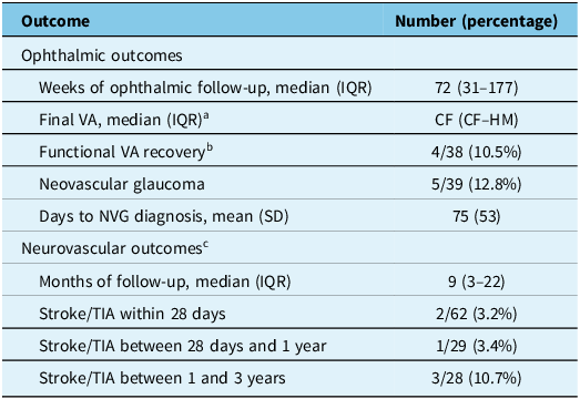
Final VA (Table 5) was available for 44 patients who had a minimum of 1 week follow-up after presentation with CRAO (median [IQR] follow-up duration in weeks: 72 [31–177]). The median final VA was counting fingers only (IQR, counting fingers – hand motions). Among 38 patients presenting with VA of 20/200 or worse, recovery to 20/100 or better occurred for 4 (10.5%). Neovascular glaucoma was diagnosed in 5 of 39 (12.8%) patients with adequate documentation of ophthalmic examination to confirm this diagnosis, at a mean (standard deviation) of 75 (53) days after vision loss.
Ophthalmic and neurovascular outcomes after central retinal artery occlusion

a VA data after a minimum follow-up of 1 week were available for 44 patients.
b Functional VA recovery is improvement from baseline visual acuity of 20/200 or worse to final VA of 20/100 or better.
c Neurovascular outcomes assessed in patients with non-arteritic central retinal artery occlusion.
VA = visual acuity; CF = counting fingers only; HM = hand motions only; IQR = interquartile range; SD = standard deviation; NVG = neovascular glaucoma.
Documentation to determine subsequent stroke or transient ischemic attack (TIA) was present in the EHR for 62 patients (median [IQR] follow-up duration in months 9.1 [2.6–21.8]). Among patients with sufficient duration of follow-up, 2 (3.2%) of 62 patients had subsequent stroke/TIA within 28 days of CRAO, 1 (3.4%) of 29 patients had stroke/TIA between 28 days and 1 year after CRAO and 3 (10.7%) of 28 patients had stroke/TIA between 1 and 3 years after CRAO.
Discussion
CRAO is a medical emergency, and the standard of care for this condition is evolving. The Canadian experience with patient presentations and practice patterns for CRAO has not been extensively described in the literature to date. In our 4-year study of CRAO in a large Canadian academic health sciences center, we found that patients seek care urgently following vision loss from CRAO and that generally patients receive timely workup; however, these patients experience poor visual outcomes and commonly have high-risk underlying conditions. Opportunities to strengthen care for this population exist.
Patients generally seek care urgently after CRAO: in our series, more than 1 in 4 first contacted the medical system within 4.5 hours, and nearly 2 out of 3 presented within 24 hours. Similar times to presentation have been reported in American and European centers. Reference Hoyer, Kahlert, Güney, Schlichtenbrede, Platten and Szabo8,Reference Lee, Tschoe and Coffman11,Reference Shah, Gilbert and Melles12 In Smith et al.’s series of patients with CRAO in Edmonton, Canada, only 3 of 27 (11.1%) presented within 4.5 hours. Reference Smith, Benson, Tennant and Jivraj13 The most common site of initial presentation was an emergency department (ED) in more than half (61.8%) of cases, similar to other series, followed by optometry and ophthalmology practices at similar rates (14.5% and 13.2%, respectively). By contrast, American studies consistently show that the second most common initial point of contact was an ophthalmologist for nearly one-third of cases. Reference Flowers, Chan, Meyer, Bruce, Newman and Biousse10–Reference Shah, Gilbert and Melles12 The comparatively lower rate of initial presentation to ophthalmology observed in our study presumably reflects the inability to self-refer to an ophthalmologist in Ontario. This is a reminder that non-ophthalmologists must be aware of this dangerous etiology of acute vision loss as they may be the first to encounter patients with CRAO.
Confirmation of diagnosis is a critical step that requires examination by a vision-care provider, typically an ophthalmologist. Among patients arriving at our tertiary care institution without a confirmed diagnosis of CRAO, the door-to-ophthalmological consultation time exceeded 12 hours in half of patients. This delay may reflect low suspicion for CRAO among providers who first encounter patients or delayed triage/assessment by ophthalmology after consultation has been requested. Delay to definitive ophthalmic diagnosis has been recognized as a major barrier to optimal care in other centers. In a recent US-based study, Shah et al. reported that only 172 (35.5%) of 484 patients with recent CRAO saw an ophthalmologist within 24 hours of vision loss. Reference Shah, Gilbert and Melles12 Fundus photography and/or optical coherence tomography platforms can be used to accurately diagnose CRAO, and data can be reviewed remotely. Reference Vasseneix, Bruce, Bidot, Newman and Biousse15,Reference Lema, De Leacy and Fara16 Implementation of a telemedicine pathway between the ED and ophthalmology that leverages ED-based ophthalmic imaging may be an optimal strategy to expedite the diagnosis (and treatment, if indicated) of CRAO.
The American Heart Association (AHA) Scientific Statement for CRAO emphasizes urgent etiologic workup including assessment for ipsilateral carotid stenosis. Reference Mac Grory, Schrag and Biousse1 Although not explicitly written to address acute retinal ischemia, CSBPR advise brain and vascular imaging be completed as soon as possible following acute stroke or TIA, ideally in an ED for those presenting within 48 hours. Reference Lindsay, Mountain, Gubitz, Dowlatshahi, Casaubon and Smith6 Nearly all patients with CRAO at our institution received acute brain and vascular imaging, and CT/CTA was the favored modality in most cases. By contrast, in the Edmonton series spanning 2018–2020, only 3 of 27 patients had any brain imaging. Reference Smith, Benson, Tennant and Jivraj13 In a large American series of 2802 patients with retinal artery occlusion presenting to EDs between 2006 and 2014, brain and carotid imaging were performed in only 20.3% and 7.1%, respectively. Reference Yousuf, Guiseppi, Katz, Nnorom and Akinyemi17 Compared to that study, a more recent series from an American center of 91 patients with CRAO between 2017 and 2020 reported increased rates of carotid imaging: CTA or MRA was obtained in 51.6% and carotid dopplers in 68.1%. Reference Lee, Tschoe and Coffman11 The increasing rates of brain and vascular imaging likely reflect an evolving standard of care with similarities between CRAO and cerebral stroke being emphasized in recent years. Reference Biousse, Nahab and Newman18
We examined rates of GCA screening in patients with recent CRAO. The American Academy of Ophthalmology Preferred Practice Pattern for CRAO advises GCA be suspected in patients over age 50. Reference Flaxel, Adelman and Bailey14 Although this is an uncommon cause of CRAO, recognition of GCA is critical given the high risk of fellow eye involvement and therefore of bilateral blindness in patients presenting with vision loss from GCA if treatment is delayed. Reference Chen, Leavitt, Fang, Crowson, Matteson and Warrington19 In our series, GCA screening was frequent but not uniform: clinical and serological assessments were each omitted in approximately 1 in 10 patients. Although this is a low proportion, this is suboptimal given the relatively high regional incidence of GCA. Reference Barra, Pope and Pequeno20 Indeed, the prevalence of GCA in our series of patients with CRAO (8.1% in those aged 50 and above) is higher than reported by other authors. Reference Mac Grory, Schrag and Biousse1
Recent clinical trials have failed to demonstrate the benefit of intravenous thrombolysis in minor nondisabling acute ischemic cerebral stroke over APT. Reference Khatri, Kleindorfer and Devlin21,Reference Chen, Cui and Zhou22 In contrast, meta-analyses of relatively small observational studies have suggested that thrombolysis may improve rates of functional recovery in selected patients with non-arteritic CRAO. Reference Huang, Wang and Zhang23,Reference MacGrory, Nackenoff and Poli24 At our center, no patient was offered thrombolytic therapy for CRAO. Smith and colleagues similarly reported no patient in their series of 27 in Edmonton, Canada, received thrombolysis. Reference Smith, Benson, Tennant and Jivraj13 Relatively low rates of thrombolysis, despite the willingness to offer this therapy by providers, have been reported in American Reference Chan, Flowers, Meyer, Bruce, Newman and Biousse9 and European Reference Hoyer, Kahlert, Güney, Schlichtenbrede, Platten and Szabo8 centers and reflect the difficulty in operationalizing this treatment for these patients in the hyperacute period after vision loss. A pathway for stroke team activation for suspected acute CRAO does not exist in our center; such pathways have been proposed and implemented in other centers as a strategy to optimize access to thrombolysis for acute CRAO. Reference Mac Grory, Schrag and Biousse1
We examined the use of antiplatelet agents after CRAO. CSBPR advise DAPT, including loading doses of both agents, for patients with acute high-risk TIA or minor ischemic stroke who are not at high bleeding risk. Reference Lindsay, Mountain, Gubitz, Dowlatshahi, Casaubon and Smith25 The AHA statement suggests a similar practice is reasonable for CRAO. Reference Mac Grory, Schrag and Biousse1 At our center, we observed significant variability in the decision to add APT and to provide a loading dose: among patients who were eligible for APT initiation or escalation, approximately one-third did not receive any antiplatelet agent, and more than half did not receive a loading dose. In Lee et al.’s series of 91 patients with acute CRAO, inconsistent use of APT was observed, with 27.5% of patients not starting any antiplatelet medication. Reference Lee, Tschoe and Coffman11 In another US study of 109 patients with CRAO between 2010 and 2020 referred to a tertiary hospital, APT was started by external providers in fewer than 10% of cases. Reference Flowers, Chan, Meyer, Bruce, Newman and Biousse10 The heterogeneity in practice patterns in our center and others highlights the importance of early involvement after CRAO of stroke experts to guide the nuanced decision around antiplatelet use.
We observed a relatively high rate of concordance between practice patterns at our institution with guidelines that emphasize the importance of managing vascular risk factors after CRAO: Reference Mac Grory, Schrag and Biousse1 neurovascular secondary prevention referrals were omitted for approximately 10% of patients with non-arteritic CRAO. This is a low proportion overall; however, it is important to note that those who experience non-arteritic CRAO are at increased long-term risk of stroke, myocardial infarction and death. Reference Wai, Knapp and Ludwig4 Presumably, some of this risk can be mitigated with appropriate workup and optimization after CRAO. We observed a higher rate of non-referral for ocular secondary prevention of closer to 20%. After CRAO, regardless of etiology, ocular follow-up serves important functions, which include optimizing residual vision, monitoring for neovascular-related complications and preserving the health of the contralateral eye. Reference Mac Grory, Schrag and Biousse1 Other institutional series have not examined rates of secondary prevention referrals to our knowledge.
With respect to the results of etiologic workup, underlying conditions included GCA in 8.1% of patients over 50, and among patients with non-arteritic CRAO, carotid stenosis was found in 25.7% and cardioembolic etiology in 10.5%. This reinforces the urgency of prompt diagnosis and workup after CRAO as these conditions are amenable to time-sensitive intervention to reduce the risk of additional morbidity.
Rates of functional visual recovery in our series (10.5%) are comparable to what has been reported in meta-analysis and demonstrate the poor natural history of vision loss from CRAO. Reference Schrag, Youn, Schindler, Kirshner and Greer26 Fortunately, we observed infrequent additional neurovascular events in our patients. Although follow-up data were not uniformly available for all patients, we interpret this as evidence of relatively effective secondary stroke prevention in the current system.
Our study is the largest Canadian series to date reporting institutional experience with CRAO; and, considering an evolving body of literature regarding the optimal management of this condition, is timely. The relatively high number of patients (for a rare condition) included over a 4-year period is also a strength as this allows us to draw meaningful conclusions. There are limitations. First, as a single-center study, there are inherent issues with generalizability. Nevertheless, The Ottawa Hospital is among the largest academic health sciences centers in Canada and serves a diverse population exceeding 1.4 million; as such, we believe our conclusions may have applicability in other Canadian centers. Supporting this, our findings overall do not deviate significantly to those of the only other Canadian series on the topic, notwithstanding the issues discussed above. Reference Smith, Benson, Tennant and Jivraj13 Second, the retrospective nature and reliance on electronic records have obligatory limitations including missing data and potential for error with time stamps; however, this is not expected to significantly alter our conclusions. Third, while we used a theoretically robust case-finding strategy that relied on pre-specified diagnosis codes that are entered in multiple areas in the chart, it is possible that some cases of CRAO during the study window were not identified. Importantly, the inadvertent exclusion of a small number of cases would not alter our findings in a meaningful way. Finally, our study window was inclusive of the onset and peak of the Covid-19 pandemic. This may have caused more patients to present to the ED as opposed to community-based providers such as optometrists; nevertheless, the distribution of sites of presentation is comparable to previous studies.
Patients with CRAO generally present within a day of vision loss, and typically present to the ED. Expediting diagnosis of CRAO should be a priority and may require telemedicine strategies to decrease the delay in ophthalmological assessment. Reference Vasseneix, Bruce, Bidot, Newman and Biousse15,Reference Lema, De Leacy and Fara16 At our institution, there are generally favorable practice patterns with respect to acute investigations and secondary prevention referrals after CRAO; these should continue to be emphasized in guidelines for CRAO, including the importance of GCA screening given Canada’s high GCA prevalence. Reference Barra, Pope and Pequeno20 Despite growing evidence for the use of thrombolytic therapy in CRAO, Reference Mac Grory, Schrag and Biousse1 our hospital system, one of the largest in Canada, has not yet adopted this within its institutional scope. The role of and strategies for implementation of thrombolysis in the management of CRAO in the Canadian context should be clarified. Patients with CRAO commonly have a high-risk underlying pathology that requires specific treatment to reduce additional morbidity and experience low rates of visual recovery. There is an unmet need for multidisciplinary Canadian guidelines for the management of this condition, especially in the acute care setting.
Data availability statement
Anonymized patient data are available on request, provided there is compliance with institutional protocols for data sharing.
Acknowledgments
N/A.
Author contributions
All authors contributed to the study conception and design, drafting and revising the study protocol, drafting and revising the manuscript and approval of the final version of the manuscript. MPQ performed the data collection.
Funding statement
This study received no funding.
Competing interests
Dr Quinn has received grant funding from Fighting Blindness Canada, the Canadian Ophthalmological Society and Bayer. Dr Quinn has received fellowship salary support from Bayer. The other authors have no disclosures.





Target article
Central Retinal Artery Occlusion at a Canadian Academic Center
Related commentaries (2)
Reviewer Comment on Quinn et al. “Central Retinal Artery Occlusion at a Canadian Academic Centre”
Reviewer Comment on Quinn et al. “Central Retinal Artery Occlusion at a Canadian Academic Centre”