Introduction
Lichens often possess complex reproductive strategies; for example, reproduction in a lichen-forming fungal species can be exclusively vegetative, strictly sexual or can combine vegetative and sexual reproduction (Seymour et al. Reference Seymour, Crittenden, Dickinson, Paoletti, Montiel, Cho and Dyer2005; Honegger & Scherrer Reference Honegger and Scherrer2008). Clonal propagation is hypothesized to be the preferred mode of reproduction during colonization of a new area as it can lead to rapid population establishment, with low energy and time expenditure (Bell Reference Bell1982; Sun & Heitman Reference Sun and Heitman2011). Furthermore, clonal reproduction might also preserve locally adapted genotypes in a stable environment (Agrawal Reference Agrawal2006; Honegger & Scherrer Reference Honegger and Scherrer2008; Sun & Heitman Reference Sun and Heitman2011). On the other hand, sexual recombination through outcrossing introduces genetic variation and is hypothesized to be the preferred mode of reproduction during changing environmental conditions (Agrawal Reference Agrawal2006; Ni et al. Reference Ni, Feretzaki, Sun, Wang and Heitman2011; Sun & Heitman Reference Sun and Heitman2011). Self-fertility (homothallism) can enable propagation and dispersal through sexual propagules in the absence of suitable mating partners, which can be advantageous over obligate outcrossing (heterothallism) especially in very small populations. Understanding an organism’s reproductive mode is of fundamental importance to conservation as it directly influences the adaptive and evolutionary potential of the organism (e.g. Charlesworth Reference Charlesworth1990; Honegger & Scherrer Reference Honegger and Scherrer2008).
Sexual reproduction of ascomycetes is controlled by the mating-type (MAT) locus (Turgeon & Yoder Reference Turgeon and Yoder2000) which codes for transcription factors required for sexual morphogenesis (Honegger & Scherrer Reference Honegger and Scherrer2008; Dyer et al. Reference Dyer, Inderbitzin and Debuchy2016). Whether a fungal species is homothallic or heterothallic depends on the composition of its MAT locus. In a heterothallic species, each haploid individual possesses one of two different forms of the MAT locus (MAT1-1 and MAT1-2; Turgeon & Yoder Reference Turgeon and Yoder2000). These two forms are referred to as ‘idiomorphs’ instead of ‘alleles’ as they contain entirely different genes, although they occupy the same physical location in the genome. The individuals of heterothallic species belong to one of two mating types and successful mating is only possible between individuals of opposite mating types (Debuchy et al. Reference Debuchy, Berteaux-Lecellier and Silar2010; Dyer et al. Reference Dyer, Inderbitzin and Debuchy2016). In a homothallic species, typically the same haploid genome either contains a copy of both idiomorphs in two separate MAT loci or all MAT genes are housed together in a combined MAT locus (Scherrer et al. Reference Scherrer, Zippler and Honegger2005; Wilson et al. Reference Wilson, Wilken, van der Nest, Steenkamp, Wingfield and Wingfield2015). However, in some atypical homothallic species (e.g. Neurospora africana, Xanthoria parietina), the genome of each haploid individual always contains the same, single mating type idiomorph, while the other idiomorph has apparently been lost (Beatty et al. Reference Beatty, Smith and Glass1994; Glass & Smith Reference Glass and Smith1994; Scherrer et al. Reference Scherrer, Zippler and Honegger2005; Gioti et al. Reference Gioti, Mushegian, Strandberg, Stajich and Johannesson2012). The genomic location of the MAT locus is highly conserved within the subphylum Pezizomycotina (i.e. Ascomycota excluding yeasts), which includes all lichenized ascomycetes. The MAT locus is usually flanked by a cytoskeleton assembly gene called SLA2 on one side and a DNA lyase gene called APN2 on the other side (Scherrer et al. Reference Scherrer, Zippler and Honegger2005; Butler Reference Butler2007). The relative orientation of these flanking genes is also conserved, as their transcriptional direction is towards the MAT locus in all reported examples. Compared with many other protein coding and rDNA loci, the MAT genes/loci are extremely variable in sequence, size and orientation even among closely related taxa (see e.g. Turgeon Reference Turgeon1998; Scherrer et al. Reference Scherrer, Zippler and Honegger2005; Martin et al. Reference Martin, Wingfield, Wingfield and Steenkamp2011). The putative DNA-binding sites known as α-box (MAT1-1) and HMG-domain (MAT1-2) contain conserved motifs at the amino acid level despite considerable variability at the nucleotide level. These conserved amino acid motifs allow homology-based searching and identification of the highly variable MAT genes in previously unstudied species.
Numerous studies examining the MAT locus and mating system have been published for non-lichenized, filamentous ascomycetes. This information has been reviewed by Butler (Reference Butler2007), Lin & Heitman (Reference Lin and Heitman2007), Debuchy et al. (Reference Debuchy, Berteaux-Lecellier and Silar2010) and Dyer et al. (Reference Dyer, Inderbitzin and Debuchy2016). In addition, a review of mating systems in lichens has been compiled by Honegger & Scherrer (Reference Honegger and Scherrer2008).
The mating system of a number of lichen mycobionts has been studied by Murtagh et al. (Reference Murtagh, Dyer and Crittenden2000), Honegger et al. (Reference Honegger, Zippler, Gansner and Scherrer2004), Scherrer et al. (Reference Scherrer, Zippler and Honegger2005), Seymour et al. (Reference Seymour, Crittenden, Dickinson, Paoletti, Montiel, Cho and Dyer2005), Honegger & Zippler (Reference Honegger and Zippler2007) and Singh et al. (Reference Singh, Dal Grande, Cornejo, Schmitt and Scheidegger2012). The methodology in these studies varied. Some of the studies have applied DNA fingerprinting techniques to single-spore cultures derived from the same ascus (i.e. the same meiotic event) to test whether the progeny descended from one or two parents (Murtagh et al. Reference Murtagh, Dyer and Crittenden2000; Honegger et al. Reference Honegger, Zippler, Gansner and Scherrer2004; Seymour et al. Reference Seymour, Crittenden, Dickinson, Paoletti, Montiel, Cho and Dyer2005; Honegger & Zippler Reference Honegger and Zippler2007). Other studies used a degenerate PCR approach to amplify the mating-type locus directly, followed by a mating-type screening of single-spore cultures or thalli with species-specific MAT primers to reveal the mating system (used e.g. by Seymour et al. Reference Seymour, Crittenden, Dickinson, Paoletti, Montiel, Cho and Dyer2005; Scherrer et al. Reference Scherrer, Zippler and Honegger2005; Singh et al. Reference Singh, Dal Grande, Cornejo, Schmitt and Scheidegger2012).
Currently, evidence for heterothallism prevails among the lichen species examined, having been found in 26 species: Anaptychia ciliaris, A. runcinata, Cladonia floerkeana, C. galindezii, C. portentosa, Diploicia canescens, Lobaria pulmonaria, Melanelixia glabra, Parmelia sulcata, Parmelina tiliacea, P. carporrhizans, P. quercina, Physcia aipolia, P. stellaris, P. tenella, Physconia distorta, Pseudevernia furfuracea, Ramalina fastigiata, R. fraxinea, Xanthoparmelia conspersa, X. stenophylla, Xanthoria calcicola, X. capensis, X. ectaneoides, X. polycarpa and X. resendei. Evidence for homothallism, by comparison, has been found in only four lichen species: Graphis scripta, Ochrolechia parella, Xanthoria elegans and X. parietina. As the vast majority of lichen mating systems were analyzed indirectly via DNA fingerprinting of single-spore cultures (i.e. without direct characterization of the MAT loci and mating types of these species), full or partial DNA sequence information of the MAT idiomorphs is available for only a small number of lichen species: Cladonia galindezii, C. grayi, Lobaria pulmonaria, Xanthoria elegans, X. flammeum, X. polycarpa and X. parietina.
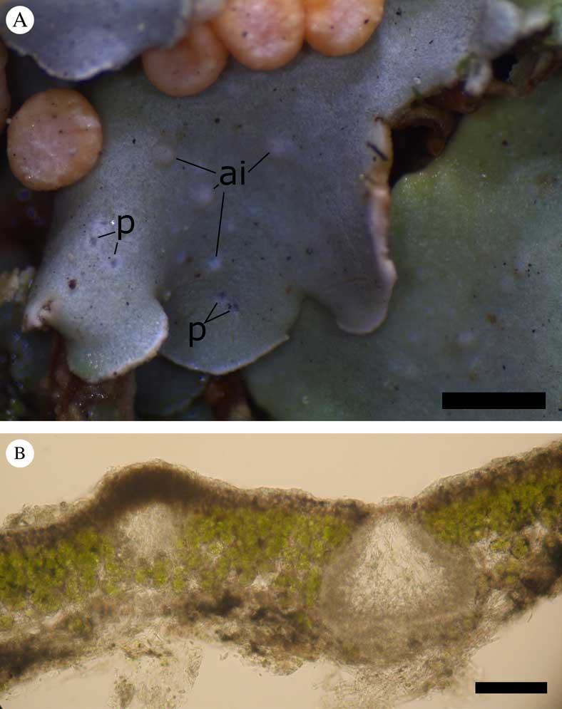
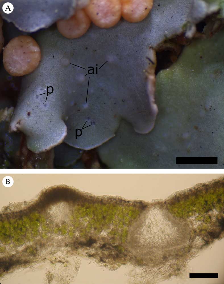
The present study examined the MAT locus and mating system of Knightiella splachnirima (Hook.f. & Taylor) Gyelnik emend. L. Ludw., a member of Icmadophilaceae endemic to southern New Zealand and the south-east Australian states of Tasmania and Victoria (for biogeographical details see Ludwig Reference Ludwig2016). It occurs mostly in endangered subalpine boggy habitats where it grows on continuously moist peaty soil, over bryophytes and plant debris, within a landscape dominated by tussock-forming grasses and low shrubs. The species has recently received the conservation status ‘Nationally Vulnerable’ in New Zealand (De Lange et al. Reference De Lange, Galloway, Blanchon, Knight, Rolfe, Crowcroft and Hitchmough2012). A recent molecular genetic reassessment of the genus Icmadophila (Ludwig Reference Ludwig2015) has shown that its transfer to this genus (Galloway Reference Galloway2000) was not correct; instead it belongs in the monotypic genus Knightiella. This was also suggested by Stenroos et al. (Reference Stenroos, Myllys, Thell and Hyvönen2002) based on their study of 18S ribosomal DNA sequences. The species can be readily recognized by the combination of eye-catching, bright pink, sessile apothecia and a small-foliose, lettuce green thallus (Galloway & Elix Reference Galloway and Elix1980; Galloway Reference Galloway2007). The occurrence of pycnidia (Fig. 1) and a previously unrecognized vegetative reproductive mode via marginal soralia were recently reported (Ludwig Reference Ludwig2011, Reference Ludwig2012).
Sexual reproductive structures of Knightiella splachnirima. A, thallus with pink apothecia, apothecial initials (ai) and pycnidia (p), which are illustrated here for the first time; pycnospores shed from pycnidia are believed to act as ‘male’ gametes during fertilization. B, young apothecial initial (left) and an immersed pycnidium (right) in a hand-cut section (unstained in water) through the same lobe as illustrated above. Scales: A=1 mm; B=100 μm.
The mating system of K. splachnirima is a crucial aspect of the species’ reproductive biology. The future survival of isolated populations of this rare species may depend on whether or not it is self-fertile and knowledge of its mating system would ideally influence the action to be taken by conservation authorities.
Materials and Methods
Materials used in this study
This study used fresh collections of K. splachnirima from New Zealand and one collection from Tasmania (see Supplementary Material Table S5, available online). We used only apothecial tissue to obtain template DNA for degenerate and inverse PCR (see below) as it is guaranteed to contain both MAT idiomorphs, even if the species is heterothallic. Moreover, the biatorine apothecia consist of mycobiont tissue only. For the mating-type screening, only minuscule samples of haploid tissue from the thalline margin were extracted to ensure the presence of only one idiomorph in case of heterothallism.
DNA isolation
DNA isolation was performed following Ludwig (Reference Ludwig2015); the detailed methodology is also provided as Online Supplementary Material. Initially a genomic DNA extract was obtained using a CTAB extraction protocol modified from Cubero et al. (Reference Cubero, Crespo, Fatehi and Bridge1999) and Summerfield (Reference Summerfield2003). This method involves cell disruption through grinding with liquid N2, and cell lysis in a CTAB (Cetyl-trimethyl ammonium bromide) extraction buffer followed by one chloroform extraction. Based on the approach of Ye et al. (Reference Ye, Ji, Parra, Zheng, Jiang, Zhao, Hu and Tu2004), the genomic DNA was purified using silica membrane spin columns (EconoSpin® All-In-One Mini Spin Columns, Epoch Life Sciences Inc.) and buffers prepared in-house (see Supplementary Material, available online).
Design of degenerate primers for conserved motifs of MAT idiomorphs
Degenerate primers for the α-box (MAT1-1) and HMG-domain (MAT1-2) sites were manually designed based on alignments of the corresponding sequences (orthologs) of several lichen-forming fungi, in order to identify regions conserved among a wider range of lichenized ascomycetes.
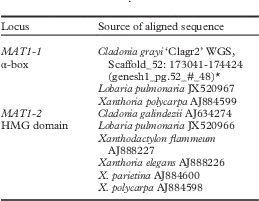
Aligned sequences were downloaded from GenBank and from the website of the Cladonia grayi whole genome sequencing project (URL1). Details of these sequences are listed in Table 1. Nucleotide alignments with translations of the respective MAT regions, including successfully used primer sites, are illustrated in Supplementary Material Figure S5 (available online). The fully assembled sequence of the MAT1-1 idiomorph (including flanking SLA2 and APN2 loci) of Dibaeis baeomyces was later provided by Dr Tami R. McDonald (Durham, USA, pers. comm. 30.04.2013) who generated it in the course of a whole genome sequencing project (McDonald et al. Reference McDonald, Mueller, Dietrich and Lutzoni2013).
Sequences which were aligned to design degenerate primers of the MAT and putative flanking loci. 8-digit codes refer to GenBank Accession numbers, the Cladonia grayi whole genome sequence is accessible through URL1

* Portion of whole genome sequence (WGS) is cited as the range of nucleotide positions of the respective scaffold number, with the systematic gene or locus name in brackets.
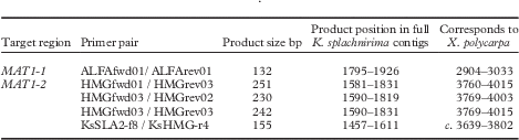
Degenerate PCR reactions consisted of 0·5 µl of each degenerate primer (at 10 pmol/µl, see Tables 2 & 3), 1 µl of genomic DNA and 18 µl of commercial ready-to-use PCR master mix (“1.1× ReddyMix PCR Master Mix (1·5 mM MgCl2)”, Thermo Scientific and “1.1× ReddyMix PCR Master Mix (2·0 mM MgCl2)”, Thermo Scientific). The PCR conditions were as follows: initial denaturation at 95°C for 5 min.; then 42 cycles with denaturation at 95°C for 45 or 60 s, annealing at 45°C, 50°C or 52°C for 45 or 60 s, elongation at 72°C for 40 to 120 s, followed by a final elongation at 72°C for 10 min.
Sequences and experimental use of the primers that were successfully used for amplification of partial sequences of Knightiella splachnirima MAT loci. IUPAC coding applies to ambiguous nucleotide positions of degenerate primers: B=C/T/G; D=A/T/G; H=A/T/C; K=T/G; M=C/A; N=A/T/C/G; R=A/G; S=C/G; V=A/C/G; W=T/A; Y=C/T

Details of degenerate PCRs. See Table 2 for primer sequences. The column ‘Product position in full K. splachnirima contigs’ refers to nucleotide positions in GenBank Accessions KP984802 for MAT1-1 and KP984801 for MAT1-2 of Knightiella splachnirima. The column ‘Corresponds to X. polycarpa’ gives the corresponding nucleotide positions of Xanthoria polycarpa GenBank sequences AJ884599 for MAT1-1 and AJ884598 for MAT1-2. Primers starting with Ks are not degenerate, but the respective PCR has extended the initial sequence prior to inverse PCRs

The PCR products were also purified with EconoSpin ® silica membrane spin columns and custom-made buffers. Direct Sanger sequencing of purified PCR products was performed by the University of Otago Genetic Analysis Service. The resulting partial sequences of the KsMAT1-1 α–box and KsMAT1-2 HMG-domain (the prefix ‘Ks’ signifies Knightiella splachnirima) were subsequently extended via inverse PCR as described below.
Inverse PCR
Inverse PCR is a method of obtaining unknown DNA sequences flanking an already sequenced section of genomic DNA (Ochman et al. Reference Ochman, Gerber and Hartl1988). Genomic DNA was digested with a restriction enzyme which cuts outside the known sequence, followed by the self-ligation of the fragments to generate a circular DNA template. Primer binding sites are near the ends of the known initial sequence but their orientation is inverted compared to a regular PCR. Because the template DNA has been circularized, a PCR product can be generated with both previously unknown flanking regions separated by the recognition sequence of the restriction enzyme.
DNA was extracted from several apothecia and digested using the restriction enzymes EcoRI, HindIII, and BamHI (Roche) at 37°C for 90 min. Each DNA digestion reaction consisted of 5 μl enzyme-specific Enzyme Buffer, 10 μl genomic DNA (c. 2400 ng), 2 μl of the restriction enzyme (20 units) and water to a final volume of 50 μl. The digestion reaction was stopped by adding 4 volumes of B2 buffer (from Invitrogen ‘PureLink Quick PCR Purification Kit’). The digested genomic DNA was then purified with the PureLink Quick PCR Purification Kit (Invitrogen) according to the manufacturer’s instructions.
The digested and purified DNA was then self-ligated using T4 ligase (Roche). Ligation reactions contained 3 μl of 10× ligation buffer, 3 μl (3 units) of T4 DNA Ligase (Roche), 30 ng (1ng/μl) or 150 ng (5ng/μl) digested genomic DNA and water to a final volume of 30 μl. Reactions were incubated at 4°C for 24 h followed by 30 min at room temperature. Ligase was heat inactivated by incubation at 65°C for 10 min. DNA was purified using buffers of the PureLink Quick PCR Purification Kit (Invitrogen) together with EconoSpin ® All-In-One Mini Spin Columns (Epoch Life Sciences Inc.). All steps of the standard Invitrogen instruction manual were performed twice in order to increase yield and purity.
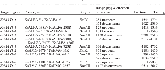
The self-ligated DNA digests were then used as a template for inverse PCR. The inverse primers specific for the K. splachnirima MAT region were designed from the sequences resulting from the degenerate PCR (K. splachnirima α-box and HMG-domain). This strategy has been used by several other authors to amplify the flanking region of the conserved regions previously sequenced with degenerate primers (Scherrer et al. Reference Scherrer, Zippler and Honegger2005; Pöggeler et al. Reference Pöggeler, O’Gorman, Hoff and Kuck2011; Singh et al. Reference Singh, Dal Grande, Cornejo, Schmitt and Scheidegger2012). The details of degenerate PCRs (primer pairs, product size and relative position) are summarized in Table 3. Primer pairs for inverse PCR (see Table 2) were designed using the Primer 3 plug-in of Geneious R7 (Biomatters Ltd, http://www.geneious.com).
Each inverse PCR reaction consisted of 0·5 µl of each primer (at 10 pmol/µl), 1 µl (c. 40 ng/µl) of circularized genomic DNA and 18 µl of a commercial ready-to-use PCR master mix (see above). Inverse PCR cycling conditions were: initial denaturation at 95°C for 5 min; then 35 to 42 cycles with denaturation at 95°C for 1 min, annealing at 52°C or 58°C for 1 min, elongation at 72°C for 1 min, followed by a final elongation at 72°C for 10 min. The details of the various inverse PCRs (primer pairs used and product size) are summarized in Table 4.
Inverse PCR details. See Table 2 for primer sequences. Restriction enzymes used for preparation of self-ligated template DNA are given in the ‘Enzyme’ column. The extension range does not include overlaps with previously generated sequences. Positions of the extensions refer to full contigs of the newly generated Knightiella splachnirima sequences deposited in GenBank, Accession numbers: KP984801 (KsMAT1-2) and KP984802 (KsMAT1-1)

Annotation of contigs
Putative coding sequences of the successively expanded contigs were identified and annotated by alignment with reference MAT sequences of other lichens, which showed the approximate position of introns and distance between start and stop codons. Exact boundaries of putative introns were manually identified by comparison with the splicing signals described in Schwartz et al. (Reference Schwartz, Silva, Burstein, Pupko, Eyras and Ast2007) and Schneider-Poetsch et al. (Reference Schneider-Poetsch, Usui, Kaida and Yoshida2010).
Mating system analysis of K. splachnirima
DNA extracts of 27 minuscule samples of haploid tissue (c. 2 mm2 or smaller, from the thallus margin) were screened for the presence of both putative KsMAT genes. Samples were randomly collected from one Tasmanian and several New Zealand localities (for details see Supplementary Material Table S5, available online) but 18 of the 27 samples represent the same locality near Dunedin (Swampy Summit and Silver Peaks region, 45°48'S, 170°29'E) which should reveal the mating-type ratio of a local population in case of heterothallism. Apothecial DNA extracts were used as a positive control since these should always contain both MAT idiomorphs.
The following primer pairs were used for mating-type screening: KsALFA-712F/KsALFA-714R (product size 1547 bp incl. primers) and KsHMG-185F/KsHMG-1888R (product size 1704 bp incl. primers). The MAT1-2 primer binding sites (KsHMG primers) are located outside the putative coding region, so the PCR product contains the whole coding region of the putative MAT1-2-1 gene.
The MAT1-1 primer binding sites (KsALFA primers) are located upstream of the putative MAT1-1-1 coding region and within the coding region, so they amplify the first c. 750 bp of the coding sequence including the whole α-box domain with conserved intron. The composition of PCR reactions was as described above for degenerate PCR, except with different primers.
PCRs containing either primer pair were run simultaneously but in separate reaction tubes (i.e. not multiplex) using the following step-down protocol: initial denaturation at 95°C for 3 min, then 10 step-down cycles with denaturation at 94°C for 30 s, annealing for 1 min at temperatures dropping from 65°C to 55°C in increments of 1°C per cycle and elongation at 72°C for 3 min. This was followed by 35 cycles with denaturation at 94°C for 30 s, annealing at 55°C for 1 min and elongation at 72°C for 3 min.
Results
Amplification of short conserved regions of the MAT idiomorphs using degenerate primers
Degenerate PCR yielded a 132 bp fragment of the MAT1-1 α-box and a 251 bp fragment of the MAT1-2 HMG domain.
Inverse PCR: KsMAT1-1, KsMAT1-2
Using the inverse PCR technique, we amplified 1794 bp upstream and 2866 bp downstream of the initial 132 bp KsMAT1-1 α-box fragment. The initial 251 bp KsMAT1-2 HMG fragment was extended upstream by 124 bp through a product resulting from amplification with the species-specific primer pair KsSLA2-f8 / KsHMG-r4, in a reaction which was an attempt to connect the SLA2 and HMG fragments through direct PCR. All subsequent inverse PCRs combined then extended the resulting 375 bp KsMAT1-2 fragment by 1456 bp upstream and 1786 bp downstream.
Continuity of the putative coding sequence of the KsMAT1-1 contig was tested and confirmed by direct PCR with the primer pairs KsALFA-712F/KsALFA-714R (product size 1546 bp) and KsALFA-749F/KsALFA-806R (product size 1595 bp) and undigested genomic DNA as the template. Likewise, continuity of the putative coding sequence of the KsMAT1-2 contig was tested and confirmed using direct PCR with the primer pairs KsHMG-185F/KsHMG-1888R (product size 1704 bp) and KsHMG-197F/KsHMG- 2397R (product size 1543 bp). For primer sequences see Table 2.
Contigs containing putative MAT genes of Knightiella splachnirima
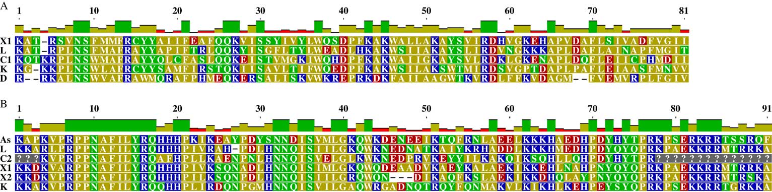
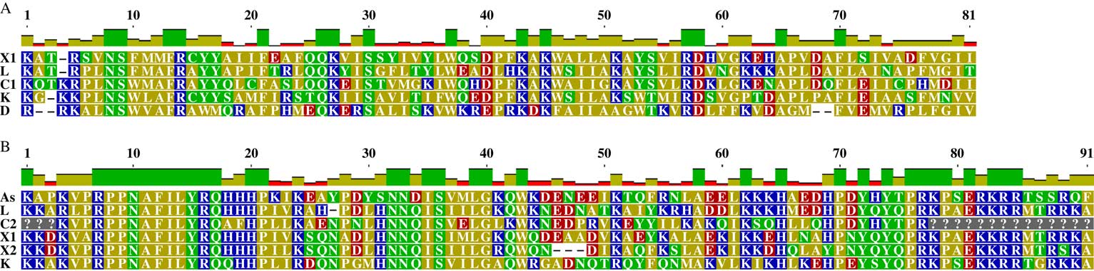
The putative K. splachnirima MAT1-1 contig (GenBank Accession KP984802) is 4792 bp long, including a 1703 bp (548 aa) putative coding sequence (CDS) which has a 56 bp intron in a conserved position within the codon of cysteine C103. This cysteine residue itself is not conserved (alignment position 15 in Fig. 3A) but the flanking FR and YY residues are highly conserved among the investigated sequences and are also conserved in Neurospora crassa (Glass et al. Reference Glass, Grotelueschen and Metzenberg1990). The putative CDS is flanked by 1475 bp upstream of the putative start methionine and a further 1614 bp downstream of the putative stop codon. These are of unknown function.
The putative K. splachnirima MAT1-2 contig (GenBank Accession KP984801) is 3617 bp long, including a 1280 bp (390 aa) putative CDS which has a 61 bp intron between amino acid residues number 62 (leucine, L) and number 63 (isoleucine, I), and a 46 bp intron within the codon of Serine (S) residue number 150 (alignment position 36 in Fig. 3B). The putative CDS is flanked by 1139 bp upstream of the putative start methionine and another 1198 bp downstream of the stop codon. These are of unknown function.
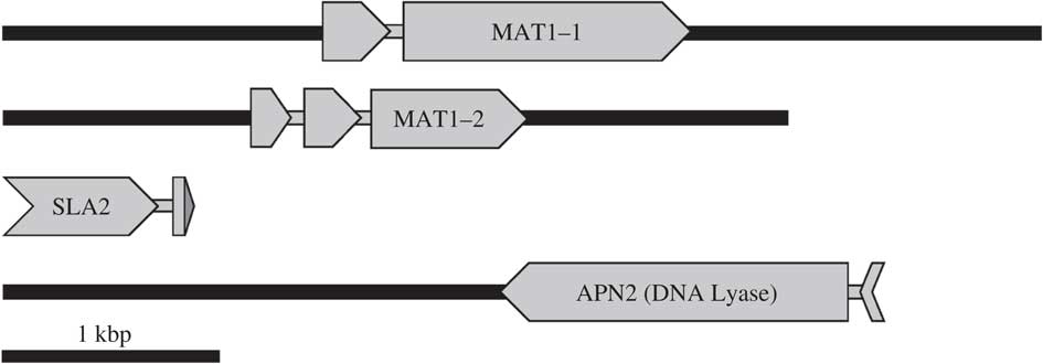
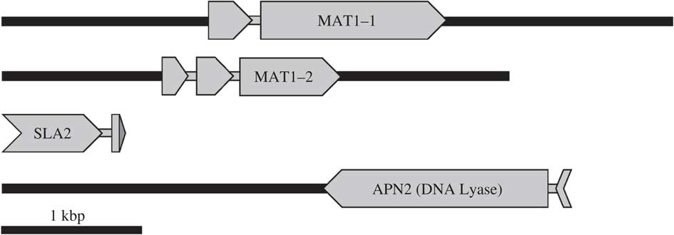
Analogous to the MAT genes, the flanking SLA2 and APN2 loci of K. splachnirima were also partially amplified and sequenced via degenerate and inverse PCR, because it was initially intended to amplify the entire MAT region from SLA2 to APN2, but these could not be united with the MAT1-1 and MAT1-2 contigs. Therefore, the general organization of the MAT region beyond the MAT1-1 and MAT1-2 fragments remains uncertain. Partial APN2 and SLA2 sequences of K. splachnirima were deposited in GenBank (KP984803 and KP984804) and the detailed methodology of this work is given in Ludwig (Reference Ludwig2015). Figure 2 shows an overview of the final contigs of the putative MAT idiomorphs as well as the potential flanking genes SLA2 and APN2 (DNA lyase) of K. splachnirima. The presumably non-coding flanking regions of all contigs do not overlap with each other, nor do they contain homologous parts of MAT regions of other lichen-forming fungi. No identifiable open reading frames were detected by BLAST searches of putative ORFs that were indicated by the Geneious software or during the process of submitting sequences to GenBank. Figure 3 shows partial amino acid alignments comparing the conserved domains of the new putative K. splachnirima mating-type genes with those of other lichen mycobionts and one non-lichenized ascomycete.
Overview of the Knightiella splachnirima mating-type region contigs produced in this study. Putative coding sequences (exons) are depicted as arrows pointing from 5’ to 3’; narrow grey bars within genes indicate introns. Narrow black bars are presumably non-coding sections of DNA. The coding sequences of KsMAT1-1 and KsMAT1-2 are complete, the start sections of KsSLA2 and KsAPN2 were not amplified and are therefore truncated. The grey terminal section attached to the KsSLA2 contig represents the last 33 bp of the coding sequence (10 amino acids plus stop codon) of the gene in Cladonia grayi, Xanthoria parietina and X. polycarpa. These last 33 bp were not amplified for K. splachnirima but the corresponding section of the other lichen-forming fungi was included to illustrate the relative position of the KsSLA2 contig. Full contig lengths and GenBank Accessions (in brackets) are: 4792 bp for KsMAT1-1 (KP984802), 3617 bp for KsMAT1-2 (KP984801), 836 bp for KsSLA2 (KP984804) and 3486 bp for KsAPN2 (KP984803).
Polypeptide alignments comparing the putative Knightiella splachnirima MAT1-1 α-box (A) and MAT1-2 HMG domain (B) with other species. Species are denoted by letters (see below). Orientation is N to C terminal (left to right). Amino acid names are given in one-letter IUPAC nomenclature and are colour-coded according to polarity. The alignment images were generated with Geneious R7. A, a conserved intron characteristic for the MAT1-1 α-box is present within the Cysteine (C) and Alanine (A) codons of alignment position number 15. Depicted sequences correspond to the given nucleotide ranges of the following Genbank Accession numbers (8-digit codes) or Whole Genome Sequence (WGS), respectively: (X1) Xanthoria polycarpa AJ884599, 3079-278; (L) Lobaria pulmonaria JX520967, 1869-2157; (C1) Cladonia grayi WGS Clagr3, scaffold 00075, 82175-82468; (K) Knightiella splachnirima KP984802, 1749-2044; (D) Dibaeis baeomyces WGS generated by McDonald et al. (Reference McDonald, Mueller, Dietrich and Lutzoni2013), scaffold 00314, 30260-30542. B, a conserved intron characteristic for the MAT1-2 HMG-domain is present within the Serine (S) codons of alignment position number 36. Depicted sequences correspond to: (As) Aspergillus fumigatus WGS Af293, Chromosome 3, 1524950-1525276; (L) Lobaria pulmonaria JX520966, 45-363; (C2) Cladonia galindezii AY634274: 1-272; (X1) Xanthoria polycarpa AJ884598, 3722-4045; (X2) Xanthoria parietina WGS Xanpa1, scaffold 3, 902642-902955; (K) Knightiella splachnirima KP984801, 1543-1861, Cited WGS data are accessible online through the following URLs: C. grayi, URL1; A. fumigatus, URL2; X. parietina, URL3; D. baeomyces, URL4. Corresponding to these polypeptide alignments, Supplementary Figures S5A & B (accessible online) illustrate the poorly conserved DNA sequences in direct comparison with their translation, along with the successfully used degenerate primer sites.
The mating system of Knightiella splachnirima
The mating-type screening showed an alternating presence of either MAT1-1 or MAT1-2 in each sample (Fig. 4). This pattern indicates that K. splachnirima is heterothallic. Of the total 27 samples examined, 13 contained only MAT1-1 and 14 contained only MAT1-2. The mating-type ratio within samples of the Dunedin population is 8:10 (lanes 11 to 28 of Fig. 4).
Mating-type screening of Knightiella splachnirima. Top row: MAT1-1 screening (product size 1547 bp). Bottom row: MAT1-2 screening (product size 1704 bp). The sample order is identical in both rows, i.e. each column shows both screening results of the same DNA extract on top of each other. Lanes 11–28 show samples from the same local population near Dunedin, New Zealand. The size markers (M) on the outermost lanes show reference bands of 1650 bp and 2000 bp. Lane 1 is a positive control with apothecial DNA extract as template, which means it should show both products. Lanes 2–10 show samples from various localities in New Zealand and Tasmania; for provenance details see Supplementary Material Table S5 (available online).

Discussion
Our results clearly demonstrate K. splachnirima to be heterothallic. The MAT-like sequences amplified from K. splachnirima in this study bear the expected degree of resemblance to MAT genes from other lichen-forming fungi and non-lichenized Pezizomycotina (Fig. 3). Conservation at the amino acid level in the α-box and HMG domains, as well as the presence of conserved introns, allows confident homology-based identification of the generated sequences as MAT genes. Furthermore, the putative gene size is in the expected range of the MAT genes and the observed alternating presence/absence pattern is typical only for the mating-type locus (in case of heterothallism). However, it was not possible to show that the K. splachnirima MAT genes were also flanked by the SLA2 and APN2 genes, as is the case in almost all previously studied Pezizomycotina (Butler Reference Butler2007).
Mating-type screening showed a consistent alternating presence of either MAT1-1 or MAT1-2 (Fig. 4) which indicates heterothallism (i.e. that K. splachnirima is most likely not capable of self-fertilization and relies on outcrossing). Mating of K. splachnirima is most likely mediated by pycnospores shed from pycnidia (Ludwig Reference Ludwig2011; Fig. 1) which are hypothesized to act as ‘male’ gametes (‘spermatia’) during fertilization (Honegger & Scherrer Reference Honegger and Scherrer2008). The use of single-spore cultures for the mating-type screening as in other studies (Scherrer et al. Reference Scherrer, Zippler and Honegger2005; Seymour et al. Reference Seymour, Crittenden, Dickinson, Paoletti, Montiel, Cho and Dyer2005) is deemed not necessary, because the observed either/or pattern indicative of heterothallism was already fully conclusive.
A balanced mating-type ratio was observed in the random sample of 27 specimens originating from various New Zealand and Tasmanian localities, as well as for the 18 samples from the same local population near Dunedin, which is consistent with the very frequent occurrence of apothecia and apothecial initials. This is in contrast to heterothallic Lobaria pulmonaria, which can also reproduce both sexually and vegetatively, but was found by Singh et al. (Reference Singh, Dal Grande, Cornejo, Schmitt and Scheidegger2012) to have a highly skewed mating-type ratio in many populations. These authors concluded that the generally rare occurrence of apothecia in L. pulmonaria could be attributed to a lack of suitable mating partners.
The widespread Xanthoria polycarpa is also a heterothallic lichen-forming fungus (Scherrer et al. Reference Scherrer, Zippler and Honegger2005) but unlike L. pulmonaria it is virtually always fertile, as is K. splachnirima. The frequently fruiting K. splachnirima and X. polycarpa both grow in more open macrohabitats compared to the rarely fertile L. pulmonaria (based on light indicator values in Wirth (Reference Wirth2010) and pers. obs. for K. splachnirima) which is a characteristic species of denser old-growth forests that provide shelter from excess light and heat (Gauslaa & Solhaug Reference Gauslaa and Solhaug1999; Jüriado & Liira Reference Jüriado and Liira2009; Gauslaa Reference Gauslaa2013). It might be that a more open habitat facilitates the transport of airborne pycnospores for fertilization. Thus a low frequency of apothecium formation in L. pulmonaria could be partly explained by low fertilization success resulting from slow wind velocities in dense forest stands, even if sexually compatible thalli are not very far apart. This idea is supported by the findings of Mikryukov et al. (Reference Mikryukov, Mikhailova and Scheidegger2010) who report that the proportion of fertile L. pulmonaria sub-populations is three times greater in deciduous forests (open canopy during wintertime) than in conifer forests (permanently closed canopy) of the Russian Ural region.
The finding that K. splachnirima does not fulfil the genetic requirements for self-fertilization has major implications for its conservation. Severe genetic bottlenecks may compromise sexual reproduction in isolated and small populations and therefore adaptation to a changing environment. Recent studies on heterothallic phytopathogenic and lichen-forming fungi (Saleh et al. Reference Saleh, Xu, Shen, Li, Adreit, Milazzo, Ravigne, Bazin, Notteghem and Fournier2012; Singh et al. Reference Singh, Dal Grande, Werth and Scheidegger2015) have found evidence that the absence of compatible mating partners in populations with skewed MAT representation is likely to have induced strict clonality in these populations, coinciding with low genetic diversity. In such a case, an affected population of K. splachnirima could probably survive temporarily via vegetative reproduction, but action from conservation authorities would be required to restore the mating-type ratio by transplantation of sexually compatible thalli in order to assure long-term viability of the population.
In conclusion, our study clearly shows that K. splachnirima is heterothallic. In addition we provide a MAT screening protocol with K. splachnirima specific MAT1-1 and MAT1-2 primers which could be used to assess the mating-type ratio in natural populations of K. splachnirima. Given the nationally vulnerable status of this species in New Zealand, the availability of such a protocol constitutes a significant conservation tool to infer the sexual reproductive potential, especially of small and isolated populations. Moreover, our newly designed degenerate MAT primers could be beneficial for future mating system analyses of other lichen species, in particular if financial constraints prevent the use of whole genome sequencing. Owing to low costs and easy application, the use of these lichen-specific degenerate MAT primers could facilitate the establishment of lichen mating system analyses as a routine practice in conservation. This is overdue, after more than a decade in which this subject was entirely restricted to basic and academic research.
The authors thank Dr Tami R. McDonald (Durham, USA) for providing the sequence of the MAT region of Dibaeis baeomyces when the WGS data was still inaccessible online. Professor Elfie Stocker-Wörgötter (Salzburg, Austria) is thanked for attempts to establish single-spore cultures of K. splachnirima, and Dr Allison Knight and Vickey Tomlinson (both Dunedin) for help with curating voucher specimens. This study was funded by a University of Otago Doctoral Scholarship awarded to the first author, along with research grants from the Miss E. L. Hellaby Indigenous Grasslands Research Trust, the Forest & Bird Dunedin branch, the British Lichen Society and Otago University’s Diane Campbell-Hunt Memorial Award. GS was supported by a fellowship from the German Academic Exchange Service (DAAD).
Supplementary Material
For supplementary material accompanying this paper visit https://doi.org/10.1017/S0024282917000214