Congenital ventricular aneurysms are rare cardiac malformations. The true incidence is unknown because they are often asymptomatic. Due to the increase in the use of fetal echocardiography in the antenatal period, cases with fetal diagnosis have started to be reported. Here, we aimed to present a case who was found to have right ventricular aneurysm in fetal echocardiography.
Case report
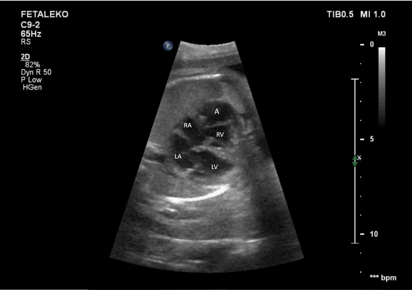
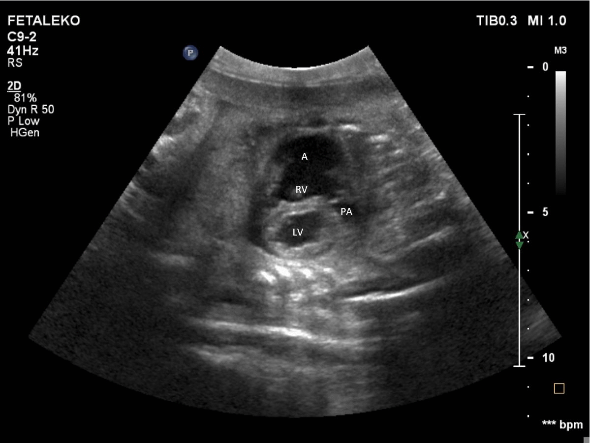
A 29-year-old woman was referred for fetal echocardiography at the 22nd week of pregnancy due to the detection of fetal cardiomegaly and fetal echocardiography showed 23 × 10 mm right ventricular outpouching originating from the lateral free wall (see Figure 1). The outpouching wall was thin, and there was no myocardial tissue. The large connection between outpouching and ventricle was measured as 12 mm (see Figure 2). There was no other accompanying cardiac pathology in fetal echocardiography. The fetus was evaluated by fetal echocardiography at intervals of 3–4 weeks until delivery, and there was no change in outpouching size. Pericardial effusion, heart failure, arrhythmia, tricuspid regurgitation, or other complications were not observed. The patient was born in the 37th week with 2450 gr by C/S. Postnatal echocardiogram showed a 23 × 13 mm ventricular outpouching with a thin and hypokinetic wall in the free wall of the right ventricle. Connection of the outpouching to the ventricle was measured as 10 mm. The tricuspid valve structure was normal, and there was no tricuspid regurgitation. We evaluated this outpouching as a ventricular aneurysm. There was no other accompanying cardiac or extracardiac abnormalities. The patient is now 2 months old, and her electrocardiogram is normal and has no symptoms.
The modified four-chamber view showing the aneurysmatic segment located anterior to the right ventricle. Due to the tricuspid anterior leaflet, the aneurysm neck was not visualized in this section. A = aneurysm; LA = left atrium; LV = left ventricle; RA = right atrium; RV = right ventricle.

Parasternal short axis of two ventricles view showing the aneurysm at the free wall of RV. The wall of the aneurysm is thin. A = aneurysm; LV = left ventricle; RV = right ventricle; PA = pulmonary artery.
Discussion
Congenital ventricular aneurysms are protrusions originating from the free wall of the ventricles or the apex. Aneurysms are mostly compared with diverticula, and the distinction between them is not clear so some authors have collected them under the term “outpouching.” Reference Williams, Collardey and Tradwell1 Congenital ventricular aneurysm and diverticula were first described in 1816 in Germany. Reference Kreysig2 Since then, it is difficult to obtain true epidemiological data because there are generally case reports in the literature. In an autopsy study of 12.924 cases in children, the prevalence was reported as 0.02%. Reference Albrecht3 It was first described as antenatal in 1990 in a fetus with ventricular extrasystole at 32 weeks of gestation. Reference Gembruch, Steil and Redel4 In later studies, the prevalence was determined as 0.5 per 100.000 live births. Reference Gonc alves, Simms and Jeanty5 We performed 16,250 fetal echocardiography between 2007 and 2020, and three cases of ventricular outpouching were detected, including one of them we reported here. The other case with left ventricular aneurysm was previously reported. Reference Demir, Ozbarlas and Gocen6
Congenital ventricular aneurysm and diverticula are both more common in the left ventricle. While congenital ventricular aneurysms are most frequently located in the apex and perivalvular area of the left ventricle (mostly the submitral region), diverticula are more frequently located in the apex of the left ventricle. Reference Ohlow, Secknus and Geller7 The number of cases reported to be located in the right ventricle is much less, and the frequency of localisation in any region of the right ventricle has not been reported. Williams et al. reported three cases of right ventricular outpouching, and in their study that included fourteen right ventricular outpouching cases previously published, they reported that nine cases were apical and eight cases were non-apical. Reference Williams, Collardey and Tradwell1 However, Zheng et al. reported that right ventricular outpouchings are more frequently located in non-apical regions, especially in the basal segment of the right ventricle. Reference Zheng, Sun and Liu8
It is important to differentiate aneurysm or diverticulum in order to plan its follow-up and predict the prognosis. Aneurysms have a wide necked connection with the ventricle and have a thin outer wall. The aneurysm wall does not contain all the wall layers of the ventricle, and the myocardial tissue has been replaced by fibrous tissue. Therefore, the aneurysm wall may be hypokinetic, akinetic, or act paradoxically with the ventricle during ventricular systole. The connection of the diverticula with the ventricle occurs through a narrow neck. Since all three ventricular layers are present in the diverticulum wall, the wall does not appear thin and its movement is normal.
Congenital ventricular outpouching can be seen with other cardiac, vascular, and extracardiac defects. Ventricular septal defect, coronary anomalies, atrial septal defect, pulmonary hypoplasia, aortic coarctation, and persistent left superior caval vein were the most common accompanying cardiac and vascular abnormalities. Reference Ohlow, von Korn and Lauer9 The most common extracardiac anomalies are midline defects such as diaphragmatic defects and sternal abnormalities. It was observed that the frequency of extracardiac defects accompanying diverticula was higher. Reference Ohlow, von Korn and Lauer9
Congenital ventricular outpouching is usually asymptomatic. However, large aneurysms and diverticula can cause heart failure due to the volume overload. This volume load is less tolerated in the fetal and neonatal period. Therefore, heart failure, pericardial effusion, and associated mediastinal shift, hydrops can be seen in younger ages, including the antenatal period. Reference Ohlow, von Korn and Lauer9 Cases that pericardiocentesis performed during antenatal period with congenital ventricular outpouching have been reported. Reference Gardiner, Wimalasundera and Pasquini10 Pericardial effusion is more common with right ventricular aneurysms. Williams et al. reported that pericardial effusion is more common in apical right ventricular aneurysms than non-apical right ventricular aneurysms. Reference Williams, Collardey and Tradwell1 At the fetal period, different clinical findings such as arrhythmia, ventricular dysfunction, and valve regurgitation are more common in non-apical right ventricular aneurysms rather than pericardial effusion, and they can be symptomatic at birth, but it has been reported that they are generally asymptomatic during follow-up. Reference Williams, Collardey and Tradwell1 Embolic events are more common in aneurysms and attributed to the aneurysm wall being akinetic or hypokinetic. Oral anticoagulants are recommended for patients with embolic events. Our case was asymptomatic in the prenatal and postnatal period.
Conclusion
The development of imaging methods and with more frequent use of fetal echocardiography in the prenatal period provide an increase in the cases diagnosed starting from the prenatal period. However, predicting the prognosis of the patients is important in terms of whether they will be treated, even if they are asymptomatic. It is also necessary to inform families, especially in cases where antenatal diagnosis is made. Therefore, more studies are needed on this subject.
Financial support
This research received no specific grant from any funding agency, commercial, or not-for-profit sectors.
Conflicts of interest
None.