INTRODUCTION
Schistosomiasis is a major poverty-related disease infecting more than 207 million people worldwide, caused by parasitic flatworms of the genus Schistosoma. In sub-Saharan Africa 112 million people are infected with S. haematobium, which causes urinary schistosomiasis, and 54 million are infected with S. mansoni, which causes intestinal schistosomiasis (Brindley and Hotez, Reference Brindley and Hotez2013). The life cycle of Schistosoma includes an asexual amplification in an intermediate snail host and sexual reproduction in a definitive human or mammal host. Schistosomiasis-related pathology is caused by the accumulation of eggs in tissues, leading to a typical inflammatory reaction, which can develop into fibrosis of the liver or urinary bladder (Gryseels et al. Reference Gryseels, Polman, Clerinx and Kestens2006). Schistosoma haematobium eggs have a terminal-spine and are excreted in the urine, whereas S. mansoni eggs have a lateral spine and are excreted in the feces. Diagnosis of schistosomiasis is mainly based on the presence and shape of parasite eggs in the urine or feces. Although today PCR-based diagnostic techniques are available, microscopy techniques are still the reference method used for disease mapping and mass drug administration programmes (Meurs et al. Reference Meurs, Brienen, Mbow, Ochola, Mboup, Karanja, Secor, Polman and van Lieshout2015; Weerakoon et al. Reference Weerakoon, Gobert, Cai and McManus2015).
Sometimes eggs with lateral spine are found in urine, or opposite, terminal-spined eggs are found in stool samples. Such eggs are called ectopic eggs. This can be due to spill-over, caused by high infection intensities (Husting, Reference Husting1965) or heterologous pairing (Ratard et al. Reference Ratard, Ndamkou, Kouemeni and Ekani Bessala1991). Heterologous pairing between different schistosome species may result in the production of parthenogenetic or hybrid eggs (Southgate et al. Reference Southgate, Jourdane and Tchuem Tchuenté1998). Studies have shown that several schistosome species easily hybridize in nature (Rollinson et al. Reference Rollinson, Southgate, Vercruysse and Moore1990; Webster and Southgate, Reference Webster and Southgate2003; Steinauer et al. Reference Steinauer, Hanelt, Mwangi, Maina, Agola, Kinuthia, Mutuku, Mungai, Wilson, Mkoji and Loker2008; Huyse et al. Reference Huyse, Webster, Geldof, Stothard, Diaw, Polman and Rollinson2009, Reference Huyse, Van den Broeck, Hellemans, Volckaert and Polman2013; Webster et al. Reference Webster, Diaw, Seye, Webster and Rollinson2013; Boissier et al. Reference Boissier, Moné, Mitta, Bargues, Molyneux and Mas-Coma2015). The detection of hybrids in human individuals is of high importance as hybrid species may have an increased transmission potential, higher virulence and different drug sensitivity (Taylor, Reference Taylor1970; King et al. Reference King, Stelkens, Webster, Smith and Brockhurst2015). Early hybridization experiments by Taylor (Reference Taylor1970), who crossed S. mansoni, S. rodhaini, S. mattheei, S. haematobium and S. bovis, showed that hybrid eggs often have a morphology intermediate to that of the parental species. Thus, the morphological analysis of eggs may have the potential to detect hybridization events. However, the morphology of eggs resulting from natural hybridization events in the field has hardly been studied.
In this study, we investigated the schistosome eggs resulting from natural hybridization between the human parasite S. haematobium and the cattle parasite S. bovis. This hybrid is common in the Senegal River basin (SRB) in Northern Senegal (Huyse et al. Reference Huyse, Webster, Geldof, Stothard, Diaw, Polman and Rollinson2009), where complex interactions between schistosome species may stem from human-induced ecological changes and migration. Today, S. mansoni and S. haematobium are co-endemic in the SRB and around the Lac de Guière, resulting in mixed infections in humans (Meurs et al. Reference Meurs, Mbow, Vereecken, Menten, Mboup and Polman2012). Schistosoma bovis, closely related to S. haematobium (Webster et al. Reference Webster, Southgate and Littlewood2006), is prevalent in the SRB where it infects livestock (Webster et al. Reference Webster, Diaw, Seye, Webster and Rollinson2013). Schistosoma haematobium × S. bovis hybrid crosses were discovered through the discordance between species identification based on a nuclear and a mitochondrial marker (Huyse et al. Reference Huyse, Webster, Geldof, Stothard, Diaw, Polman and Rollinson2009). This hybrid has also been found in other parts of Senegal (Webster et al. Reference Webster, Diaw, Seye, Webster and Rollinson2013), Niger (Brémond et al. Reference Brémond, Sellin, Sellin, Nameoua, Labbo, Theron and Combes1993), Mali (Soentjens et al. Reference Soentjens, Cnops, Huyse, Yansouni, De Vos, Bottieau, Clerinx and Van Esbroeck2016) and Corsica (Moné et al. Reference Moné, Holtfreter, Allienne, Mintsa-Nguéma, Ibikounlé, Boissier, Berry, Mitta, Richter and Mouahid2015). In Senegal, the majority of hybrid eggs have typically a S. haematobium nuclear (ITS rDNA) and a S. bovis mitochondrial [cytochrome c oxidase subunit I (cox1)] profile. They are found in urine and stool samples, together with ‘pure’ eggs, from which both the nuclear and the mitochondrial profiles are identified as S. haematobium.
Crossing experiments between S. haematobium and S. bovis by Taylor (Reference Taylor1970) and Brémond et al. (Reference Brémond, Sellin, Sellin, Nameoua, Labbo, Theron and Combes1993) resulted in eggs with intermediate sizes and shapes. Previous field observations of a limited number of eggs, found in patients infected with S. haematobium × S. bovis hybrids, suggested that different egg morphotypes are present (Huyse et al. Reference Huyse, Webster, Geldof, Stothard, Diaw, Polman and Rollinson2009; Moné et al. Reference Moné, Holtfreter, Allienne, Mintsa-Nguéma, Ibikounlé, Boissier, Berry, Mitta, Richter and Mouahid2015; Soentjens et al. Reference Soentjens, Cnops, Huyse, Yansouni, De Vos, Bottieau, Clerinx and Van Esbroeck2016). However, despite being widely distributed, the egg morphology of this hybrid has never been thoroughly investigated. Here we studied the egg morphology of S. haematobium × S. bovis hybrids from a community in Northern Senegal. We address the question whether hybrids have indeed distinct egg morphotypes that can be recognized during microscopy and used for hybrid detection. The egg morphology of terminal-spined eggs found in the SRB is discussed in relation to S. haematobium and S. bovis egg morphologies from literature and compared with the egg morphology described in the experimental crosses by Taylor (Reference Taylor1970) and Brémond et al. (Reference Brémond, Sellin, Sellin, Nameoua, Labbo, Theron and Combes1993). In addition, we assessed whether the morphology of the hybrid terminal-spined schistosome eggs with a S. bovis mitochondrial DNA (mtDNA) is different from the ‘pure’ eggs with a S. haematobium mtDNA profile. Finally, we discuss the usefulness of egg morphology as a characteristic to study and diagnose hybridization events, in comparison with traditional molecular markers.
MATERIAL AND METHODS
Study area
This study was performed in the village Pakh, Northern Senegal (16°24′9″N; 15°48′42″W). Data collection was conducted between February and June 2014 in the framework of a larger molecular-epidemiological study on schistosomiasis in this village. Approval was obtained from the review board of the Institute of Tropical Medicine (Antwerp, Belgium), the ethical committee of the Antwerp University Hospital (Antwerp, Belgium) and the Comité National d'Ethique de la Recherche en Santé (Dakar, Senegal).
Host information
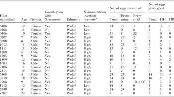
Demographic parameters (gender, age and ethnicity) and the infection status (S. haematobium infection intensity and co-infection with S. mansoni) of participants were defined (see Table 1). From each participant at least two urine and two stool samples were collected. Schistosoma haematobium infection intensity was determined after urine filtration using a filter of 12 µm pore-size (Isopore, USA) and expressed as the number of eggs per 10 mL of urine (ep 10 mL). According to WHO standards, >49 eggs/10 mL corresponds to a high infection intensity, while <50 eggs/10 mL is considered a low infection intensity. Co-infection with S. mansoni was determined using the Kato-Katz technique (Katz et al. Reference Katz, Chaves and Pellegrino1972). The presence or absence of ectopic eggs (S. haematobium eggs in stool samples or S. mansoni in urine samples) was recorded during microscopy.
Host individual information (demographic factors and infection status) and sample information concerning the number of eggs measured and genotyped per host

HH eggs (ITS + cox1 = S. haematobium); HB eggs (ITS = S. haematobium; cox1 = S. bovis).
The number of eggs analysed per host is not related to infection intensity.
a WHO Categories: <50 eggs/10 mL = low infection intensity; >49 eggs/10 mL = high infection intensity.
b Five lateral spined eggs.
Collection of schistosome eggs
Schistosoma eggs from urine samples were isolated by filtering urine through a filter of 12 µm pore-size (Isopore, USA). Schistosoma eggs from stools were isolated by first separating out large debris with water and a metal sieve and then filtering the remaining aqueous solution through a Pitchford and Visser funnel (Pitchford and Visser, Reference Pitchford and Visser1975). Isolated eggs were put in Petri dishes filled with clean water and exposed to sunlight to induce hatching. Unhatched eggs were transferred to a 1·5 mL microtube filled with 95% ethanol.
Egg morphology
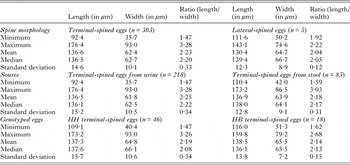
For morphological analysis 303 eggs were randomly selected from 19 individuals infected with S. haematobium. Eggs were temporarily mounted in milli-Q water under a coverslip. Photographs were taken using a Leica DMLB microscope and a Qimaging camera (Fast 1394 or MicroPublisher 5·0 RTV). Measurements were taken using the software programs Auto-Montage Pro (Synoptics Ltd.) or Surveyor (Objective Imaging Ltd.). The egg size (length and width, in μm) and the spine morphology (terminal or lateral) were recorded. As a measure of egg shape, the ratio of egg length divided by egg width was also calculated. For each measurement the mean, median, standard deviation (SD), minimum and maximum was calculated (Table 2) and recorded separately per spine morphology (terminal-spined vs lateral-spined) and per source (eggs from urine vs stools) (Table 2). Lateral-spined eggs were excluded for further analysis. For terminal-spined eggs, the length and length/width ratio per host individual were reported as boxplots, for individuals with minimum ten eggs measured (see Fig. 1). Density histograms were made to visualize the distributions of length and width in terminal-spined eggs and a scatter plot with regression line to illustrate their association (Figs 2 and 3). Analysis of variance (ANOVA) was used to test whether measurements of terminal-spined eggs differed according to source (urine or stool) or host individual. The R package lme4 (Bates et al. Reference Bates, Mächler, Bolker and Walker2015) was used to perform a linear mixed effect analysis of the relationship between egg length, width and ratio with host characteristics. As egg measurements are grouped per host, the variable host individual was included as a random effect in a random intercept model.
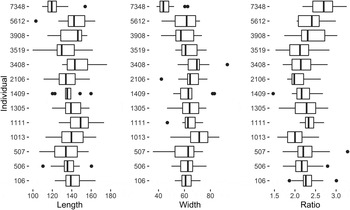
Boxplots for egg length, width and ratio per host individual. Only individuals with at least ten measured eggs are shown. The boxplots are based on median values.

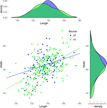
Scatter plot showing the length and width of terminal-spined eggs. The relation between egg length and egg width is indicated by a regression line. Kernel density histograms are shown for egg length and egg width. Eggs originating from urine samples (UE) are shown in green, eggs originating from stool samples (SE) are in blue.

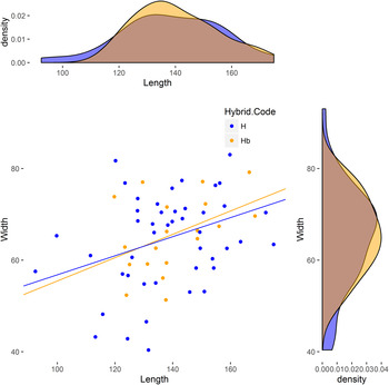
Scatter plot showing the length and width of genotyped, terminal-spined eggs. The relation between egg length and egg width is indicated by a regression line. Kernel density histograms are shown for egg length and egg width. Eggs genotyped as HH (S. haematobium) are shown in blue, HB eggs (S. haematobium × S. bovis) in orange.

Egg measurements from urine and stool samples

HH eggs (ITS + cox1 = S. haematobium); HB eggs (ITS = S. haematobium; cox1 = S. bovis).
Molecular analysis
DNA extraction was performed on 207 out of the 303 eggs following the proteinase K extraction method described in Van den Broeck et al. (Reference Van den Broeck, Geldof, Polman, Volckaert and Huyse2011). Eggs were genotyped with a mitochondrial (mtDNA) and a nuclear marker (ITS rDNA).
mtDNA genotyping: To discriminate between S. haematobium, S. bovis and S. mansoni a rapid diagnostic ‘multiplex’ one-step polymerase chain reaction (RD-PCR) was performed (Webster et al. Reference Webster, Rollinson, Stothard and Huyse2010; Van den Broeck et al. Reference Van den Broeck, Geldof, Polman, Volckaert and Huyse2011). Parasite DNA was subjected to PCR amplification of a partial cox1 mtDNA fragment in a total reaction volume of 25 µL consisting of 1 × PCR buffer, 1·5 mm MgCl2, 200 µ m of each dNTP, 0·4 µ m Asmit 1 (Eurogentec, forward primer TTTTTTGGTCA-TCCTGAGGTGTAT, Bowles et al. Reference Bowles, Blair and McManus1992), 0·2 µ m of each reverse primer [Eurogentec; S. haematobium (Sh.R.) TGATAATCAATGACCCTGCAATAA 543 bp (Webster et al. Reference Webster, Rollinson, Stothard and Huyse2010), S. bovis (Sb.R) CACAGGATCAGACAAACGAGTACC 306 bp (Webster et al. Reference Webster, Rollinson, Stothard and Huyse2010), S. mansoni (Sm.R) TGCAGATAAAGCCACCCCTGTG 375 bp (Van den Broeck et al. Reference Van den Broeck, Geldof, Polman, Volckaert and Huyse2011)], 1 unit Taq polymerase (Eurogentec), milli-Q water and 2 µL of DNA. PCR parameters during thermal cycling were 3 min at 95 °C, followed by 40 cycles of 30 s at 94 °C, 1 min 30 s at 58 °C and 1 min 30 s at 72 °C followed by a final cycle at 72 °C for 7 min. PCR products were visualized on a 1% Midori Green agarose gel.
ITS rDNA genotyping: Samples were subjected to PCR amplification of the complete ITS rDNA region [981 bp; primers ITS4: TCCTCCGCTTATTGATATGC and ITS5: GGAAGTAAAAGTCGTAACAAG (Barber et al. Reference Barber, Mkoji and Loker2000)]. PCR amplification was performed in a volume of 25 µL containing 1 × PCR buffer, 2 mm MgCl2, 200 µ m of each dNTP, 2 mm of each primer, 1 unit Taq polymerase (Eurogentec), milli-Q water and 2 µL of DNA. Thermal cycling started with 3 min at 95 °C, followed by 40 cycles of 30 s at 94 °C, 45 s at 54 °C and 1 min 30 s at 72 °C followed by a final cycle at 72 °C for 7 min. PCR products were analysed by restriction fragment length polymorphism (RFLP) and visualized on a 1% Midori Green agarose gel. Digestion of the ITS fragment with MboI (Fermentas) distinguishes S. haematobium from S. bovis and S. mansoni. Schistosoma haematobium PCR products are digested in fragment sizes of (in bp) 280, 147, 130, 100, 85 and 36; S. bovis in fragments sizes of 380, 174, 167, 130, 83 and 36; and S. mansoni in sizes of 380, 176, 174, 130, 81, 16 and 9 bp. Samples showing a S. bovis or S. mansoni MboI pattern were subsequently digested with AluI (Fermentas), which differentiates between S. bovis (no cut; 981 bp) and S. mansoni (fragments of 545 and 421 bp). To confirm the genotype based on restriction enzymes, for a random subset of eggs (n = 11) the ITS rDNA region was sequenced by Sanger sequencing. The PCR products were purified using the E.Z.N.A Cycle Pure kit (Omega Bio-tek, Norcross, USA). Sequencing was done using a Big Dye Chemistry Cycle Sequencing kit (version 3.1) in a 3130 Genetic Analyser (Applied Biosystems). Sequences were assembled and manually edited using Geneious® 6.1.6. (http://geneious.com/).
Egg morphology in relation to genotype
The measurements of terminal-spined eggs were related to their genotype. Genotype classes were based on the genotype of cox1 mtDNA [S. haematobium (H), S. bovis (B) or S. mansoni (M)] and the ITS rDNA genotype (H and/or B and/or M). A scatter plot and density histograms were generated to visualize the length and width of genotyped eggs per genotype-class (Fig. 3). ANOVA was used to compare the length, width and ratio of eggs among genotype groups. R version 3.1.3 (R Development Core Team, 2015) was used for all statistical analyses.
RESULTS
Dataset and host information
We measured 303 eggs from 19 different individuals. 218 eggs originated from urine of 16 patients, 85 eggs were extracted from stool samples of eight patients (see Tables 1 and 2).
Table 1 provides host demographic information and infection status. Individuals were between four and 22 years old, nine males and ten females. The majority of individuals (13) had a high S. haematobium infection intensity (>49 eggs/10 mL) whereas six individuals had a low infection intensity (<50 eggs/10 mL). Nine individuals were co-infected with S. mansoni; the S. mansoni infection intensities were low (1–99 epg). In eight of the 19 selected individuals ectopic S. haematobium eggs were reported from stools during microscopic analysis of Kato–Katz slides.
Egg morphology
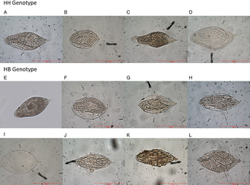
All eggs extracted from urine samples were terminal-spined, i.e. no ectopic S. mansoni eggs were present in urine samples. Five eggs originating from one stool sample had a lateral spine typical for S. mansoni and were excluded from further analyses. All other eggs from stools (94%) were ectopic, i.e. terminal-spined. The eggs showed only minor variations in shape. Most eggs were round-to-oval, resembling typical S. haematobium eggs (e.g. Fig. 4B, F–J andL). A few eggs appeared very slightly spindle-shaped (e.g. Fig. 4C). However, no distinct classes of egg shape could be distinguished. Egg size showed a high variability. Terminal-spined eggs had a length ranging from 92·4 to 176·4 µm (mean 136·6 µm) and a width ranging from 35·7 to 93·0 µm (mean 62·4 µm). The length/width ratio ranged from 1·47 to 3·28 (see Table 2). The distributions of egg length and egg width were continuous and unimodal as shown by density histograms (Figs 2 and 3).
Photographs of a selection of terminal-spined eggs measured in this study. Eggs A–D were genotyped as HH (S. haematobium) based on ITS and cox1, the eggs E–L were genotyped as HB (S. haematobium × S. bovis).

The mean size of terminal-spined eggs from stool samples (n = 85) was not significantly different from those from urine samples (n = 218). The mean size of eggs was significantly different between host individuals (ANOVA, P < 0·001). Linear mixed models (LMM) indicated that a correlation was present of egg size within individuals. On average 25% of the variation in egg length was present between individuals, 75% of the variation in egg length was present within an individual. The correlation of egg width within individuals was slightly higher than egg length: 30% of the variation was present between individuals, 70% of the variation within individuals. For the length/width ratio 23% was present between individuals and 77% within individuals. Figure 1 shows a visual representation of egg length, egg width and the length/width ratio for each host individual with at least ten eggs measured. We did not find a consistent relation between egg size and host characteristics (age, gender, ethnicity, infection intensity and co-infection with S. mansoni).
Molecular analysis
We performed DNA extraction and molecular analysis on 166 terminal-spined eggs from urine, 38 terminal-spined eggs from stool and three lateral-spined eggs from stool. The success rate of the PCR reactions was rather low, especially for eggs from stools. In total, 66 terminal-spined eggs (32%; 59 from urine, seven from stool) were successfully genotyped for both cox1 and ITS rDNA. Based on the RFLP profiles of ITS rDNA and cox1, 46 eggs were identified as S. haematobium (HH; ITS = haem, cox1 = haem) and 19 eggs (29%) as S. haematobium × S. bovis hybrids (HB; ITS = haem, cox1 = bovis). No eggs were found with a reverse profile (BH; ITS = bovis, cox1 = haem), nor with a pure S. bovis profile or a hybrid ITS profile with fragment sizes of both species. To confirm the RFLP results the ITS region was sequenced for 11 randomly chosen eggs. All 11 sequences were identical to the S. haematobium isolate from Northern Senegal (Accession no. FJ588861) described in Huyse et al. (Reference Huyse, Webster, Geldof, Stothard, Diaw, Polman and Rollinson2009). HB eggs were found in 10 out of 15 individuals with at least one egg genotyped.
Egg morphology in relation to genotype
Hybrid S. haematobium × S. bovis eggs (HB) were slightly larger and varied less in size and shape (length/width ratio) than ‘pure’ S. haematobium eggs (HH) (Table 2). Differences were however not significant (Fig. 3). HB were present in stools (=ectopic eggs) and urine. Seven ectopic eggs were successfully genotyped; four of these had a HH genotype and three had a HB genotype. From the 59 terminal-spined eggs in urine 16 eggs were HB and 42 were HH. Egg measurements were not associated with egg genotype (Fig. 3), nor did egg shape correlate with egg genotype. Several HB eggs showed the round-to-oval shape typical for pure S. haematobium (Fig. 4E–L), and one HH egg was somewhat spindle-like shaped, more typical for S. bovis (Fig. 4C) (Pitchford, Reference Pitchford1965).
DISCUSSION
The morphology of terminal-spined eggs from the SRB
This study is the first to thoroughly study the egg morphology of S. haematobium × S. bovis hybrids collected from human samples. Although the eggs of both parental species, S. haematobium and S. bovis, are terminal-spined, they differ in several important aspects. Typically, S. haematobium eggs are deposited in urine, are round-to-oval in shape and range in length from 83 to 187 µm (Pitchford, Reference Pitchford1965; Table S1). In contrast, S. bovis eggs are deposited in stool and are spindle-shaped, consisting of a broad middle portion and drawn-out rod-like ends, one bearing a well differentiated spine, the other evenly rounded (Taylor, Reference Taylor1970; Touassem, Reference Touassem1987). Schistosoma bovis eggs are also larger than those of S. haematobium, their length usually exceeding 200 µm and reaching up to 300 µm (Taylor, Reference Taylor1970; Touassem, Reference Touassem1987; Table S1). The egg sizes obtained in this study (136·6 ± 14·6 µm × 62·4 ± 10·1 µm) correspond very well with S. haematobium egg sizes but not with S. bovis egg sizes (Table S1, Khalil, Reference Khalil1924; MacHattie et al., Reference MacHattie, Mills and Chadwick1933; Alves, Reference Alves1949; Taylor, Reference Taylor1970; Loker, Reference Loker1983; Southgate et al., Reference Southgate, Rollinson, Ross, Knowles and Vercruysse1985 and Touassem, Reference Touassem1987). The range of the egg lengths (92·4–176·4 µm) falls within the range of reported S. haematobium egg lengths. The mean egg width obtained in this study (62·4 ± 10·1 µm) was rather large in comparison with other studies. However, only few authors have reported the width of S. haematobium eggs from humans. De Clercq et al. (Reference De Clercq, Rollinson, Diarra, Sacko, Coulibaly, Landouré, Traoré, Southgate, Kaukas and Vercruysse1994) reported a mean width of 55·0 ± 5·0 µm from the Bankass district in Mali, and Pitchford (Reference Pitchford1959) found a mean of 51·4 µm for eggs from South Africa. Vercruysse et al. (Reference Vercruysse, Southgate and Rollinson1984) measured the egg width of a strain that originated from children from Guédé Chantier (SRB) and that was subsequently cycled in mice. These eggs had almost the same width (62 ± 12 µm) as those examined in this study but the length was larger (153 ± 11 µm).
The mean egg length/width ratio found in this study (2·23) also corresponds with the mean ratios of S. haematobium eggs reported in other studies (2·47 to 2·53). The range of the ratio of the eggs was rather broad (1·47–3·28). A ratio of 3·28 approaches the mean ratio of S. bovis eggs, which are more elongated than S. haematobium eggs (3·41–3·85, Table S1). This suggests that some eggs in this study, despite being smaller in size, may have a shape resembling S. bovis. However, we found only one egg with a somewhat spindle-like shape (Fig. 4C). Evidently, the morphological methods used in this study (visual inspection of photographs and calculation of the length/width ratio) can capture only some aspects of the egg shape. In theory, the egg shape could be determined more precisely by taking additional measurements or by using geometric morphometrics. However, such methods are time-consuming and hence would be impractical in the context of the high-throughput screening of samples, especially in the field. Besides, egg size and shape (length/width ratio) were highly variable; about 70% of this variation was present within a single patient. Intraspecific variability is known to impede species identification (Almeda et al. Reference Almeda, Ascaso, Marçal, Corachan, Southgate and Rollinson1996) and therefore complicates the differentiation between pure and hybrid species. Finally, it should be noted that none of the studies mentioned above used molecular methods to verify species status. Hence, some of these measurements could include HB rather than pure S. haematobium or S. bovis eggs.
The HB eggs from Senegal do not closely resemble the eggs from previous experimental hybridization studies. Taylor (Reference Taylor1970) performed experimental crosses between S. bovis ♀ (from Iran) and S. haematobium ♂ (from Iran and Nigeria). Parental S. bovis eggs were bigger and had always a spindle shape in contrast to S. haematobium eggs, which suggests that Taylor probably used pure parental species for his experiments. First generation hybrid (F1) eggs of the crosses were typical of S. bovis in morphology. This is in line with expectations as the egg shape of schistosomes is supposed to be determined by the female ootype (Smyth and Clegg, Reference Smyth and Clegg1959). In this large, egg-shaped chamber that is part of the oviduct, the eggshell is formed. F2 eggs were intermediate between S. bovis and S. haematobium, and similar in size to S. intercalatum (length 175·0 ± 8·5 µm; width 61·7 ± 2·6 µm) or S. mattheei (173·0 ± 14·0 µm; 52·6 ± 4·5 µm) (Taylor, Reference Taylor1970). F3 eggs were similar in shape to those of the F2 generation but less viable. Similar results were obtained by Brémond et al. (Reference Brémond, Sellin, Sellin, Nameoua, Labbo, Theron and Combes1993) who performed experimental crossings between S. bovis and S. haematobium from Niger, resulting in hybrids with intermediate egg morphology. It should be noted that both Taylor (Reference Taylor1970) and Brémond et al. (Reference Brémond, Sellin, Sellin, Nameoua, Labbo, Theron and Combes1993) only studied F1–F3 crosses. Such crosses cannot be directly compared with the HB from this study, which are probably the result of back-crossing with parental species as suggested by the dominance of S. haematobium ITS profiles.
Recently an outbreak of human urinary schistosomiasis was reported from Corsica (France). Moné et al. (Reference Moné, Holtfreter, Allienne, Mintsa-Nguéma, Ibikounlé, Boissier, Berry, Mitta, Richter and Mouahid2015) sequenced eggs isolated from infected tourists and identified, based on partial cox1 and ITS rDNA sequencing, both ‘pure’ S. haematobium and hybrid crosses between S. haematobium and S. bovis. They described two morphotypes of eggs, typical round-to-oval eggs (four out of 15 eggs) and more elongated eggs referred to as ‘non-typical’ S. haematobium eggs. The 15 eggs measured 106·5 ± 6·2 µm × 42·8 ± 5·4 µm, which is smaller than the mean egg sizes we found in Senegal (136·6 ± 14·6 µm × 62·4 ± 10·1 µm) and almost half the length of ‘typical’ S. bovis eggs (Table S1). Unfortunately, morphological and molecular analyses were not performed on the same eggs, so the link between morphotype and genotype could not be studied. Similar to our study, eggs resembling typical S. bovis shape were not found (Moné et al. Reference Moné, Holtfreter, Allienne, Mintsa-Nguéma, Ibikounlé, Boissier, Berry, Mitta, Richter and Mouahid2015). Soentjens et al. (Reference Soentjens, Cnops, Huyse, Yansouni, De Vos, Bottieau, Clerinx and Van Esbroeck2016) studied a case of S. haematobium × S. bovis infection in travellers from Mali. Eggs were suspected to be hybrids because of the lozenge shape, which was intermediate between the typical round-to-oval S. haematobium eggs and the spindle-shaped S. bovis eggs. The length of the eggs varied between 110 and 175 µm. DNA sequencing of two eggs confirmed their hybrid constitution. The eggs in these two studies show the same discordance between the nuclear (S. haematobium) and mitochondrial (S. bovis) genotype Although the reported egg sizes differ in the study of Moné et al. (Reference Moné, Holtfreter, Allienne, Mintsa-Nguéma, Ibikounlé, Boissier, Berry, Mitta, Richter and Mouahid2015) the descriptions of atypical egg shapes show resemblance with the few atypical eggs observed in this study (Fig. 4C).
Link between egg morphology and genotype?
The genetic constitution of the eggs confirmed the presence of natural crosses between S. haematobium and S. bovis in Pakh. Almost 30% of the eggs (n = 19) had a S. bovis cox1 genotype, while the ITS genotype was always identical to S. haematobium. Eggs with a ‘pure’ S. bovis genotype were absent in this study, as were eggs with a typical S. bovis size and spindle shape. In our study, the egg size showed a high variability. This variation was not linked to the genotype (ITS and cox1), neither to the origin of the eggs (urine or stool). Although 70% of the variation was present within individuals, egg sizes differed significantly between individuals and 30% of the variation in egg size was attributable to differences between individuals. We could however not link this variation to specific host characteristics (age, sex, infection intensity), neither to the presence of hybrid eggs.
Different schistosome species or strains have typically different mean egg sizes or different maxima when plotting density curves (Pitchford, Reference Pitchford1965; Almeda et al. Reference Almeda, Ascaso, Marçal, Corachan, Southgate and Rollinson1996). The unimodal distribution of egg-measurements in this study and the absence of morphological differences between HH eggs and HB eggs suggest that both genetic classes of eggs belong to one random mating schistosome population, without mating restrictions in the next generation. The observation that HH and HB eggs were found together in almost all studied host individuals, supports the idea of random mating. This leads to the hypothesis that the majority of parasite stages found in Pakh stem from a (historical) hybridization event. Repeated backcrossing with S. haematobium not only results in a dilution of the introduced S. bovis mitochondrial genes, but also in a mixing and spreading of these genes into the population. As a consequence, a varying degree of introgression with S. bovis genes is likely to be present at the nuclear level of both the hybrid and the so-called ‘pure’ eggs, which is not detectable with traditional ITS barcoding. This is also suggested by the HH genotype of the egg with the spindle-shape, typical of S. bovis eggs (Fig. 4C).
The strengths and limitations of molecular markers for hybrid detection
In combination with a mtDNA marker, the internal transcribed spacer rDNA (ITS) is considered a good marker to detect hybridization as it retains both parental copies for several generations (Sang et al. Reference Sang, Crawford and Stuessy1995; Steinauer et al. Reference Steinauer, Hanelt, Mwangi, Maina, Agola, Kinuthia, Mutuku, Mungai, Wilson, Mkoji and Loker2008). However, due to concerted evolution, a phenomenon that removes paralogous sequences (for example ITS copies) from the genome and eliminates intra-individual polymorphism (Garcia et al. Reference Garcia, Silva, Carniello, Veldman, Rossi and de Oliveira2011) a biased homogenization towards one of the parental sequences can already occur in F2 hybrids (Fuertes Aguilar et al. Reference Fuertes Aguilar, Rosselló and Nieto Feliner1999). As a consequence, in case of unequal back-crossing with only one of the parental species (e.g. S. haematobium), the signal of the other parental species (e.g. S. bovis) will fade out. When both the mtDNA and the ITS rDNA sequences are identified as S. haematobium we can therefore not reliably assign them as ‘pure’ since we cannot rule out crossing with S. bovis in previous generations, nor can we rule out introgression of other nuclear S. bovis genes (e.g. maintained through positive selection). As such, the current method (combining cox1 barcoding and ITS1) is limited in regard to detecting historical hybridization events and will therefore underestimate the presence of hybrids.
The presence of a S. bovis cox1 profile easily reveals (historical) hybridization and we therefore advocate the use of the rapid diagnostic PCR assay used in this study. We do need however more sensitive nuclear markers to identify hybridization at the nuclear level. Microsatellite markers will simultaneously sample more genomic regions and therefore increase the chance to pick up introgressed regions, but they too appeared limited in detecting hybrids between S. mansoni and S. rodhaini (Steinauer et al. Reference Steinauer, Hanelt, Mwangi, Maina, Agola, Kinuthia, Mutuku, Mungai, Wilson, Mkoji and Loker2008). The best method until now appears to be cloning different ITS copies to detect both parental ITS variants, or single-stranded conformational polymorphism (SSCP) analyses (see Webster et al. Reference Webster, Tchuem Tchuenté and Southgate2007; Steinauer et al. Reference Steinauer, Hanelt, Mwangi, Maina, Agola, Kinuthia, Mutuku, Mungai, Wilson, Mkoji and Loker2008). While the former method is very labour-intensive, the latter method only consists of PCR analysis and gel electrophoresis. SSCP analysis can pick up different ITS variants because single base-pair changes affect the migration on a gel.
Concluding remarks
Due to the absence of distinct egg morphotypes and the large intra-individual variation in egg sizes, egg morphology appears of limited value to detect hybridization events between S. haematobium and S. bovis in natural field isolates. Despite being efficient for high-throughput screening of field samples, traditional barcoding cannot detect introgression of S. bovis genes at the nuclear level. As this may result in an underdetection of hybrid schistosomes, other detection methods are needed. The detection of hybrids is essential to control the transmission of schistosomiasis and to select the best treatment for infected communities (Leger and Webster, Reference Leger and Webster2016). Whole genome analysis of natural field stages is therefore needed to understand the introgression of genes from one species into the other in natural conditions (Twyford and Ennos, Reference Twyford and Ennos2012), while hybridization experiments are needed to study the phenotype and fitness of different hybrid crosses compared to the pure parental strains.
SUPPLEMENTARY MATERIAL
The supplementary material for this article can be found at https://doi.org/10.1017/S0031182017000087.
ACKNOWLEDGEMENTS
We thank the field team of Richard Toll, Abdoulaye Yague, Mankeur Diop, Magette Ndao and Safall Boh for assisting with the sample collection and microscopic analysis and Bart Hellemans for assisting with DNA sequencing. Finally, a special thanks to the villagers of Pakh for their kind participation in the study.
FINANCIAL SUPPORT
N.B. benefited from a VLADOC Ph.D. fellowship of Flemish Interuniversity Council (VLIR-UOS). Research was supported by a Research Grant of the Research Foundation – Flanders (contract G.0552.10) and a Krediet to navorsers (contract 1516913N) to T.H.