Significant outcomes
• Emotion and cognitive processes appears to contribute to slowing of movement in bipolar depression.
• The functional deafferentiation theory for bradykinesia in Parkinson's disease seems not to be applicable to bipolar depression.
Limitations
• Small sample size.
• Medication effects may bias cortical and subcortical activation patterns.
• No control for task performance.
Introduction
Motor retardation (RET) is a classic feature of depression and has been the focus of numerous investigations over the years. RET displays as a general slowing of movement, slumping in posture, diminished facial expression and decreased rate and lower tone of speech. The presence of RET is important for diagnostic assessment of depression subtypes and is more common in the bipolar depression subtype, where episodes of mania alternate or co-exist with periods of depression Reference Mitchell and Malhi1, Reference Mitchell, Wilhelm, Parker, Austin, Rutgers and Malhi2. RET also seems to predict beneficial treatment response to pro-dopaminergic drugs Reference Austin, Mitchell and Hadzi-Pavlovic3, Reference Schrijvers, Hulstijn and Sabbe4, Reference Buyukdura, McClintock and Croarkin5, Reference Calugi, Cassano and Litta6, Reference Mah, Zarate, Nugent, Singh, Manji and Drevets7.
The mediators of RET in depression have, however, been difficult to single out Reference Sobin and Sackeim8. A review of investigations into motor disturbances in depression shows deficits and decreased processing speed in both cortical brain regions, primarily in the frontal cortex and in subcortical motor systems involved in motor control Reference Schrijvers, Hulstijn and Sabbe4. Functional magnetic resonance imaging (fMRI) studies using manual reaction time tasks have shown how cortical and subcortical brain regions are activated differently in patients with bipolar depression compared to healthy controls Reference Caligiuri, Brown and Meloy9, Reference Caligiuri, Brown and Meloy10. However, it remains unclear how cortical brain regions that are involved in the preparation and execution of movement activate in relation to subcortical regions involved in motor control.
Parkinson's disease is a neurodegenerative movement disorder in which dopamine neurons deteriorate in the midbrain, leading to low levels of dopamine. This impairs movement facilitation by subcortical structures, resulting in observable slowing and delayed initiation of movement: bradykinesia and akinesia. RET in depression shares these motor features with Parkinson's disease. Moreover, pro-dopaminergic drugs that alleviate bradykinesia and akinesia in Parkinson's disease have the same effect on RET in depression Reference Dewey, Hutton, LeWitt and Factor11, Reference Kano, Ikeda and Kiyozuka12.
In Parkinson's disease, the functional deafferentiation theory has been a successful theoretical framework to explain the genesis of motor disturbances Reference DeLong and Wichmann13, Reference DeLong and Wichmann14. This theory predicts changes in the cortico-striatal-thalamo-cortical motor circuit (hereafter referred to as the ‘motor circuit'), where decreased dopamine release in the putamen and pallidum leads to decreased subcortical output to pre- and primary motor cortices. This impairs the preparation, control and execution of movement, resulting in bradykinesia and akinesia. These features resemble those seen in bipolar depression, which led us to adopt the functional deafferentiation framework in examining motor symptoms in bipolar depression. The functional deafferentiation theory is supported by a growing body of neuroimaging data Reference Dagher and Nagano-Saito15.
Using the framework of the functional deafferentiation theory, we hypothesised that RET in bipolar depression is mediated by disrupted activation of subcortical structures, leading to decreased activation of cortical motor areas during finger tapping, which is a simple measure of motor speed.
Aims of the study
We used fMRI to investigate the motor circuit during self-paced continuous finger tapping and studied whether regions that mediate preparation, control and execution of movement are activated differently in subjects with bipolar depression compared to healthy controls.
Material and methods
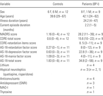
We recruited nine patients with a bipolar I diagnosis and a current episode of depression from The Affective Disorders Unit at Psychiatry Southwest at Karolinska University Hospital in Huddinge, Sweden. Twelve healthy controls without psychiatric diagnoses were recruited. The Karolinska University Hospital and Stockholm City Council Ethics Committee approved the protocol. Each subject gave oral and written informed consent to participate in the study. All participants were right handed, had no history of neurological illness and had good visual acuity. All patients were on medications and all controls were drug free. When patient files were reviewed, we did not find any notes indicating secondary Parkinsonism around the time of scanning. We confirmed the clinical diagnoses in patients and the absence of diagnoses in controls with a structured clinical interview (SCID-I, computerised version). No patient fulfilled the diagnostic criteria for concurrent mania, hypomania or rapid cycling. We rated depression severity with the Montgomery Åsberg Depression Rating Scale (MADRS) Reference Asberg, Montgomery, Perris, Schalling and Sedvall16. We rated motor retardation with the CORE-scale, where retardation (CORE-R) and total score (CORE) were calculated Reference Hickie, Mason and Parker17. Subjects self-rated their affective state using the AS-18 scale. Subject characteristics are presented in Table 1. B. L. and M. A. performed all interviews.
Sample characteristics

BP-I bipolar disorder, type I; F, female; M, male.
The finger tapping test measures motor speed Reference Sweeney, Slade, Ivins, Nemeth, Ranks and Sica18. All participants performed finger tapping while in the MRI-scanner, following instructions on a computer screen shown to them using a mirror. Before the experiment started, participants were shown how to perform finger tapping, defined as a thumb-index finger opposition of the right hand. Participants were asked to tap as quickly as possible. The experiment had an on/off design consisting of 20 s of finger tapping followed by 20 s of rest. During the first second of each on-period the instruction ‘tap’ was presented. The screen then turned black and the task was performed followed by rest. The stimulus cycle was repeated seven times. The total functional scanning time was 280 s.
A Siemens 1.5 T scanner (Erlangen, Germany) was used to acquire blood-oxygen-level-dependent (BOLD) sensitive T2*-weighted echo planar images. Each echo planar image comprised 22 axial slices with a resolution of 3.75 × 3.75 × 5 mm, and an inter-slice interval of 1 mm. Volumes were acquired with a repetition time (TR) of 2.5 s and the first six (dummy) volumes of each run were discarded to allow for T1 equilibration effects. A total of 112 volumes were acquired during one run. After the functional scans had been collected, a T1-weighted anatomical image (MP-RAGE, 128 slices, TR 2400 ms, TE 3.44 ms, with a voxel size of 1.3 × 1.3 × 1.3 mm) was acquired for all subjects. M. K-W., a consultant in neuroradiology, did a radiological assessment of the anatomical scans of each subject to rule out radiological signs of pathology.
We did three analyses. The first two were whole-brain analyses. In the first analysis, we used a cluster forming threshold (Z = 3.1) and a corrected cluster significance threshold (p = 0.05) Reference Worsley, Jezzard, Matthews and Smith19. In the second analysis, we used an arbitrary, less conservative and uncorrected statistical threshold (p = 0.001). The third analysis was a hypothesis driven region-of-interest (ROI) investigation into restricted sets of voxels. These voxels corresponded to the motor circuit: the putamen, pallidum, thalamus, medial and lateral premotor cortex, and the primary motor cortex, respectively. The ROI masks were derived from the probabilistic Harvard-Oxford Cortical Atlas (‘Juxtapositional lobule/Formerly supplementary motor area’ corresponding to the mesial premotor cortex) Reference Desikan, Segonne and Fischl20 and the probabilistic Jülich histological Atlas (Brodmann area 4a, 6 corresponding to the primary motor and premotor cortex) supplied with FSL Reference Geyer, Ledberg and Schleicher21, Reference Geyer22. Masks for the putamen, pallidum and thalamus were derived from a mean template of all subjects. The subcortical structures were automatically segmented using FIRST Reference Patenaude, Smith, Kennedy and Jenkinson23. We set a corrected significance threshold (p = 0.05) in the third analysis Reference Worsley, Jezzard, Matthews and Smith19.
We used FSL 4.1.5 software (FMRIB, Oxford University, UK) to analyse imaging data. We processed data with FEAT (FMRI Expert Analysis Tool) Version 5.98. We corrected each subject's run for head motion Reference Jenkinson, Bannister, Brady and Smith24. To account for time difference in slice acquisition, we performed slice-timing correction using Fourier-space time-series phase-shifting. We removed non-brain tissue Reference Smith25. We smoothed functional data to compensate for anatomical variability after registration, and to permit application of Gaussian random field theory for the corrected statistical inference. We used a Gaussian kernel set to FWHM 8 mm. Also, we normalised the grand-mean intensity of the entire 4D dataset by a single multiplicative factor, and filtered out physiological noise using high-pass temporal filtering set to 100 s. We calculated the parameter estimates (PE) for all brain voxels using the general linear model, comparing each condition with rest (i.e. the baseline). We used local autocorrelation correction to do the time-series statistical analysis Reference Woolrich, Ripley, Brady and Smith26. Then we used nonlinear registration to further refine registration from high-resolution structural to standard space Reference Jenkinson and Smith27, Reference Andersson28. We then used the subject-specific images of the contrast (finger-tapping > rest) in the second level group analysis.
To do the first higher level analysis, we used FLAME (FMRIB's local analysis of mixed effects) stages 1 and 2 with automatic outlier detection Reference Woolrich, Jbabdi and Patenaude29, Reference Woolrich, Behrens, Beckmann, Jenkinson and Smith30, Reference Woolrich31, Reference Beckmann, Jenkinson and Smith32. Z (Gaussianised T/F) statistic images were thresholded using clusters determined by Z > 3.1 and a (corrected) cluster significance threshold of p = 0.05 Reference Worsley, Jezzard, Matthews and Smith19. We did the second higher level analysis using FLAME stages 1 and 2 with automatic outlier detection. In this analysis, the Z statistic images were thresholded using an uncorrected significance threshold of p = 0.001. We did the third, ROI, analysis using FLAME stage 1. FLAME stage 2 and automatic outlier detection were not used in the ROI analyses due to being computationally exhaustive. The Z statistic images in the ROI analyses were thresholded using a corrected significance threshold of p = 0.05.
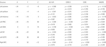
We correlated the patients' clinical rating scores (MADRS, CORE, CORE-R and AS-18-R) with peak PE values in patients' anatomical structures pertaining to the left motor circuit, the dorsolateral prefrontal cortex and the ventromedial prefrontal cortex (Table. 3). We searched for linear associations between changes in the BOLD signal and clinical ratings using the Python based statistical software SciPy. We used a non-parametric test for the correlation analysis: Spearman's rho. We set the significance threshold to p = 0.05.
Results
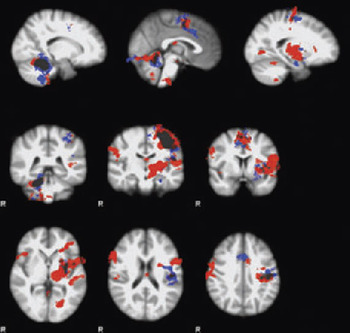
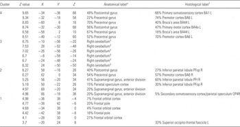
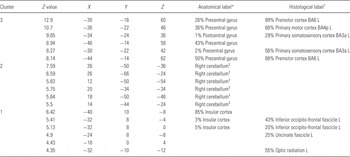
Both patients and controls significantly activated the left motor circuit and right cerebellum while performing the task with their right hand. However, there were differences in the activation patterns of brain structures pertaining to preparation, control and execution of movement between patients and controls (Table 2, Table. 3, Fig. 1).
Activated voxels during self-paced finger-tapping (cluster forming threshold Z = 3.1, corrected p = 0.05). The cortico-striatal-thalamo-cortical motor circuit was significantly activated in both groups. Accordingly, left sensorimotor cortex, premotor areas, posterior putamen, globus pallidus, thalamus and ipsilateral cerebellum were activated. Subcortical activations are less widespread in the patient group. In patients, there was no significant activation of the insula or premotor areas on the right side. Left is right in the image. Red clusters represent controls. Blue clusters represent patients.

Co-ordinate list of activated clusters and their local maxima during finger tapping in controls (cluster-forming threshold Z = 3.1, corrected p = 0.05)

Empty space means no label was found.
* Harvard-Oxford Cortical Atlas.
† Julich Histological Atlas.
‡ MNI Atlas.
Co-ordinate list of activated clusters and their local maxima during finger tapping in patients (cluster-forming threshold Z = 3.1, corrected p = 0.05)

Empty space means no label was found.
* Harvard-Oxford Cortical Atlas.
† Julich Histological Atlas.
‡ MNI Atlas.
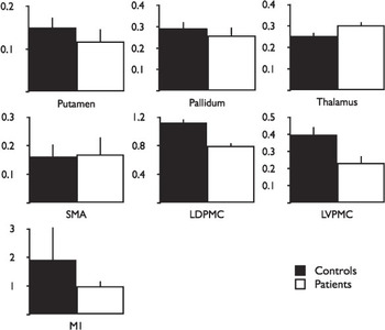
In our first, stringent analysis of mean activation in patients (cluster-forming threshold Z = 3.1, corrected p = 0.05) we saw non-significant signal decreases in the lateral dorsal and ventral premotor cortex, as well as in the primary motor cortex, compared to controls (Fig. 2). In this analysis, the activation of the supplementary motor area proper was increased in patients compared to controls. In patients, the medial premotor activations also extended rostrally, and voxels approximating the posterior rostral cingulate zone bilaterally were more anteriorly widespread compared to controls. There were no significant activations in the right insula, right primary motor cortex or subcortical structures (Fig. 1). The whole-brain analysis subtracting control activation from patient activation did not reveal any significant between-group differences (cluster-forming threshold Z = 3.1, corrected p = 0.05).
Brain areas involved in preparation, control and execution of movement is affected in depressed bipolar patients with motor retardation. Activity in the putamen and pallidum indicates disrupted signals, which lead to an increased activation of the thalamus and supplementary motor area. Both dorsal and ventral lateral premotor areas and the primary motor cortex display decreased activity in patients. The image illustrates percentage blood oxygen level dependent (BOLD) signal change compared to rest in brain areas comprising the motor circuit during right hand self-paced finger tapping. Black bars represent controls. Values represent the mean from the peak voxel and the 26 surrounding ones. Error bars represent the standard deviation. SMA, supplementary motor area; LDPMC, lateral dorsal premotor cortex; LVPMC, lateral ventral premotor cortex; M1, primary motor cortex.

In our stringent analysis of mean activation in controls, the activations appeared more widespread in all of the structures that make up the motor circuit, although these differences were non-significant (cluster-forming threshold Z = 3.1, corrected p = 0.05, Fig. 1). In addition, the control group showed bilateral activations of the insula and right-side activations of the lateral premotor cortex. The right pre-supplementary motor area was significantly activated and the cluster stretched beyond the ventral anterior commissure line to y = 20 in a region classified as premotor cortex and BA6. Also, the left lateral orbitofrontal cortex was significantly activated. However, the whole-brain analysis subtracting patient activation from control activation did not reveal any significant between-group differences (cluster-forming threshold Z = 3.1, corrected p = 0.05).
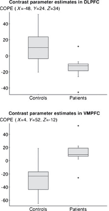
Our less stringent whole-brain analysis showed significant group differences in the activation of the frontal cortex (Fig. 3). Controls activated their left dorsolateral prefrontal cortex significantly more than patients (p ≤ 0.001, uncorrected). Patients showed negative mean activation in the dorsolateral prefrontal cortex, whereas controls showed positive mean activation. Patients activated their right medial frontal cortex and left parietal operculum significantly more than controls (p ≤ 0.001, uncorrected). Patients showed positive mean activation in the ventromedial prefrontal cortex while controls showed negative mean activation.
Activation in two prefrontal brain areas involved in cognition and emotion. These contained the largest clusters with differential activation between patient and controls in the less stringent analysis (p ≤ 0.001, uncorrected). The image illustrates the peak contrast PE (COPEs, finger tapping > rest) and the standard space coordinates in the right ventromedial and left dorsolateral prefrontal cortex. The COPE values reflect the degree of activation explained by the modelled time course of signal change during the experimental task. DLPFC, dorsolateral prefrontal cortex; VMPFC, ventromedial prefrontal cortex.

The hypothesis driven ROI analysis showed non-significant differences in the activation patterns in the respective groups. Only in the control group were all structures pertaining to the left motor circuit activated. The left and right putamens were activated only in the control group (Z ≥ 2.5). The left and right pallidum were activated in both groups (Z ≥ 2.0). The left thalamus was activated in both groups, but the right thalamus only in the control group (Z ≥ 2.9). The left supplementary motor area was activated in both groups (Z ≥ 3.5). In controls, there was more widespread activation in the regions that were activated in both groups. The right lateral premotor cortex was only activated in controls (Z ≥ 3.2). The left premotor cortex was activated in both groups (Z ≥ 3.2). The left primary motor cortex was significantly activated in both groups (Z ≥ 3.5). There were fewer significantly active voxels in the patient group whenever both groups activated the same region. However, none of the ROI analyses revealed any significant differences between patient and controls (control > patients, patient > controls, corrected p = 0.05).
There was a trend towards higher correlation between peak activations and the AS-18-R compared to other rating scales. However, none of the clinical rating scales correlated significantly with any of the activations in structures pertaining to the left motor circuit, the dorsolateral or ventromedial prefrontal cortex (Table. 4).
Correlations between clinical ratings and the peak voxel parameter estimate in the patient group

DLPFC, dorsolateral prefrontal cortex; M1, primary motor cortex; MFC, medial frontal cortex; ρ, rho; p, p value; PMC, premotor cortex; SMA, supplementary motor area.
Discussion
We hypothesised in this study that RET in bipolar depression is mediated by disrupted subcortical activation, leading to decreased activation of cortical motor areas during self-paced finger tapping. This study does not support the functional deafferentiation theory implicating the motor circuit as a single explanation for RET in bipolar depression. Using fMRI, we found statistically significant differences between patients and controls in the brain regions that mediate cognition and emotion, with patients showing negative mean activation of the left dorsolateral prefrontal cortex and increased activation in the ventromedial prefrontal cortex compared to controls. We also found discernible but not statistically significant differences between patients and controls in brain regions that mediate preparation, control and execution of movement during self-paced continuous finger tapping. These differences indicate a trend of altered signalling in the motor circuit in patients compared to controls. Patients show decreased activation of the basal ganglia, increased activation of the thalamus, decreased activation in lateral pre- and primary motor areas, and increased activation in the supplementary motor area proper. Patients lacked activation of the pre-supplementary motor area that were present in controls and showed a bilateral activation of the rostral cingulate area. Finally, we found a trend in patients towards higher correlation between left side activations of the motor circuit, right ventromedial or left dorsolateral prefrontal cortex and self-rated RET than observer ratings.
Prior studies have correlated major depression with changes in prefrontal and paralimbic metabolism, altered dopamine transmission in subcortical structures, and differential activation of the basal ganglia and motor cortex during manual reaction time tasks. However, these studies have either correlated clinical retardation with metabolism at rest Reference Dolan, Bench and Liddle33, Reference Mayberg, Lewis, Regenold and Wagner34, Reference Bench, Friston, Brown, Frackowiak and Dolan35, Reference Videbech, Ravnkilde and Pedersen36 with indirect measures of regional dopamine transmission Reference Meyer, Kruger and Wilson37, Reference Meyer, McNeely and Sagrati38 or with transient motor performance Reference Caligiuri, Brown and Meloy9, Reference Caligiuri, Brown and Meloy10, Reference Caligiuri, Brown, Meloy, Eberson, Niculescu and Lohr39, Reference Marchand, Lee and Thatcher40, Reference Marchand, Lee, Thatcher, Thatcher, Jensen and Starr41. In this study, we focused instead on global activation patterns during continuous motor performance – a known clinical marker of motor retardation that can be measured with the finger tapping test.
Our study indicates that there may be interplay between the motor circuit and cognitive areas that contributes to motor retardation in bipolar depression. While we could not establish any between-group differences in our more stringent analysis of the motor circuit, we found significant differences in the analysis with an uncorrected statistical threshold. These differences were confined to the dorsolateral and ventromedial prefrontal cortex, which we did not expect to be activated by our simple motor task. The dorsolateral prefrontal cortex is activated during cognitively demanding tasks and the ventromedial prefrontal cortex during affective tasks with reward incentives Reference Miller and Cohen42, Reference Keedwell, Andrew, Williams, Brammer and Phillips43, Reference Kringelbach44.
We did not fully reproduce the activation pattern in the motor circuit predicted by the functional deafferentiation theory. There were non-significant trends towards decreased activation of the putamen in patient group, and this is consistent with data on Parkinson's disease Reference Brooks45. However, this did not lead to an increase in pallidal activity compared to controls as expected because the putamen has inhibitory projections to the pallidum. Instead, there was a decrease in pallidal activation compared to controls. The increased activation of the thalamus is consistent with decreased inhibitory pallidal activation and increased activation in the supplementary motor area. However, an inconsistency in our data is the decreased activation of lateral premotor cortex in relation to the increased activation of thalamus, which has excitatory projections to the cortex. In general, the lateral premotor cortex displays a compensatory increase in activation in Parkinson's disease. We also found a decreased activation of the left primary motor cortex contrary to some other studies of Parkinson's disease showing that it is preserved or increased Reference Dagher and Nagano-Saito15, Reference Ceballos-Baumann46, Reference Grafton47.
fMRI is a good technique for investigation of RET in bipolar depression because the dynamics in the preparation, control and execution of movement involve the motor circuit, containing both surface and deep-brain structures, can be visualised. The finger tapping task also has several advantages because it allows for separation of localised brain pathology when clinical phenotypes overlap Reference Shimoyama, Ninchoji and Uemura48. Different variations of the finger tapping test in imaging research activate the motor circuit that mediates movement Reference Witt, Laird and Meyerand49. In this loop, the lateral and mesial premotor cortex project onto the posterior putamen, the globus pallidus, the thalamus, and back to the premotor cortex. We found significant activation in both groups that complies with earlier data Reference Allison, Meador, Loring, Figueroa and Wright50. This validates our choice of experimental design. A previous study implicated a retardation factor (AS-18-R) in a clinical self-rating scale that estimates affective state Reference Adler, Liberg, Andersson, Isacsson and Hetta51. Our study raised the possibility that self-rated RET may be more sensitive than observer ratings and may show higher correlation with the neural substrates approximated with fMRI. The low number of participants in our study is a methodological weakness. We used an established and well evaluated statistical threshold technique for finding active voxels in the brain, but there is a chance that our results contain false negatives (type II errors). During the scanning session, we recorded the frequency of finger tapping by participants, but more than half of those recordings were considered non-analyzable due to glitches in optical transmission and signal interference. We decided to exclude that incomplete behavioural parameter from the analysis but therefore cannot control for individual differences in performance. Another important confounding factor that may affect the degree of activation and contribute to the lack of significant differences is the heterogeneous medication regime in our rather small clinical sample of nine patients. Three patients were treated with atypical antipsychotics that modulate dopamine transmission in the basal ganglia. Four patients were treated with mood stabilisers that affect both excitatory and inhibitory transmission in both the motor loop and the areas that mediate cognition. This may affect both cortical and subcortical activation. Possible medication effects limit the generalisability of our results even though our findings correspond to previous studies investigating RET in both bipolar depression and Parkinson's disease. Another weaknesses in our study is the unequal distribution of gender in the patient group. However, we added gender and neuroleptics as a co-variates in the regression model and this did not change our results.
In conclusion, this study indicates that not only the motor circuit but also the involvement of emotion and cognitive systems may be important in the mediation of RET in bipolar depression.
We suggest a way forward where preparation, control and execution of movement in bipolar disorder are studied separately and in relation to cognition and extended self-rating of motor disturbance.
Acknowledgements
Mrs Kerstin Eriksson for clinical assistance. Dr Caroline Wachtler for comments and language revisions of the manuscript. B. L. has recieved honoraries from Eli Lilly for one lecture during 2010.