Introduction
The recent trend in biomaterials research is focused towards the nanotechnology, which offers a distinctive approach to overcome the shortcomings of bulk materials due to their high surface area and quantum confinement effects.[ Reference Dorozhkin 1 , Reference Suchanek and Yoshimura 2 ] Nanocrystalline hydroxyapatite (HAp) is well-known biomaterial owing to its excellent bioactivity, biocompatibility, osteoconductivity, and similarity to the inorganic constituent of calcified tissues. Hence, it is widely used for several biomedical applications such as fillers for bone defects, scaffold for tissue engineering and coating on metallic implants to improve the biocompatibility, and carrier for drug/protein delivery.[ Reference Dorozhkin 1 , Reference Suchanek and Yoshimura 2 ] One of the most important problems in orthopedics is a bone infection caused by infective micro-organisms.[ Reference Lew and Waldvogel 3 ] Hence, HAp in combination with antibiotics is very useful for the development of biomaterial for filling bone defects.[ Reference Kumar, Govindan and Girija 4 – Reference Kumar and Girija 6 ] Moreover, HAp substituted with silver ions has been developed to address this problem.[ Reference Iqbal, Kadir, Malek, Mahmood, Murali and Kamarul 7 ]
On the other hand, green synthesis of nanoparticles using plant leaf extracts has opened a new era in research and many researchers have reported the green synthesis of metal nanoparticles using plant leaf extracts and its antibacterial activity.[ Reference Iravani 8 – Reference Arunachalam, Dhanasingh, Kalimuthu, Uthirappan, Rose and Mandal 11 ] However, green synthesis of HAp nanoparticles using plant leaf extract is rarely reported.[ Reference Gopi, Bhuvaneshwari, Indira, Kanimozhi and Kavitha 12 ] Consequently synthesis of HAp nanoparticles with antibacterial properties using different plant extracts is important from the viewpoint of materials processing.
Azadirachta indica is commonly available plant in India and each part of this tree has been used from ancient times as a homemade remedy against various viral, bacterial, and fungal infections.[ Reference Nazeruddin, Prasad, Waghmare, Garadkar and Mulla 9 , Reference Ahmed, Ullah, Ahmad, Swami and Ikram 10 ] Similarly, Coccinia grandis is abundantly available plant in India and it is also well known for antibacterial, antidiabetic, anti-inflammatory, antipyretic, analgesic, and expectorant activities.[ Reference Arunachalam, Dhanasingh, Kalimuthu, Uthirappan, Rose and Mandal 11 , Reference Tamilselvan, Thirumalai, Elumalai, Balaji and David 13 ] To the best of our knowledge, there are no reports in the scientific literature on the green synthesis of HAp nanorods using A. indica and C. grandis leaf extracts. Hence, the current study is aimed to synthesize HAp nanorods using A. indica and C. grandis leaf extract as a solvent and to evaluate its antibacterial activity.
Experimental procedure
Green synthesis of HAp nanorods
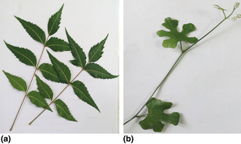
Chemicals such as calcium chloride (CaCl2), di-sodium hydrogen phosphate (Na2HPO4), and sodium hydroxide (NaOH) were obtained from Merck, Mumbai, India. A. indica and C. grandis leafs (Fig. 1) were collected and they were cleaned with double distilled water and air dried at room temperature. About 50 g of finely cut leaves was kept in two beakers each containing 250 mL double distilled water and was boiled for 2 h separately. After 2 h the extracts were cooled down, filtered, and stored at 4 °C for further use. For the synthesis of HAp nanorods using leaf extract, 1 M CaCl2 and 0.6 M Na2HPO4 were prepared using leaf extract as the solvent and separately brought to pH above 10 using 0.8 M NaOH solution. Then CaCl2 solution was stirred vigorously using a magnetic stirrer at room temperature, and then Na2HPO4 solution was added drop wise into it to produce a gelatinous precipitate. The precipitation of HAp can be described as follows:
 $$\eqalign{{\rm 1}0{\rm CaCl}_{\rm 2} + {\rm 6Na}_{\rm 2} {\rm HPO}_{\rm 4} + {\rm 8NaOH} \to &{\rm Ca}_{{\rm 1}0} ({\rm PO}_{\rm 4} )_{\rm 6} ({\rm OH})_{\rm 2} \cr&+ {\rm 2}0{\rm NaCl} + {\rm 6H}_{\rm 2} {\rm O}.} $$
(a) Azadirachta indica and (b) Coccinia grandis leafs.

The reaction mixture was then stirred for 1 h and kept at room temperature for 24 h. The obtained precipitate was centrifuged to remove byproduct and then dried at 150 °C for 6 h in hot air oven. Finally, the dried cakes were crushed to obtain powders. HAp synthesized using A. indica and C. grandis leaf extract were named as NHA and GHA, respectively. Moreover, HAp without leaf extract was prepared and named as control (pure HAp) for comparison.
Characterization
X-ray diffraction (XRD) patterns were obtained (Rigaku MiniFlex II powder X-ray diffractometer) using Cu K α monochromatic radiation (1.5406 Å) as a source in the range between 20° ≤ 2θ ≤ 60°. The lattice parameters (a and c) of HAp were calculated by the method of least squares using the following equation:
 $$\displaystyle{1 \over {d^2}} = \displaystyle{{4(h^2 + hk + k^2 )} \over {3a^2}} + \displaystyle{{l^2} \over {c^2}}, $$
where d is the spacing between the planes in the atomic lattice. The volume V of the hexagonal unit cell was determined by using the following equation:
 $$V = \displaystyle{{\sqrt 3} \over 2} \times a^2 \times c.$$
The average crystallite size of HAp along the c-axis was calculated from the XRD pattern using the Debye–Scherrer formula.[ Reference Patterson 14 ]
 $$D_{hkl} = \displaystyle{{K\lambda} \over {\beta _{1/2} \cos \theta}}, $$
where D hkl is the average crystallite size, K is the broadening constant, λ is the wavelength of Cu K α radiation (1.5406 Å), β 1/2 is the full-width at half-maximum of (002) peak (in radian), and θ is the diffraction angle (in degree). The diffraction peak at 2θ = 25.8° corresponding to (002) Miller's plane of HAp, was selected for calculation of the crystallite size since it was sharper and isolated from others. Fourier transform infrared (FTIR) spectra were obtained using Perkin Elmer RX1 FTIR spectrometer in the range of 400–4000 cm−1. Thermogravimetric (TG) analysis was performed using SDT Q600 thermal analyzer (TA Instruments, New Castle, DE, USA) under an argon atmosphere. The morphological feature was analyzed using JEM JEOL-2100 transmission electron microscope (TEM).
Antibacterial activity test
Antibacterial activity of prepared samples was analyzed by standard disk diffusion method.[ Reference Kumar, Govindan and Girija 4 , Reference Kumar and Girija 6 ] For antibacterial activity test, 100 mg of synthesized samples were pressed at ~24 MPa to form circular disk, each nominally 14 mm diameter and 1 mm thick. Then, micro-organisms such as Escherichia coli and Staphylococcus aureus were spread on the surface of nutrient agar plates. After the spreading of individual bacteria in each plate, disks of each sample was placed on the surface of the culture plate and incubated for 24 h at 37 ± 0.5 °C. The microbial inhibition zone including disk was measured after the incubation period and its images were documented.
Results and discussion
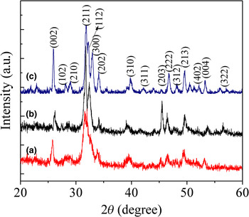
XRD pattern of NHA, GHA and control samples are shown in Fig. 2. XRD patterns of all the samples are well matched with JCPDS data for HAp, which confirmed that prepared samples are pure HAp having hexagonal crystal structure. The broad peak in the region of 30°–34° can be ascribed to (211), (112), and (300) Miller's planes of HAp. The resolution of these peaks depends upon crystallinity and crystallite size. NHA and GHA shows broad diffraction peaks when compared with control sample (HAp prepared without leaf extract), which indicates that they are poorly crystalline in nature. The calculated lattice parameter, unit cell volume and average crystallite size for all the samples are given in Table I. Significant variation in the lattice parameters were observed between the samples. Moreover, the average crystallite size of the control sample is high when compared with the NHA and GHA. This illustrates that the biomolecules present in leaf extracts play an important role on the crystallization process and inhibiting the crystallization of HAp.
XRD pattern of synthesized samples (a) NHA, (b) GHA, and (c) control (pure HAp).

The calculated lattice parameter, unit cell volume, and average crystallite size of prepared samples.

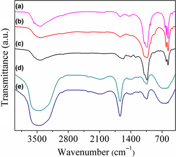
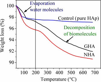
Different vibrational modes of the phosphate PO4 3− and hydroxyl OH− groups can be used to identify the HAp. FTIR spectrum (Fig. 3) of pure HAp (Control) shows vibrational modes of phosphate at 462, 565, 605, 961, 1032, and 1108 cm−1, and hydroxyl group at 3569 and 632 cm−1.[ Reference Suchanek and Yoshimura 2 , Reference Kumar, Govindan and Girija 4 ] FTIR spectrum of NHA and GHA shows vibrational modes of phosphate at 460, 565, 605, 961, 1041, and 1102 cm−1.[ Reference Suchanek and Yoshimura 2 , Reference Kumar, Govindan and Girija 4 ] However, vibrational modes of the hydroxyl group are not evidently seen in the FTIR spectrum of NHA and GHA samples due to poor crystalline nature of the samples. The band at 1642 cm−1 and band extending from 3000 to 3600 cm−1 are attributed to the O–H vibration modes of water molecule and N–H stretching vibrations of amides and amines.[ Reference Suchanek and Yoshimura 2 , Reference Kumar, Govindan and Girija 4 ] The peak at 2925 cm−1 is attributed to the C–H stretching of polyols.[ Reference Nazeruddin, Prasad, Waghmare, Garadkar and Mulla 9 – Reference Arunachalam, Dhanasingh, Kalimuthu, Uthirappan, Rose and Mandal 11 ] The presence of above functional groups shows that biomolecules such as flavonoids, terpenoids, and protein compounds may be present in the NHA and GHA, which are incorporated during the synthesis process. Moreover, FTIR spectra of NHA show additional peaks at 1475 and 1294 cm−1, which are due to C = O stretching of carboxylic acid. It reveals that the carboxyl group strongly interacts with HAp crystallites. FTIR spectra of A. indica and C. grandis leaf extracts were also included in Fig. 3 for comparison, which showed the vibrations of biomolecules such as flavanoids and terpenoids. TG analysis of the synthesized samples is shown in Fig. 4. The weight loss observed up to 200 °C in all samples is due to the desorption of adsorbed water molecules.[ Reference Kumar, Govindan and Girija 4 ] With the increase in temperature, there is significant weight loss that was observed between 200 and 600 °C for NHA and GHA due to the burning of biomolecules such as flavonoids, terpenoids, and protein compounds. From TG analysis, we have determined the amount of biomolecules in NHA and GHA as 4.90 and 4.79%, respectively.
FTIR spectra of (a) control, (b) GHA, (c) NHA, (d) Coccinia grandis, and (e) Azadirachta indica leaf extract.

TG analysis of synthesized samples.

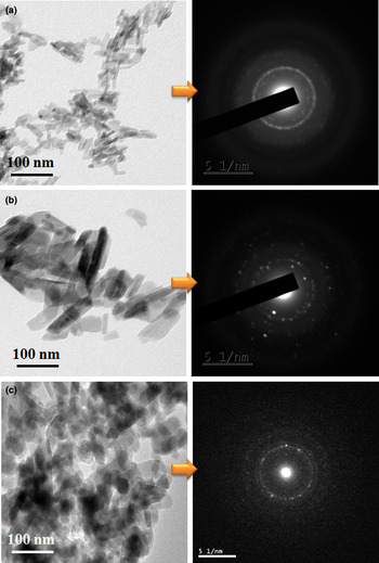
TEM image and selected area electron diffraction (SAED) pattern of synthesized samples are shown in Fig. 5. NHA consist of nanorods having 10–20 nm width and 40–80 nm length, whereas GHA consist of nanorods of 30–50 nm wide and 80–150 nm length. On the other hand, control sample (pure HAp) consist of aggregated elongated spherical particles having 30–40 nm width and 70–80 nm length. The SAED pattern of all the samples exhibits continuous rings around the sharp spot, which shows the polycrystalline nature of prepared samples. When A. indica leaf extract was used as a solvent, the formation of HAp with the reduced particle size is due to the strong interaction between the carboxyl groups and HAp, which effectively controlled the growth of HAp particles.[ Reference Kumar, Govindan and Girija 4 , Reference Gopi, Bhuvaneshwari, Indira, Kanimozhi and Kavitha 12 ] The strong carboxyl band appearing in the FTIR spectrum of NHA confirmed that there is a strong interaction between carboxyl group and HAp crystallites, which support the results obtained from TEM results. It is important to mention that the particle size of NHA and GHA obtained from the TEM observation is slightly higher than that of crystallite size of HAp obtained from XRD data, which may be due to chemical heterogeneity of NHA and GHA. FTIR spectrum of NHA and GHA shows that biomolecules such as flavonoids, terpenoids, and protein compounds are present along with HAp, which confirms its chemical heterogeneity.
TEM image and SAED patterns of (a) NHA, (b) GHA, and (c) control (pure HAp).

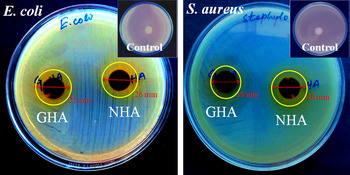
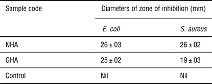
E. coli and S. aureus are the most common bacteria found in bone infection such as osteomyelitis.[ Reference Lew and Waldvogel 3 ] Antibacterial activity of the prepared samples against E. coli and S. aureus are shown in Fig. 6. The diameters of inhibition zone of synthesized samples against E. coli and S. aureus are given in Table II. NHA showed significant inhibition zone against both the tested bacteria. However, the GHA showed higher inhibition zone against E. coli when compared with S. aureus. The observed antimicrobial activity may be due to the presence of biomolecules such as flavonoids, terpenoids, and protein compounds in the samples, which inhibit the enzymes required for bacterial growth and replication. It is reported that tetratriterpenoids, including azadirachtin compound from A. indica leaf extract may be able to inhibit the production of polysaccharide intercellular adhesion for the growth of S. aureus.[ Reference Ahmed, Ullah, Ahmad, Swami and Ikram 10 , Reference Arunachalam, Dhanasingh, Kalimuthu, Uthirappan, Rose and Mandal 11 ] Hence, NHA showed higher antibacterial activity against S. aureus when compared with GHA. Gopi et al. reported green synthesis of HAp nanorods using banana, grapes, and tamarind extracts with excellent antibacterial properties.[ Reference Gopi, Bhuvaneshwari, Indira, Kanimozhi and Kavitha 12 ] The difference in the antibacterial activity depends on the particles size, nature of the particles, and types of bacteria used for the antibacterial test.
Antibacterial activity of the prepared samples against E. coli and S. aureus.

Zone of inhibition of as-synthesized samples against E. coli and S. aureus.

Conclusion
The phase pure HAp nanorods with different particle sizes were prepared by green synthesis using A. indica and C. grandis leaf extracts as a solvent. The obtained HAp nanorods exhibit excellent antibacterial activity against E. coli and S. aureus. Hence, green synthesis can be a potential method to develop HAp nanorods with antibacterial properties for filling bone defects.
Acknowledgments
Dr. Govindan Suresh Kumar would like to express his sincere thanks to the University Grant Commission, India for financial support through minor research project scheme [File No: 4-4/2015-16 (MRP/UGC SERO)]. The work was carried out with financial support from the Ministry of Education and Science of the Russian Federation in the framework of Increase Competitiveness Program of NUST “MISiS”, implemented by a governmental decree dated 16th of March 2013, N 211.