Case presentation
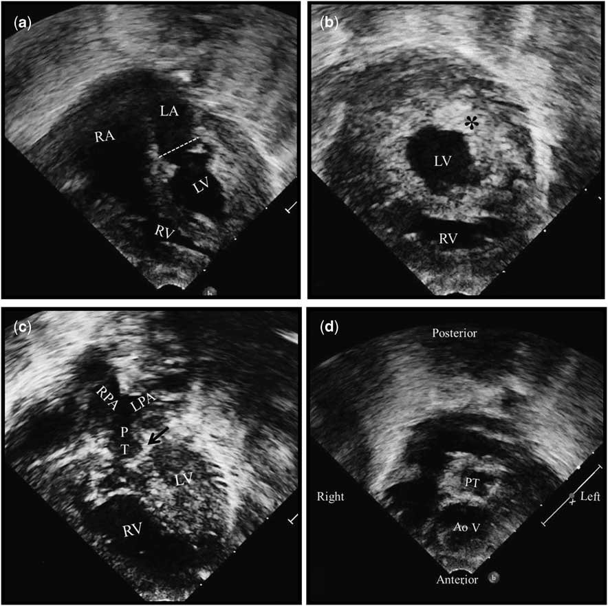
The patient, a male neonate, was 14 days old when referred for evaluation of cyanotic congenital heart disease. He was born full term in hospital, weighing 2.3 kg. Central cyanosis was apparent on the day of birth. Examination revealed central cyanosis, with systemic saturation measured at 75%, and a systolic murmur that was audible over the left sternal border. The chest radiograph showed pulmonary oligaemia. The electrocardiogram showed tall P waves, a normal P and QRS axis, prolonged QT interval, and non-specific ST–T changes. The transthoracic echocardiogram revealed usual atrial arrangement, with normal drainage of the systemic and pulmonary veins. A communication of 4-mm diameter was found in the oval fossa. The atrioventricular connections were concordant, with right hand ventricular topology. The mitral valve, which was mildly regurgitant, was markedly hypoplastic, measuring only 7 mm in diameter, equivalent to a z score of –2.25 (Fig 1a and online Supplementary Video 1). The left ventricular cavity was globular and hypoplastic, being lined by a thickened, hyperintense endocardium, which reduced its systolic function (Fig 1b and online Supplementary Video 2). The right ventricle was apex forming, and the ventricular septum was intact. Left ventricular systolic pressure, as estimated on the basis of the mitral regurgitation, was suprasystemic. The ventriculo-arterial connections were discordant, but the pulmonary valve was imperforate, blocking access from the left ventricle to the pulmonary trunk, which arose leftward and posterior to the aortic valve (Fig 1c, d and online Supplementary Video 3). The diameter of the pulmonary ventriculo-arterial junction was measured at 5.5 mm, giving a z score of –2.3. The pulmonary arteries were confluent and of good size, each having a diameter of 4 mm. Pulmonary flow was through a patent arterial duct, which had a restrictive pulmonary end. Owing to worsening of cyanosis, he was started on intravenous infusion of prostaglandin E1. This improved both flow across the duct and systemic saturation.
The apical four-chamber view of the transthoracic echocardiogram ( a ) shows usual arrangement of atria, right hand ventricular topology, and hypoplasia of the left ventricle and mitral valve. The parasternal short-axis view ( b ) shows a globular and hypoplastic left ventricular cavity, with a thickened and hyperintense endocardium (*). The apical four-chamber view with anterior tilt ( c ) shows an imperforate pulmonary valve (arrow), and hypoplasia of the pulmonary trunk and its right and left branches. The arrow shows the potential origin of the atretic pulmonary trunk from the left ventricle. The modified parasternal short-axis view ( d ) shows the atretic pulmonary valve to be leftward and posterior relative to the aorta. AoV=aortic valve; LA=left atrium; LPA=left pulmonary artery; LV=left ventricle; PT=pulmonary trunk; RA=right atrium; RPA=right pulmonary artery; RV=right ventricle.

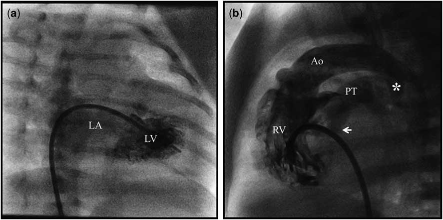
Cardiac catheterisation permitted balloon atrial septostomy. Angiography at this time confirmed the hypoplastic nature of the left ventricle. No evidence was found, however, of fistulous connections with the coronary arteries (Fig 2a and online Supplementary Video 4). The injection in the right ventricle confirmed right ventricular origin of aorta, showing also filling of the pulmonary arteries through patent arterial duct, and confirming the atretic nature of the pulmonary valve (Fig 2b and online Supplementary Video 5). Although we recommended urgent construction of a modified Blalock–Taussig shunt, the parents declined consent for any surgical intervention. The child was discharged home against medical advice.
The left ventricular angiogram, seen in the right anterior oblique view ( a ) shows a severely hypoplastic left ventricular cavity, with moderate mitral regurgitation but absence of ventriculo-coronary connections. The right ventricular angiogram, in lateral view ( b ) shows the aorta arising from the right ventricle. The pulmonary trunk is seen filled through patent arterial duct (*). The imperforate pulmonary valve (arrow) is seen posterior to the interventricular septum and the aorta and is lying close to the left ventricular outflow. Ao=aorta; LA=left atrium; LV=left ventricle; PT=pulmonary trunk; RV=right ventricle.

Discussion
Pulmonary atresia with an intact ventricular septum is typically found in the setting of concordant atrioventricular and ventriculo-arterial connections.Reference Knight, Litovsky, Pearce and Petersen 1 Although rare in the setting of discordant ventriculo-arterial connections, when found the atrioventricular connections are usually also discordant, thus producing a variant of congenitally corrected transposition.Reference Knight, Litovsky, Pearce and Petersen 1 To the best of our knowledge, there has been but one prior description of the lesion in the setting of discordant ventriculo-arterial connections combined with concordant atrioventricular connections.Reference Hayes, Arya and Klieinman 2 The effect of this latter combination is to produce a variant of hypolastic left heart, rather than hyoplastic right heart, as seen in the more usual examples of pulmonary atresia with intact ventricular septum.Reference Daubeney 3
When found with concordant ventriculo-arterial connections, the evidence from foetal cardiology suggests that the problem originates with pulmonary valvar stenosis, which progresses during foetal life to become atresia. The earlier in foetal life the original insult is, the more hypoplastic is the right ventricle.Reference Daubeney 3 When associated with transposition, however, the situation is more akin, in terms of ventricular anatomy, to the hypoplastic left heart syndrome. It was not unexpected, therefore, to find a thick fibroelastotic lining within the hypoplastic left ventricular cavity. Fistulous communications with the coronary arteries are a frequent finding when the atretic pulmonary trunk arises from the right ventricle, but fibroelastosis is then not as severe as in the hypolastic left heart syndrome. Fistulous communications can also be found in hypoplastic left heart syndrome, but they tend to be microscopic rather than overt. Obvious fistulous communications were seen in the previously reported case,Reference Hayes, Arya and Klieinman 2 but were not obvious in our patient.
When found in the setting of concordant ventriculo-arterial connections, biventricular repair can be achieved when the right ventricle is of reasonable size. This is unlikely to be a possibility in the setting of transposition when, as in our case, the left ventricle was not only hypoplastic, but also lined by fibroelastosis.
Author Contribution
All authors contributed to the manuscript preparation. S.K.G. reviewed and finalised the manuscript.
Financial Support
This research received no specific grant from any funding agency, commercial or not-for-profit sectors.
Conflicts of Interest
None.
Supplementary material
To view supplementary material for this article, please visit http://dx.doi.org/10.1017/S1047951113002205.