Epidemiological studies revealed diabetic ketoacidosis prevalence in children with type 1 diabetes mellitus is of 30% at the time of diagnosis.Reference Dabelea, Rewers and Stafford1 Diabetic ketoacidosis is one of the most severe and common complications of diabetes, with between 15 and 70% of a new-onset type 1 diabetes mellitus worldwide presented with diabetic ketoacidosis.Reference Craig, Jefferies, Dabelea, Balde, Seth and Donaghue2–Reference Rewers, Klingensmith and Davis4 According to a previous study evaluating mortality rates in children with insulin-dependent diabetes, 69 deaths (out of 83 diabetic deaths) were due to diabetic ketoacidosis and 7 due to hyperglycaemia, where cerebral oedema, a complication of diabetic ketoacidosis, was the most common cause of death in children under 12 years.Reference Edge, Ford-Adams and Dunger5 A small proportion succumbs to a "dead in bed" syndrome, where nocturnal hypoglycaemia has been hypothesised to cause dysrhythmias, especially if the patient has recently started on insulin.Reference Wasag, Gregory, Dayan and Harvey6
Arrhythmia, an abnormal heart rhythm, is a rare complication of diabetic ketoacidosis. Supraventricular tachycardia is the most common childhood arrhythmia with an incidence of 35 per 100,000 person per year in the general population and a prevalence of 0.1–0.4% in the paediatric population.Reference Ganz and Friedman7–Reference Garson9 In children, supraventricular tachycardia is most commonly caused by either an atrioventricular re-entrant tachycardia (72–73%) or an atrioventricular nodal re-entrant tachycardia (9–13%).Reference Ko, Deal, Strasburger and Benson10,Reference Tanel, Walsh, Triedman, Epstein, Bergau and Saul11 In turn, atrioventricular reentrant tachycardia is due to an accessory pathway bypassing the AV node and is considered the most common mechanism in infants.Reference Gandhi and Suvarna12,Reference Chung and Chung13 This can be a visible accessory pathway such as Wolff−Parkinson−White syndrome.Reference Ganz and Friedman7,Reference Delacrétaz8 Furthermore, atrioventricular nodal re-entrant tachycardia is confined to the AV node with both fast and slow anatomical pathways and surrounding myocardium.Reference Ganz and Friedman7,Reference Delacrétaz8 The electrical impulse moves through the fast pathway to reach the ventricles but to prevent a re-entry circuit, the impulse meets the impulse coming from the slow pathway and they cancel each other out. However, a premature atrial contraction may enter the slow pathway, whereas the fast pathway is still refractory following a sinus discharge. By the time the impulse reaches the end of the slow pathway, the fast pathway is no longer refractory, and it may ascend the fast pathway. This slow-fast atrioventricular nodal re-entrant tachycardia recycles around the AV node resulting in supraventricular tachycardia. Supraventricular tachycardia may also fall under the category of ectopic tachycardia, where enhanced atrial automaticity arises from disturbed electrolytes and produces atrial extrasystoles.Reference Ganz and Friedman7,Reference Delacrétaz8
Case report
A previously healthy 2-year-old girl was brought to the emergency room with a history of polyuria and polydipsia for 1 week. Three days before admission, she developed a fever, abdominal pain, and vomiting. She had no significant previous history of any medical or surgical illnesses. No family history of type 1 diabetes mellitus was stated. On admission, Glasgow Coma Scale reported a score of 15/15, indicating full consciousness. She was pale, tachypnoeic with Kussmaul breathing, and showed signs of shock (poor perfusion and tachycardia), yet no evidence of low blood pressure. Vital signs on arrival were recorded: a temperature of 38.4°C, heart rate of 142 BPM, respiratory rate of 48/minute, and blood pressure of 110/65 mmHg. Chest examination revealed good air entry bilateral with no added sounds. A focused assessment of the cardiac system reported normal first and second heart sounds, soft ejection systolic murmur, grade 2/6, with maximum intensity at the left upper sternal border. Further examination showed a soft, non-tender abdomen with no hepatosplenomegaly. The patient recorded normal tone, power, and reflexes.
Initial laboratory investigations revealed hyperglycaemia with a random blood sugar level of 400 g/dl. Venous blood gas showed a pH of 6.91 (7.35–7.45), pCO2 of 19 (35–45), and HCO3 of 2.5 (22 ± 4); all these findings were consistent with a severe diabetic ketoacidosis, including ketonuria in the urine. Further blood sample tests were recorded; Na: 140, K: 4.1, Cl: 118, Urea: 11, Cr: 0.2. WBC: 14.85, 69% were neutrophils, 20% were lymphocytes, 10% were monocytes. Hb: 12.5 g/dl, PLT: 244, HbA1c: 12.9%, AST: 36, ALT: 71, magnesium: 1.6, phosphorus: 3.8. She was then admitted to the Paediatric Intensive Care Unit. She was tachycardic, but no electrocardiogram changes were noted.
A resuscitation 10 ml/kg bolus of normal saline was given, and maintenance fluids with a replacement for the deficit were started as well, calculated to be corrected over 48 hours. Insulin was started at 0.1 units/kg/hour as per hospital guidelines for diabetic ketoacidosis management.
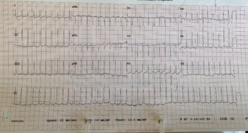
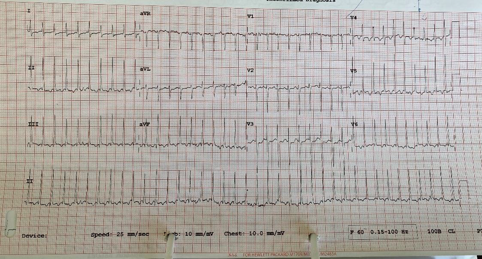
After facing some difficulties, the acidosis was gradually corrected. Subsequently, brief runs of supraventricular tachycardia were noted on the cardiac monitor 12 hours after admission with a heart rate of 234, the longest of 10 minute duration with no haemodynamic compromise, and spontaneously converted to sinus rhythm with vagal stimulation. It was confirmed by a 12-lead electrocardiogram (Fig 1). During the attack, venous blood gas was evaluated indicating a pH of 6.99, a CO2 of 12.7, and HCO3 of 6.1, with normal serum electrolytes including potassium, magnesium, and phosphorus levels.
A 12-lead electrocardiogram showing supraventricular tachycardia with HR of 234 BPM.
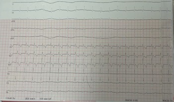
Echocardiography showed a small patent ductus arteriosus with normal cardiac function, and the supraventricular tachycardias were attributed to the preceding metabolic disturbances associated with diabetic ketoacidosis. A sustained episode of supraventricular tachycardia lasting more than 30 minutes. VBG during the episode showed PH: 6.95, pCO2:11.2, HCO3: 7.4, and the electrolytes were normal with no haemodynamic compromise. The vagal stimulation was unsuccessful. She became more tachypnoeic with respiratory distress, and then she was intubated and attached to a mechanical ventilator. She received one dose of adenosine resulting in conversion to sinus rhythm with the heart rate decreasing from 234 to 140 beats per minute. The repeated 12-lead electrocardiogram showed normal sinus rhythm with HR 100 BPM, no pre-excitation (Fig 2).
A 12-lead electrocardiogram showed normal sinus rhythm with HR 100 BPM, no pre-excitation.
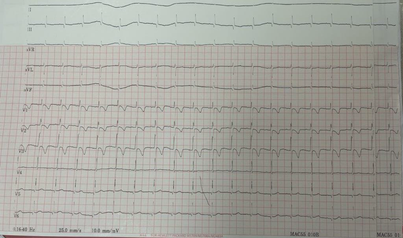
The electrocardiogram was closely examined for signs of an accessory pathway consistent with Wolff−Parkinson−White syndrome, but none were evident. No further recurrence of supraventricular tachycardia was noted. The patient was stabilised, extubated, and monitored in the PICU for 3 days with no further episodes of supraventricular tachycardia. The diabetic ketoacidosis has been completely resolved, and she was started on the standard management for patients with newly diagnosed type 1 diabetes mellitus: subcutaneous insulin using Novorabid (short-acting) and Lantus (long-acting), and she was discharged to home with no anti-arrhythmic medications and careful education for diabetes and possible episodes of arrhythmia. She returned for the cardiology follow-up after 2 weeks with no symptoms. Both the electrocardiogram and the Holter monitor were normal.
Discussion
Diabetic ketoacidosis is one of the serious metabolic complications of diabetes, which consists of the triad of hyperglycaemia, ketosis, and acidosis; the diagnostic criteria being blood glucose level > 250 mg/dl, arterial pH < 7.3, bicarbonate level < 15 mEq/L, and a moderate degree of ketonaemia and ketonuria. There has been little change in the mortality rate associated with diabetic ketoacidosis despite great improvements in our understanding of its pathophysiology and treatment. During diabetic ketoacidosis, cardiovascular damage can occur as a form of (a) arrhythmias due to electrolyte imbalance, (b) adverse effects of acidosis, (c) acute myocardial infarction, and (d) pulmonary oedema.Reference Wasag, Gregory, Dayan and Harvey6,Reference Gandhi and Suvarna12
Fluid and electrolyte imbalance is very common in diabetic ketoacidosis cases and manifested typically by a loss of 5–8 L of water, 400–700 mEq of sodium, 250–700 mEq of potassium, and 30–50 mEq of magnesium. However, as water loss is greater relative to electrolytes, most patients will have normal or even elevated sodium, potassium, and magnesium levels at the time of presentation, although their body stores of these elements are almost always lower than normal.Reference Gandhi and Suvarna12 Potassium deficit, one of the essential electrolyte imbalances seen in diabetic ketoacidosis, can lead to fatal arrhythmias. The most common cardiac arrhythmias are atrial premature contractions, atrial tachycardia with or without AV block, supraventricular tachycardia, and ventricular premature contractions. Less common are AV junctional tachycardia or escape rhythm and AV block.Reference Chung and Chung13 However, multiple additional factors can also cause arrhythmias, including re-entry circuits, abnormal automaticity, or triggered activity occurring within myocardial focus.Reference Cubbon and Kearney14 A common cause of re-entry circuits within the myocardium is electrolyte abnormalities involving calcium and potassium.Reference Cubbon and Kearney14
Additionally, hypomagnesaemia and hypophosphataemia have also been associated with tachyarrhythmia such as supraventricular tachycardia.Reference Klevay and Milne15 During the treatment of diabetic ketoacidosis, phosphate serum levels subsequently drop profoundly as a consequence of insulin administration.Reference Shen and Braude16 The greater the initial acidosis, the greater the subsequent hypophosphataemia.Reference Shen and Braude16 Our patient had profound serum hypophosphataemia suggests an even greater total body phosphate depletion. Moreover, phosphate supplementation may precipitate hypocalcaemia.Reference Becker, Brown, Steranka and Drash17
Acidosis resulting from the production of ketoacidosis is a key clinical feature found in diabetic ketoacidosis that may precipitate arrhythmias. The junctional resistance between myocardial cells is increased by about 30% if the pH falls by 0.4 units.Reference Kagiyama, Hill and Gettes18,Reference Marrannes, de Hemptinne and Leusen19 This reduction in conduction velocity can enable a re-entrant arrhythmia to form in a structurally normal heart in which an acidic region is present.Reference Garson9 However, as the acidosis was resolved in this case, this suggests that electrolyte shifts and hypophosphataemia and hypomagnesaemia are more likely to cause supraventricular tachycardia.
As almost all cases in these previous studies had a normal echocardiogram, the likely cause is an atrioventricular nodal re-entry tachycardia without an accessory pathway and can be triggered by hypo/hyperkalaemia.Reference Goyal, Senst, Bhyan and Zeltser20 An accessory pathway with antegrade conduction or WPW may be less likely in the differential with a normal electrocardiogram, although not entirely ruled out as antegrade conduction may be heart rate dependent. Re-entry is defined as a closed-loop circuit that develops within the myocardium and allows the action potential to self-sustain.Reference Goyal, Senst, Bhyan and Zeltser20 It may also fall under the category of ectopic tachycardia with atrial extra systoles, where enhanced atrial pacemaker automaticity arises from disturbed electrolytes, for example, potassium.Reference Ko, Deal, Strasburger and Benson21 The atrial extra systoles may have originated from metabolic derangements and then precipitated the supraventricular tachycardia.
In this case, hypokalaemia would explain both the premature atrial contractions and the supraventricular tachycardia through enhanced atrial automaticity. However, the potassium was only at the lower limits of normal at its nadir. It is important to mention that diabetic ketoacidosis can result in very rapid potassium derangements, which may be undetected where potassium levels are typically “normal” at presentation.Reference Ko, Deal, Strasburger and Benson21 Although serum potassium levels were generally well-controlled, intracellular potassium homoeostasis was likely to be seriously disturbed and was probably a major contributor to the genesis of the arrhythmia. Furthermore, with appropriate insulin and fluid therapy, as the acidosis reverses, potassium is concomitantly forced into the cells, resulting in a rebound of hypokalaemia.Reference Ko, Deal, Strasburger and Benson21 A possible hypothesis is that our patient’s low magnesium levels partook in the occurrence of supraventricular tachycardia.Reference Winter, Harris, Phillips and Green22 ATPase pumps in the myocardium are dependent on magnesium, and reduced magnesium alters membrane potential, irritates the myocardium, and results in an increased frequency of supraventricular beats.Reference Rother and Schwenk23
Our patient was responding to adenosine as acute treatment and discharged with no anti-arrhythmic medications. Inverse, while the patient with same presentation did not respond to adenosine, flecainide was given and showed abortion of supraventricular tachycardia.Reference Finn, Fraser and O'Connell24
Although diabetic ketoacidosis can cause life-threatening arrhythmias, it is usually reversible with appropriate treatment consisting of insulin and IV fluids. During treatment, it is imperative to monitor patients' electrolytes and acid–base status closely. It is recommended that electrolytes be replaced with a solution with tonicity greater than 0.45% normal saline, such as the one administrated to our patient, with the addition of potassium chloride, potassium phosphate, or potassium acetate.Reference Klevay and Milne25 In profoundly unwell patients with severe diabetic ketoacidosis, cardiac monitoring forms a routine part of intensive care.
Conclusion
Supraventricular tachycardia observed in the reported case was most likely due to electrolyte abnormalities and ketoacidosis. Deviations in electrolyte levels from normal reference ranges and acidosis have been associated with the development of arrhythmias due to a variety of mechanisms, such as re-entry circuits, after-depolarisation, and alterations of AP conduction, among other factors. However, a multifocal aetiology resulting in the onset of the arrhythmia is also plausible. Diabetic ketoacidosis and arrhythmias are rare associations. It would be interesting to see other reports of a similar clinical picture.
Acknowledgements
We would like to thank the patient who gave us the chance for participation to publish such case in the literature.
Financial support
This research received no specific grant from any funding agency, commercial or not-for-profit sectors.
Conflicts of interest
The authors declare that there is no conflict of interest.