Introduction
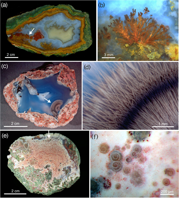
The variety and structural complexity of texture, structure and forms displayed by agates have always been a source of fascination and frequent discussions. Textural specifics such as ‘wall-lining banding’ (‘fortification banding’), ‘gravitational banding’ (or ‘Uruguay banding’) as well as ‘infiltration channels’ (or ‘escape tubes’) are not only characteristic growth phenomena in agates, but have also resulted in a lot of discussions about the general processes of agate formation (e.g. Moxon, Reference Moxon1996; Walger et al. Reference Walger, Mattheß, von Seckendorff and Liebau2009; Götze, Reference Götze and Zenz2011; Moxon et al., Reference Moxon, Petrone and Reed2013). Moreover, numerous other minerals, as well as silica phases, can be formed pre-, syn- and post-genetically in agates resulting in spectacular forms such as ‘moss agate’, ‘plume agate’, intergrowths or pseudomorphs (Götze, Reference Götze2014). Their dimensions can range from microscopic inclusions up to several centimetres in size (Fig. 1b,d). Curious structures and specific features led to the assumption that micro-organisms might be involved in the creation of some of these phases (e.g. Ferris et al., Reference Ferris, Beveridge and Fyfe1986; Parenteau and Cady, Reference Parenteau and Cady2010; Götze et al., Reference Götze, Müller, Polgári and Pál-Molnár2011; Thewalt and Dörfner, Reference Thewalt and Dörfner2012).
Examples of curious micro-structures in agates; (a,b) plant-like structures (so called ‘plumes’) in an agate from Kardzhali, Bulgaria formed by inclusions of oxide/silicate minerals in the silica matrix; (c,d) agate from the Darrell Friend Ranch near Ashwood, Oregon/USA (sample courtesy G. Schiecke, photo A. Gabert) with bundle-like structures resembling the shape of natural sheaf of wheat; the unusual shape is probably caused by calcite traced in silica gel (compare Fig. 2 in Dominguez-Bella and Garcia-Ruiz, Reference Dominguez-Bella and Garcia-Ruiz1986); (e,f) agate from Berschweiler near Idar-Oberstein (Germany) with inclusions of spiral forms (arrows indicate the position of each close-up).

In general, the morphologies of mineral phases can typically be used to differentiate between those formed in inorganic, non-living environments and others originating from activities of living organisms (biominerals). In contrast to the typical shapes of inorganically formed minerals with angular crystal faces and a distinct symmetry, biominerals can exhibit elaborate architectures (biosignatures) and rounded shapes without sharp edges. Therefore, such filigree rounded shapes are associated commonly with the activity of living matter.
However, recent studies have shown that several chemical pathways exist to produce inorganic mineral matter resembling living forms (e.g. Yang et al., Reference Yang, Coombs and Ozin1997; Glaab et al., Reference Glaab, Kellermeier, Kunz, Morallon and Garcia-Ruiz2012; Noorduin et al., Reference Noorduin, Grinthal, Mahadevan and Aizenberg2013; Haudin et al., Reference Haudin, Cartwright, Braua and De Wit2014). The processes resulting in such crystal architectures include complex chemical interactions such as complexation of ions, selective adsorption onto certain faces of growing crystals, and/or temporary stabilisation of an amorphous precursor phase. These interactions might result in control over solution supersaturation, nucleation, growth and polymorphism (Kellermeier, et al., Reference Kellermeier, Cölfen and García-Ruiz2012). In conclusion, biogenic origin of mineral matter cannot be identified on the basis of morphological aspects alone.
Observed unusual shapes of minerals in agate structures can also be related to such inorganic chemical processes, for instance the well-known phenomenon of silica gardens – plant-like structures formed upon addition of metal salts to sodium silicate solutions (Fig. 1a,b). These examples of inorganic self-assembly form by a combination of osmosis, buoyancy and chemical reaction (Glaab et al., Reference Glaab, Kellermeier, Kunz, Morallon and Garcia-Ruiz2012; Haudin et al., Reference Haudin, Cartwright, Braua and De Wit2014). Furthermore, alkaline-earth carbonates can develop highly unusual shapes when crystallising in a silica-rich environment (Kellermeier et al., Reference Kellermeier, Cölfen and García-Ruiz2012). Accordingly, the growth behaviour of calcite traced in silica gel was found to be highly variable, for instance, resulting in finger-like structures or bundles resembling the shape of a natural sheaf of wheat (Dominguez-Bella and Garcia-Ruiz, Reference Dominguez-Bella and Garcia-Ruiz1986 – Fig. 1c,d). Detailed investigations revealed that this specific form consists of cleaved calcite rhombohedra that were aligned along their c axis and self-assembled to build laminar aggregates with rhythmical banded patterns.
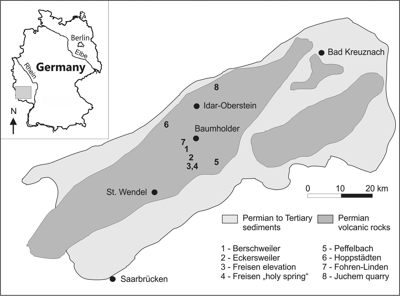
Agates with spectacular features were found recently in volcanic rocks at several occurrences in the Saar-Nahe region, Germany (Schäfer, Reference Schäfer2002, Fig. 2). These agates include spirals of several tens of μm up to several hundreds of μm in size within the silica matrix (Fig. 1e,f). Up to now such specific agate features are unique and not known from other agate occurrences in Germany or worldwide. Because of their specific structure, the conspicuous creations first led to a controversial discussion, whether they possibly have a biogenic origin (microfossils such as foraminifera or snails) or result from inorganic processes (Lenz and Schäfer, Reference Lenz and Schäfer2008). However, in light of the above mentioned results, the proposed biological origin of these spiral forms in agates has to be discussed with caution and several facts contradict an organic formation.
Schematic map of the Saar-Nahe region (Germany); the numbers indicate the agate occurrences with spiral textures investigated in the present study.

First, it is unlikely that microfossils such as foraminifera with similar morphology would reach cavities within the Permian volcanic rocks where the agates formed. Permian foraminifera are benthic, i.e. they are marine, stenohaline organisms with a narrow (normal marine) salinity window (Elicki and Breitkreuz, Reference Elicki and Breitkreuz2016). Furthermore, the spirals in agates show a circular shape without chambers, and sometimes show the intergrowth of two or more aggregates (compare Fig. 1f) that is uncommon for organic forms (Elicki and Breitkreuz, Reference Elicki and Breitkreuz2016). The size of the spirals is also not adequate: several hundreds of μm is too small for benthic Permian foraminifera and even more for snails (pers. comm. O. Elicki). In conclusion, a microfossil origin is very unlikely for the spirals in agates because of morphological and facies criteria and would demonstrate an inorganic creation.
The objective of the present study is to determine the origin of the spirals in agate/chalcedony by various methods of microanalysis. The Saar-Nahe region is one of the most famous agate locations in Western Germany and agates from the Permian volcanic rocks in the area have been investigated. The investigations combine several analytical techniques such as polarising microscopy, Nomarski DIC microscopy, scanning electron microscopy (SEM), cathodoluminescence (CL) microscopy and spectroscopy as well as electron back-scatter diffraction (EBSD) to elucidate the chemical and mineralogical composition of the spirals in agates and their crystal structural features. Moreover, the analytical data provides information for the reconstruction of possible processes leading to the formation of the spirals. This knowledge can also provide additional arguments concerning general aspects of agate genesis.
Materials and methods
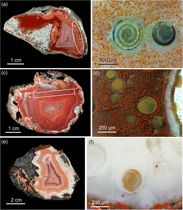
The sample material consists of agates from eight occurrences in the Saar-Nahe region, Western Germany. The agates occur in basic Permian volcanic rocks, where they were formed in amygdales of the volcanics (Schmitt-Riegraf, Reference Schmitt-Riegraf1996). Polished sections of the agates were examined regarding their micro-structural features under a ZEISS stereo microscope. Samples exhibiting features of spiral growth (Figs 3, 4) were documented and pieces of selected areas, as well as polished thin sections, were prepared with abrasive diamond powders for further investigations with polarising microscopy, CL, SEM and EBSD measurements.
Images of polished agate halfs and related micrographs of magnified areas with spiral growth (see marked boxes in a, c, e) in samples from different locations of the Saar-Nahe region (Germany) investigated in the present study: (a,b) Freisen; (c,d) Hoppstädten; and (e,f) Fohren Linden.

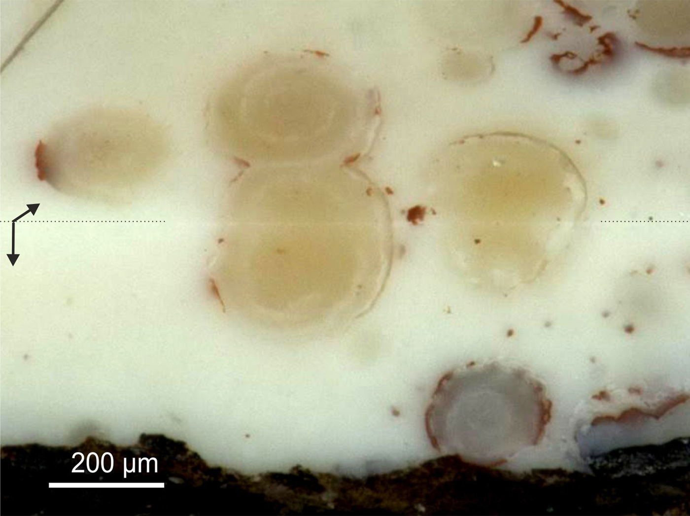
Micrograph of an agate from Fohren Linden showing a cutting edge from preparation (see arrows) that reveals the three-dimensional structure of the spirals.

Polarising microscopy of polished thin sections was performed using a ZEISS Axio Imager A1m microscope. In addition, Nomarski differential interference contrast (DIC) imaging in reflected light was carried out with the same equipment using specific Nomarski DIC prisms (Götze, Reference Götze2009a).
Cathodoluminescence microscopy and spectroscopy were performed on carbon-coated thin sections using a hot-cathode CL microscope HC1-LM (Neuser et al., Reference Neuser, Bruhn, Götze, Habermann and Richter1995). The system was operated at 14 kV and 0.2 mA (~10 mA/mm2) with a defocused electron beam of ~5 mm diameter. Luminescence images were captured using a Peltier cooled digital video-camera (OLYMPUS DP72). CL spectra in the wavelength range 370 to 920 nm were recorded with an Acton Research SP-2356 digital triple-grating spectrograph with a Princeton Spec-10 CCD detector that was attached to the CL microscope by a silica-glass fibre guide. CL spectra were measured under standardised conditions (wavelength calibration by an Hg-halogen lamp; spot width = 30 µm; measuring time = 5 s).
Scanning electron microscopy measurements were performed on carbon-coated thin sections and polished sample pieces with a JEOL JSM-7001F (20 kV, 2.64 nA) using secondary electron (SE), back-scattered electron (BSE) and energy-dispersive analyses (EDX) techniques.
In addition, small blocks (20 mm x 20 mm x 2 mm) from the same sample pieces were prepared for EBSD measurements. The samples were embedded in epoxy resin and polished stepwise with grinding powder having grain-sizes down to 1 µm. The final polishing was done with VibroMet2 (BUEHLER) vibration polishing equipment to produce a perfect, undisturbed surface. The surface was afterwards sputtered with ~0.1 nm PtPd20 in a Turbo Sputter Coater 208HR (CRESSINGTON) to achieve sufficient electric conductivity. EBSD measurements were performed on a PHILIPS XL30 and FEI Versa3D equipment with integrated EDAX EDS and EBSD systems.
Results
Polarising microscopy
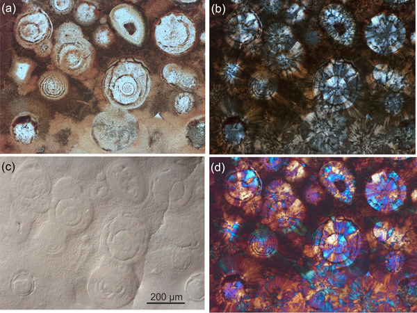
The appearance of spiral growth is detectable in separate zones within the agates, which exhibit no distinct banding (compare Figs 1e/f, 3). In contrast, spirals occur both in clear, colourless areas and coloured areas with strong pigmentation (Fig. 3). In the latter case, the fine-grained surrounding matrix contains dispersed separate micro-particles (e.g. Fe compounds according to SEM-EDX), whereas the area of the spirals is much brighter and almost free of impurities (Fig. 5). Sometimes, the spiral shape and surface is marked by a thin layer of strong pigmentation.
Micrographs of areas with spiral growth within an agate from Freisen (Germany); (a) transmitted light (plane polarised); (b) transmitted light (crossed polars); (c) reflected light Nomarski DIC mode; and (d) transmitted light (crossed polars) with nγ/λ-compensator.

In polarised light (Fig. 5b) the micro-texture of the spirals is characterised by radiating SiO2 aggregates (chalcedony spherulites). The surrounding silica matrix is extremely fine-grained and partially optically-isotropic (dark in polarised light, i.e. non- or purely crystalline). The application of an additional nγ/λ-compensator in polarised light (Fig. 5d) reveals the varying orientation of the crystallites due to the different interference colours. Most of the spirals show continuous, radiating spherulite fibres with concentric orientation. However, sometimes irregular segmentations are visible with sub-areas of different optical orientation.
A photomicrograph of the sample area in reflected light with Nomarski interference contrast is shown in Fig. 5c. This technique enables visualisation of the surface relief at the micro-scale. The varying resistance of different phases to grinding and polishing results in a micro-topography (in the order of 0.2–1 µm) which is thrown into sharp relief by the equipment. Thus, the shape and the interfaces of the spiral loops as well as areas with varying consistency are distinguishable. However, the relief of the spirals is only visible if the cut face of the spirals is at the sample surface.
Cathodoluminescence microscopy and spectroscopy
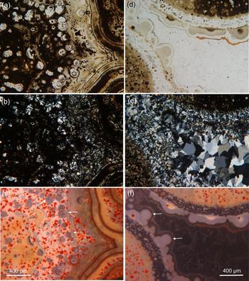
Polished thin sections of the agates were investigated by CL to compare areas with spiral growth with those showing common agate textures (Fig. 6). Quartz and other minerals of the SiO2 group are very interesting phases for CL studies, as their luminescence properties show a significant diversity which is mostly not visible in transmitted and polarised light. Different structural defects cause a range of CL emission bands in the UV, visible and infrared region (Ramseyer et al., Reference Ramseyer, Baumann, Matter and Mullis1988; Stevens-Kalceff, Reference Stevens-Kalceff2009, Götze, Reference Götze2009b). Accordingly, the occurrence of specific luminescence emissions typically can be related to specific SiO2 phases and/or to specific conditions of formation (Götze, Reference Götze2009b).
Micrographs of an agate from Freisen (Germany) with areas of spiral growth (a–c) and central agate (d–f). (a,d) Transmitted, plane polarised light; (b,e) transmitted light, crossed polars; and (c,f) cathodoluminescence; note the common pale blue CL of spiral regions and spherolitic domains (see arrows).

The regions with spirals are generally characterised by pale blue CL (Fig. 6c). Other crystallisation domains such as ring-like aggregates (holospherulites – Schäfer, Reference Schäfer2002) or spherulitic domains in the marginal parts of the agates (Fig. 6f) show similar luminescence behaviour. This is in contrast to the cryptocrystalline and non-crystalline silica matrix, which dominantly exhibits orange CL (Fig. 6c).
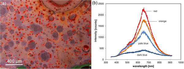
The spiral loops are traced by a thin (several μm) layer with orange CL. Sometimes, there is also a thin orange luminescent margin around the spiral form. Within the spirals, small segments with dark blue luminescence exist (Fig. 7), which have similar CL characteristics to the macro-crystalline quartz in the centre of the agate. Areas with local or extensive accumulation of Fe compounds exhibit reddish CL colour (Fig. 6, 7).
CL micrograph of an agate from Freisen (Germany) showing (a) spiral growth and (b) related CL spectra of the differently luminescent areas shown in Figs. 6 c,f and 7a.

The differently luminescing regions were investigated separately by locally-resolved spectral CL measurements. The spectra in general show identical emission bands but have varying intensities (Fig. 7b). These intensity variations cause the different visible luminescence colours. The main CL emission is a broad band centred at ~650 nm. This 650 nm (1.9 eV) emission is attributed to the so-called non-bridging oxygen hole centre, which can be formed from different precursor defects (Stapelbroek et al., Reference Stapelbroek, Griscom, Friebele and Sigel1979; Stevens-Kalceff, Reference Stevens-Kalceff2009).
Accordingly the increase of the 650 nm band characterises an increase in the number of disordered Si–O bonds resulting in decreasing regular networking of SiO4 tetrahedra in the structure. Thus, the macro-crystalline quartz in the agate centre and the spirals have the highest crystalline order, whereas the surrounding silica matrix is highly disordered. In particular the areas with high amounts of impurities (inclusions of Fe compounds) in the silica matrix are characterised by a high defect density with disturbance of the regular three-dimensional network.
Scanning electron microscopy
The investigations by SEM provided detailed information concerning the micro-texture and composition of the spirals in the agates. Figure 8 illustrates that the circular forms might consist of spirals or simple rings (holospherulite). Despite a homogeneous composition, the spirals are even visible in SE imaging because of the existence of a thin layer of high porosity between the spiral loops (Fig. 8a, c). The surrounding silica matrix also exhibits a much higher porosity than the crystalline spirals.
SEM micrographs of an agate from Freisen (Germany) in secondary (SEI – a) and back-scatter electron imaging (BSE – b) showing circular forms with spiral growth (left) or simple rings (right - holospherulite); (c) close up of the marked area in (a) showing high porosity between the spiral loops; (d) marginal part of the spiral under higher magnification (see marked rectangular in (b) illustrating the accumulation of iron compounds and the interface to the surrounding highly porous silica matrix.

Back-scatter electron imaging (Fig. 8b, d) reveals a bright rim in the marginal part of the spiral. Micro-chemical EDX showed that this crust consists exclusively of Fe and O. Considering the fact that hydrogen is not detectable by EDX, this bright rim is made up of iron compounds (Fe oxides and probably Fe hydroxides), which are intergrown with SiO2. The same feature is visible in the marginal part of the holospherulite.
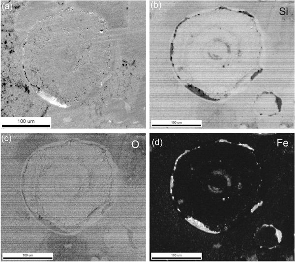
Element mapping data of selected spirals confirmed and extended these results. The element distribution maps reveal that both the spirals and the surrounding matrix are composed of silicon dioxide. Inclusions of iron oxide (and probably hydroxide) compounds occur especially in the marginal parts of the spirals (Fig. 9).
EBSD micrographs of spiral growth in an agate sample from Freisen (Germany): (a) forward scatter detector (FSD) image; (b–d) element distribution maps of Si (b), O (c) and Fe (d) (brightness correlates with increasing element concentration).

EBSD
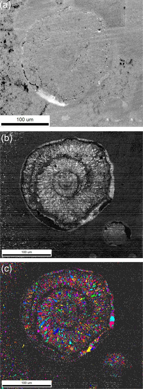
Selected spirals within two agate samples from Freisen and Fohren Linden, respectively, were chosen for EBSD measurements. An example is presented in Fig. 10. The band contrast, which characterises the crystalline order of the phases, reveals that the spirals are composed of numerous micro-crystals of α-quartz with high crystalline order (Fig. 10b). The interspace between the spiral loops as well as the surrounding SiO2 matrix exhibit a low crystalline order (strongly disordered or partially amorphous). The same applies for the Fe compounds, which also seem to be amorphous.
EBSD micrographs showing a spiral in the agate from Freisen (Germany): (a) FSD image; (b) band contrast (increasing crystalline order is related to increasing brightness); (c) orientation map (identical crystallographic orientation is marked by identical colours).

The circular arrangement of the quartz crystallites is clearly visible in Fig. 10. The small quartz crystallites show a systematic rotation of the crystal orientation perpendicular to the direction of the spiral loops. Both images of the band contrast (Fig. 10b) and the crystal orientation (Fig. 10c) indicate a helical growth of the quartz crystals.
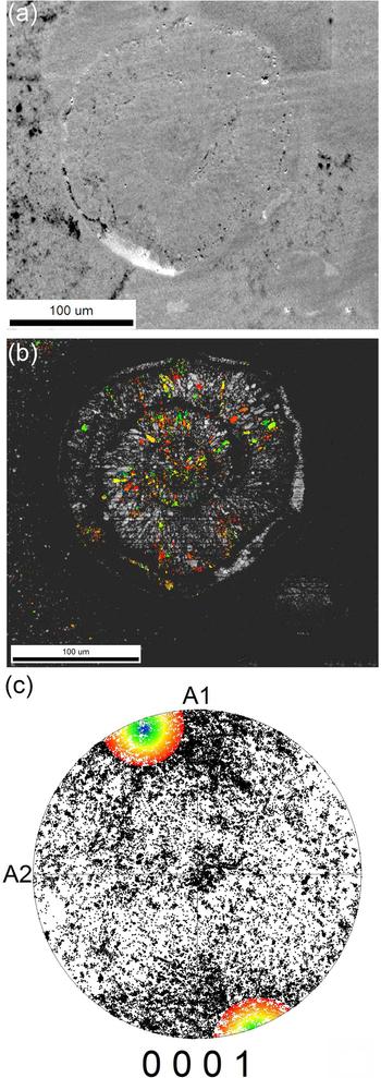
The general orientation of the quartz grains in the pole figure (Fig. 11c) is more or less random and only a slight texture is noticeable. Only few crystals exhibit a <0001 > orientation, whereas more crystals exist, which are ~90° tilted to the <0001 > orientation. A couple of quartz crystals with similar orientation are marked within the pole figure and the related grains are visible according to their analogous colours in the micrograph of the band contrast (Fig. 11b). These grains show a nearly radial distribution.
EBSD micrographs showing a spiral in the agate from Freisen (compare Fig. 8): (a) FSD image; (b) band contrast with crystallographic orientation of selected grains marked by different colours; (c) pole figure with indication of the marked micro-crystals in (b) (compare related colours in b and c).

Discussion
Micro-structure and chemical composition of the spirals
The results of the present investigations showed that spirals occur both in strongly coloured agate areas with strong pigmentation by different iron compounds and bright areas almost free of impurities. However, the development of spiral structures seems to be related to specific zones within the agates. Whereas in zones without banding, spirals and other circular structures such as holospherulites are common, other areas with typical agate banding or macro-crystalline quartz do not contain these spectacular features. Furthermore, the microscopic studies on cut edges revealed the three-dimensional structure of the spirals, which can present different shapes depending on the two-dimensional cutting face. Because of this three-dimensional structure, the name spiral coving should probably be used.
Chemical and crystallographic analyses provide evidence that the spirals consist of well-ordered trigonal α-quartz, whereas the surrounding matrix is composed of strongly disordered or amorphous SiO2 phases. The spiral loops are traced by a thin interlayer of high porosity and sometimes residues of iron compounds. The outer margin of the spirals is commonly marked by a rim of amorphous Fe compounds (probably Fe oxides and/or Fe hydroxides). Considering the fine-dispersed iron compounds in the surrounding porous silica matrix of the spirals, a kind of self-purification mechanism during the formation of the quartz crystallites in the spirals can be assumed. The fine-grained Fe particles were probably released from the silica matrix during crystallisation, moved together with the growth front and accumulated along the outer margin of the spirals.
The results of polarising microscopy studies show a spherulitic internal texture of the spirals indicating radial growth and the development of chalcedony fibres. However, EBSD measurements clearly show that chalcedony fibres do not really exist. The spirals are composed of quartz micro-crystals showing a systematic rotation of the crystal orientation perpendicular to the direction of the spiral loops. Therefore, a helical growth of the spirals must be supposed. The fibrous chalcedony texture is only an optical effect due to the crystallographic orientation and arrangement of the fine quartz crystallites.
Origin of spiral growth
The details and specific physico-chemical conditions of spiral growth in agate are still not fully understood, but the results of the present study have provided first indications for the formation process. The locally limited occurrence of spirals in non-banded areas of the agates could be explained by crystallisation domains in amorphous or highly disordered SiO2. They start with punctual crystallisation processes in an amorphous silica matrix, similar to the growth of chalcedony spherulites at the cavity interface during the first step of agate formation.
Heaney (Reference Heaney1993) showed that spiral growth of chalcedony can be initiated by a screw dislocation (Burgers vector b = n/2 [110]). He explained the formation of the fibrous structure of chalcedony with the formation of dislocations in the crystal structure during crystal growth. This form of ‘dislocation growth’ is favoured if the silica concentration in the mineralising fluids is high and it is present in polymer forms of non-branched chains of silica (Heaney, Reference Heaney1993). The atoms/molecules are packed into spiral layers, which is energetically favoured in comparison with the incorporation into plane crystal faces. A similar mechanism, a defect induced spiral growth, can probably be assumed for the spirals in the investigated agates.
Spiral growth in crystals or at crystal surfaces has been known for a long time (Verma, Reference Verma1951). Dislocations with a screw component can initiate crystal growth and develop into spirals centred on such dislocations. The crystallisation is dependent on the symmetry of the crystal and the orientation of the spiral depends on the chirality of the screw. In contrast to crystals, the lacking symmetry of an amorphous matrix can initiate a curved development. Investigations by Ozin et al. (Reference Ozin, Yang, Sokolov and Coombs1997) and Yang et al. (Reference Yang, Coombs and Ozin1997) illustrate that specific shapes develop from the polymerisation, curvature and growth of a silica liquid crystal seed, probably initiated by a screw dislocation in a system far from equilibrium. Such types of self-organisation growth and polymerisation initiated by a screw dislocation can produce variable spiral morphologies, which are completely different from defect-induced spiral growth at the surface of crystals.
Whereas crystal growth is in general related to interfaces of solids or liquids because of the reduced nucleation barriers, a crystallisation front developing from a fractal precursor far from a foreign interface will more-or-less immediately fold back onto itself to use its own, already existing surface as a growth substrate (Kellermeier et al., Reference Kellermeier, Cölfen and García-Ruiz2012). This growth results in ‘winding’ of distinct growth segments around one another and may explain the formation of complex spiral morphologies and the residues of the porous pre-existing silica matrix between the spiral loops. Spiral growth and the occurrence of curved shapes would accordingly be the direct consequence of an amorphous precursor and the lack of a foreign surface to grow on.
The chemical experiments of Noorduin et al. (Reference Noorduin, Grinthal, Mahadevan and Aizenberg2013) provided evidence that a great variety of extreme shapes of carbonate-silica micro-structures can be produced by reaction-diffusion processes with dynamic feedback between the reaction front and the solution (complex spatial field of overlapping chemical gradients). Discrete modulations in temperature, pH, or concentration can modify the local chemical field (microenvironment) and directly influence the growth front and structure evolution. Other factors such as inorganic and organic additives and epitaxy can also initiate and influence the growth processes by self-organisation into higher ordered structures (Glaab et al., Reference Glaab, Kellermeier, Kunz, Morallon and Garcia-Ruiz2012; Noorduin et al., Reference Noorduin, Grinthal, Mahadevan and Aizenberg2013; Haudin et al., Reference Haudin, Cartwright, Braua and De Wit2014).
Several details of the origin of spirals in the investigated agates are not completely revealed. It is obvious that the formation of spirals seems to be related to specific zones within the agates, which haven't undergone the process of band formation. Therefore, an amorphous silica precursor is probably necessary for the development of spiral forms. The question concerning a specific temperature or pH cannot be answered at present. From a general point of view concerning conditions of agate formation it can be assumed that the processes were at temperatures of ≤ 100°C (Moxon, Reference Moxon1996; Götze, Reference Götze and Zenz2011). This would be in accordance with data from the literature, where high curvature mesoporous silica morphologies were synthesised in the laboratory at 80°C for one week at quiescent conditions (Yang et al., Reference Yang, Coombs and Ozin1997).
Conclusions
The present investigation is the first to examine examples of spirals (three-dimensional spiral covings) within agates of volcanic host rocks of the Saar-Nahe region (Germany). The spectacular forms resemble living forms, so that an organic origin (e.g. microfossils) was first taken into consideration. However, certain morphological and facies criteria exclude a microfossil origin of the spiral creations and an inorganic origin is supposed.
Integrated studies by polarising microscopy, SEM, CL and EBSD showed that spirals developed exclusively in agate areas that do not exhibit structural banding. The spiral loops consist of trigonal α-quartz and are surrounded mostly by a thin layer of highly disordered Fe compounds (Fe oxides/hydroxides). Therefore, it is assumed that the spirals develop from an amorphous silica precursor by self-organisation growth and polymerisation initiated by a screw dislocation (dislocation growth). The lacking symmetry of the amorphous matrix initiates a curved development of the growth front and causes winding of distinct growth segments around one another. This might explain the formation of complex spiral morphologies. During the crystallisation process, fine-grained Fe particles were probably released from the silica matrix, moved together with the growth front and accumulated along the outer margin of the spirals.
The results of the study emphasise that agate formation runs through several structural states of SiO2 with amorphous silica as the first solid phase. In general, crystallisation processes start with the formation of spherulites and then crystallisation proceeds inwards to the centre of the nodule. Spherulitic growth is a characteristic feature of crystallisation processes from amorphous (non-crystalline) precursors (Gránásy et al., Reference Gránásy, Pusztai, Tegze, Warren and Douglas2005). Crystallisation and stepwise self-organisation of SiO2 are the most important processes in the formation of the agate micro-structure (e.g. Ortoleva et al., Reference Ortoleva, Chen, Chen and Kruhl1994). The visible agate banding appears to be due to periodic changes of the microscopic sub-particles, i.e. it is a result of variations in the type of silica phases, their crystal size and habit, as well as porosity (Moxon, Reference Moxon1996; Götze, Reference Götze and Zenz2011). When banding is lacking, punctual crystallisation can produce circular (holospherulites) or helical (spiral covings) domains such as in the case of the agates in the present study.
Acknowledgements
The authors thank C.G. Aneziris, G. Geyer, M. Magnus and A. Obst (Freiberg, Germany) for providing analytical facilities, assistence during the analytical work and with sample preparation. G. Schiecke and A. Gabert (Leipzig, Germany) are acknowledged for the allocation of sample material and photographs. The reviews of T. Moxon and Masahiro Kayama improved the quality of the paper significantly.