INTRODUCTION
Nucleoside triphosphate diphosphohydrolases (NTPDases) (gene family ENTPD, GDA1_CD39 protein superfamily) are a predominantly eukaryotic family of enzymes characterized by the ability to hydrolyse a wide range of nucleoside tri- and di-phosphates such as ATP and UDP. In particular a defining feature is the presence of 5 ‘apyrase conserved regions’ (ACRs), which are conserved short stretches of amino acids containing residues essential for enzyme function. Enzyme activity is usually dependent on the presence of divalent cations such as magnesium or calcium (Handa and Guidotti, Reference Handa and Guidotti1996; Robson et al. Reference Robson, Sevigny and Zimmermann2006). In mammals NTPDases are divided into 8 subclasses, termed NTPDase1-8, although for historical reasons human NTPDase1 is also usually referred to as CD39. This family of proteins is commonly found anchored in the membrane with the active site facing either the extracellular space or the lumen of the organelle in which they are located. In lower eukaryotes NTPDases are often secreted from the cell (Sansom et al. Reference Sansom, Robson and Hartland2008b). Broadly speaking mammalian NTPDases can be divided into 3 groups: cell surface-located – comprising CD39/NTPDase1, NTPDase2, NTPDase3 and NTPDase8, intracellular but able to be secreted – NTPDase5 and NTPDase6, and organelle located (and not known to be secreted) – consisting of NTPDase4 (Golgi apparatus) and NTPDase7 (intracellular membrane compartment) (Zimmermann et al. Reference Zimmermann, Beaudoin, Bollen, Goding, Guidotti, Kirley, Robson, Sano, Vanduffel and Lemmens2000). It is important to note, however, that secretion of NTPDase5 and 6 was only observed following heterologous expression, and the significance of NTPDase secretion in mammals is unknown.
In mammals, cell surface-located NTPDases are thought to play key roles in a process known as purinergic signalling. First described nearly 40 years ago (Burnstock, Reference Burnstock1972), purinergic signalling describes a system in which ATP and other nucleoside triphosphates (NTPs) and nucleoside diphosphates (NDPs) act as the signalling molecules, stimulating specific purinergic (P2) receptors. Two classes of P2 receptors exist, namely P2X receptors, comprising 7 subtypes and activated specifically by ATP, and P2Y receptors, consisting of 8 subtypes and stimulated by ATP but also by ADP, UTP, UDP, ITP and nucleotide sugars. A second receptor class of purinergic receptors, P1 receptors, is stimulated by adenosine, which is generated by 5′-ectonucleotidases (or ecto-5′-nucleotidases) from AMP, which in turn can be produced from ATP or ADP by the action of NTPDases (Burnstock, Reference Burnstock2007; Robson et al. Reference Robson, Sevigny and Zimmermann2006).
Purinergic signalling plays important roles in a number of processes, including vascular haemostasis, inflammation and the immune response. CD39, the prototypic and most well-characterized member of the NTPDase family, has been shown to play a key role in blood clotting (Dwyer et al. Reference Dwyer, Robson, Nandurkar, Campbell, Gock, Murray-Segal, Fisicaro, Mysore, Kaczmarek, Cowan and d'Apice2004; Atkinson et al. Reference Atkinson, Dwyer, Enjyoji and Robson2006) and is also expressed on the surface of regulatory T cells (Treg) where it is thought to play a role in the inflammatory response (Borsellino et al. Reference Borsellino, Kleinewietfeld, Di Mitri, Sternjak, Diamantini, Giometto, Hopner, Centonze, Bernardi, Dell'Acqua, Rossini, Battistini, Rotzschke and Falk2007; Deaglio et al. Reference Deaglio, Dwyer, Gao, Friedman, Usheva, Erat, Chen, Enjyoji, Linden, Oukka, Kuchroo, Strom and Robson2007). Cell surface-located NTPDases are also thought to play a role in purine salvage. In the case of organelle-located NTPDases, the main role of these enzymes appears to be in nucleotide sugar transport into the organelles in which they are located (as reviewed by Robson et al. Reference Robson, Sevigny and Zimmermann2006).
In this review the function of NTPDases in higher eukaryotes is discussed only as it relates to potential NTPDase function in parasites, as further detail is available in several recent and excellent reviews (Robson et al. Reference Robson, Sevigny and Zimmermann2006; Deaglio and Robson, Reference Deaglio and Robson2011; Knowles, Reference Knowles2011). Instead the focus here is on the evidence for the presence of this enzyme family in parasites, and what data exist on the role(s) of this family of enzymes in both parasite biology and pathogenesis. In addition an attempt is made to clarify the rather confusing nomenclature currently used in the parasite field for NTPDases, and bring the terminology used into a unified framework consistent with that used by the mammalian NTPDase field.
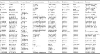
In microbial organisms, it is only in the bacterium Legionella pneumophila that NTPDases have definitively been shown to play a role in disease pathogenesis. L. pneumophila is a gram-negative bacterium and the causative agent of Legionnaires Disease, a systemic disease characterized by pneumonia. Although very unusual for a prokaryotic organism, the genome of L. pneumophila encodes 2 NTPDases (Chien et al. Reference Chien, Morozova, Shi, Sheng, Chen, Gomez, Asamani, Hill, Nuara, Feder, Rineer, Greenberg, Steshenko, Park, Zhao, Teplitskaya, Edwards, Pampou, Georghiou, Chou, Iannuccilli, Ulz, Kim, Geringer-Sameth, Goldsberry, Morozov, Fischer, Segal, Qu, Rzhetsky, Zhang, Cayanis, De Jong, Ju, Kalachikov, Shuman and Russo2004). Inactivation of these genes by insertion of an antibiotic resistance cassette leads to a decreased ability of the strains to replicate within macrophages and to cause disease in the mouse model of pathogenesis. Importantly, for at least 1 of the NTPDases, it was demonstrated that the NTPDase activity was essential for disease, as complementation of the mutant with an inactive form of the protein did not restore virulence (Sansom et al. Reference Sansom, Riedmaier, Newton, Dunstone, Muller, Stephan, Byres, Beddoe, Rossjohn, Cowan, d'Apice, Robson and Hartland2007, Reference Sansom, Riedmaier, Newton, Dunstone, Muller, Stephan, Byres, Beddoe, Rossjohn, Cowan, d'Apice, Robson and Hartland2008a, Reference Sansom, Robson and Hartlandb; Vivian et al. Reference Vivian, Riedmaier, Ge, Le Nours, Sansom, Wilce, Byres, Dias, Schmidberger, Cowan, d'Apice, Hartland, Rossjohn and Beddoe2010). In parasites, however, there is a distinct lack of molecular studies in which genes encoding potential NTPDases have been inactivated. Instead, as will be discussed here, evidence for the function of NTPDases in virulence relies primarily on the use of antibody and inhibitor studies. These studies, while not definitive, do suggest roles in both parasite biology and disease pathogenesis. Further complicating the study of NTPDases in parasites is the wide range of nomenclature used for the NTPDases. Terms such as ‘apyrase’, ‘NDPase’ and ‘ATPDase’ are in general misleading, as they do not accurately reflect the differing substrate range of this enzyme family, and are not necessarily specific for the GDA1_CD39 superfamily of proteins. The nomenclature in the mammalian field was standardized around 10 years ago (Zimmermann et al. Reference Zimmermann, Beaudoin, Bollen, Goding, Guidotti, Kirley, Robson, Sano, Vanduffel and Lemmens2000) and it is the suggestion of this author that the parasite field follow suit, to enable clear identification of NTPDases between studies, in particular when referring to NTPDases from parasites that encode multiple potential NTPDases. The proposed nomenclature is outlined in Table 1 and discussed in more detail in the following sections.
Putative and known NTPDases encoded on the genomes of parasites

NTPs, nucleoside triphosphates.
NDPs, nucleoside diphosphates.
SP, signal peptide.
TMD, transmembrane domain.
PV, parasitophorous vacuole.
* Putative domains detected using simple modular architecture research tool (SMART) (http://smart.embl-heidelberg.de/).
** Terminology used here to indicate a homologue of the S. cerevisiae protein.
# Terminology used here for convenience to indicate a third putative NTPDase in C. parapsilosis.
MOLECULAR EVIDENCE FOR NTPDASES IN PARASITES
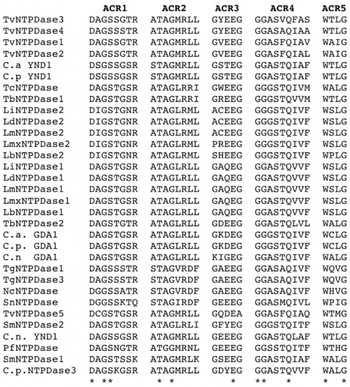
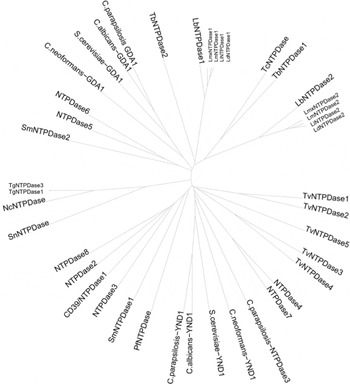
From a molecular standpoint, the defining characteristic of an NTPDase is the presence of the 5 ACRs (Handa and Guidotti, Reference Handa and Guidotti1996; Vasconcelos et al. Reference Vasconcelos, Ferreira, Carvalho, Souza, Kettlun, Mancilla, Valenzuela and Verjovski-Almeida1996). In this section the evidence for the presence of genes encoding NTPDases in a range of parasites is reviewed, as well as the results of any biochemical studies examining the substrate range and specificity of such NTPDases. This evidence is also summarized in Table 1 for ease of reference, and Fig. 1 details the amino acid alignment of the ACRs of all parasite NTPDases discussed here. Figure 2 details the phylogenetic relationship between the parasitic NTPDases and the mammalian and yeast NTPDases, as these relationships may provide clues to the function of the uncharacterized NTPDases.
Amino acid alignment of putative and known NTPDases of parasites demonstrating the presence of the 5 crucial ‘apyrase conserved regions’ (ACRs) in all predicted protein sequences. Absolutely conserved residues are starred. Sequence alignment was performed using the ClustalW sequence alignment tool (http://www.ebi.ac.uk/Tools/msa/clustalw2/) (Larkin et al. Reference Larkin, Blackshields, Brown, Chenna, McGettigan, McWilliam, Valentin, Wallace, Wilm, Lopez, Thompson, Gibson and Higgins2007; Goujon et al. Reference Goujon, McWilliam, Li, Valentin, Squizzato, Paern and Lopez2010). Identifying numbers for amino acid sequences used in the alignment are listed in Table 1. C. a., C. albicans; C. p., C. parapsilosis; C. n., C. neoformans.

Phylogenetic tree constructed from amino acid sequences of human, yeast and parasite NTPDases (sequence identifying numbers are listed in Table 1 or below). The phylogenetic tree was generated from an alignment of sequences using the ClustalW sequence alignment tool, with the resulting tree viewed and edited using the Interactive Tree of Life web tool (http://itol.embl.de/index.shtml) (Letunic and Bork, Reference Letunic and Bork2007, Reference Letunic and Bork2011). Human CD39/NTPDase1:NP_001767, human NTPDase2:NP_982293, human NTPDase3:NP_982293, human NTPDase4:NP_004892, human NTPDase5:NP_001240, human NTPDase6:NP_001238, human NTPDase7:NP_065087, human NTPDase8:NP_001028285, S. cerevisiae GDA1:NP_010872, S. cerevisiae YND1:EDN62971.

Apicomplexan parasites
Toxoplasma gondii, the cause of the zoonotic disease toxoplasmosis, a disease of serious significance in pregnant women and the immunocompromised, encodes 3 NTPDases on its genome. However, analysis of cDNA demonstrated that only 2 of these genes are translated. Complicating the nomenclature in this species, 2 research groups simultaneously identified the NTPDases, naming them NTPase-I and NTPase-II, or NTPase3 and NTPase1 respectively (Bermudes et al. Reference Bermudes, Peck, Afifi, Beckers and Joiner1994; Sibley et al. Reference Sibley, Niesman, Asai and Takeuchi1994). Both amino acid sequences contain all 5 ACRs, demonstrating membership of the GDA1_CD39 superfamily, suggesting that it is more accurate to refer to the enzymes as NTPDases rather than simply NTPases. Thus, proposed new nomenclature referring to NTPase I (or 3) as TgNTPDase1 and NTPase II (or 1) as TgNTPDase2 will be used here (see Table 1). The 2 T. gondii enzymes are very similar (97% identity), and biochemical techniques have been used to examine substrate range and specificity. Specifically, GST-tagged proteins were expressed and used to raise polyclonal antibodies, which were then used to pull down native NTPDases from T. gondii. This enabled characterization of the substrate specificity, revealing interesting differences despite the similarity of the proteins. Both NTPDases hydrolyse ATP, GTP, CTP and UTP, but TgNTPDase1 has less than 1% of the relative activity of hydrolysis of ADP, GDP, CDP and UDP. However, it has higher NTPase activity, being 4·5 times more efficient at hydrolysing ATP than TgNTPDase2 (Asai et al. Reference Asai, Miura, Sibley, Okabayashi and Takeuchi1995). Intriguingly, it is an area outside the ACRs which appears to dictate substrate specificity – namely a 12-residue block of amino acids in the C-terminus of the proteins. Synthesis of protein chimeras has demonstrated that a 12-residue block of amino acids FITGREMLASID and IVTGGGMLAAIN near the C-terminus of TgNTPDase2 and TgNTPDase1 respectively (residues 488–499) alters specificity for NTPs and NDPs (Nakaar et al. Reference Nakaar, Beckers, Polotsky and Joiner1998a). Sera from a small fraction of T. gondii patients can discriminate between TgNTPDase1 and TgNTPDase2 on the basis of these 12 residues demonstrating that the difference is antigenically distinct, although the significance of this finding is not clear (Johnson et al. Reference Johnson, Broady and Johnson1999).
The related parasite Neospora caninum causes disease such as abortion in domestic animals, but is not thought to infect humans. The genome of N. caninum is not yet published, but 1 study used Southern blotting to demonstrate the presence of multiple genes encoding NTPDases (Asai et al. Reference Asai, Howe, Nakajima, Nozaki, Takeuchi and Sibley1998). However, analysis of cDNA clones indicated that at least 2 genes were nearly identical, as well as confirming that the predicted proteins contain all 5 ACRs. Recombinant NcNTPDase was produced from one cDNA clone, and native NTPDase was also partially purified. Analysis of the enzyme activity demonstrated an ability to hydrolyse a wide variety of NTPs, but little activity against NDPs, consistent with the observation that NcNTPDase is most similar (69% identity) to TgNTPDase1. Given the large difference in the range of substrates of the 2 T. gondii NTPDases (despite being very similar in amino acid sequence) it would be interesting, once the complete genome is published, to identify which genes are actually transcribed, and if recombinant forms of each NTPDase do indeed only hydrolyse NTPs.
More recently another apicomplexan parasite Sarcocystis neurona, a cause of encephalitis in horses, was shown to possess a gene encoding an NTPDase (Zhang et al. Reference Zhang, Gaji and Howe2006). A recombinant form of this protein was produced and tested for ability to hydrolyse both ATP and ADP. Similar activity was seen against both substrates, although other NTPs and NDPs were not tested.
Finally, the genome of the important apicomplexan parasite Plasmodium falciparum, the major cause of human malaria, encodes a single NTPDase (Gardner et al. Reference Gardner, Hall, Fung, White, Berriman, Hyman, Carlton, Pain, Nelson, Bowman, Paulsen, James, Eisen, Rutherford, Salzberg, Craig, Kyes, Chan, Nene, Shallom, Suh, Peterson, Angiuoli, Pertea, Allen, Selengut, Haft, Mather, Vaidya, Martin, Fairlamb, Fraunholz, Roos, Ralph, McFadden, Cummings, Subramanian, Mungall, Venter, Carucci, Hoffman, Newbold, Davis, Fraser and Barrell2002). Interestingly this NTPDase appears evolutionarily distinct from the NTPDases of other apicomplexan parasites (Fig. 2), (Sansom et al. Reference Sansom, Robson and Hartland2008b), and instead more closely related to the cell surface-located NTPDases of humans and Schistosoma mansoni. Currently no information exists as to substrate preference or activity levels of the predicted protein.
Trypanosomatids
Of the trypanosomatid parasites only the NTPDase of T. cruzi, the causative agent of Chagas disease, has been characterized on a molecular level. The genome of T. cruzi encodes a single predicted NTPDase containing all 5 ACRs. A recombinant form of this enzyme has been expressed and purified from bacteria, and the substrate specificity examined. TcNTPDase has activity against ATP and ADP, with an ATPase:ADPase ratio similar to NTPDase1/CD39 (Santos et al. Reference Santos, Possa, Bastos, Guedes, Almeida, Demarco, Verjovski-Almeida, Bahia and Fietto2009). The genome of Trypanosoma brucei, the cause of African sleeping sickness, encodes 2 predicted NTPDases (Berriman et al. Reference Berriman, Ghedin, Hertz-Fowler, Blandin, Renauld, Bartholomeu, Lennard, Caler, Hamlin, Haas, Bohme, Hannick, Aslett, Shallom, Marcello, Hou, Wickstead, Alsmark, Arrowsmith, Atkin, Barron, Bringaud, Brooks, Carrington, Cherevach, Chillingworth, Churcher, Clark, Corton, Cronin, Davies, Doggett, Djikeng, Feldblyum, Field, Fraser, Goodhead, Hance, Harper, Harris, Hauser, Hostetler, Ivens, Jagels, Johnson, Johnson, Jones, Kerhornou, Koo, Larke, Landfear, Larkin, Leech, Line, Lord, Macleod, Mooney, Moule, Martin, Morgan, Mungall, Norbertczak, Ormond, Pai, Peacock, Peterson, Quail, Rabbinowitsch, Rajandream, Reitter, Salzberg, Sanders, Schobel, Sharp, Simmonds, Simpson, Tallon, Turner, Tait, Tivey, Van Aken, Walker, Wanless, Wang, White, White, Whitehead, Woodward, Wortman, Adams, Embley, Gull, Ullu, Barry, Fairlamb, Opperdoes, Barrell, Donelson, Hall, Fraser, Melville and El-Sayed2005), but no biochemical characterization has been performed. Similarly, all 5 species of Leishmania for which genome sequence information is available possess 2 predicted NTPDases (Peacock et al. Reference Peacock, Seeger, Harris, Murphy, Ruiz, Quail, Peters, Adlem, Tivey, Aslett, Kerhornou, Ivens, Fraser, Rajandream, Carver, Norbertczak, Chillingworth, Hance, Jagels, Moule, Ormond, Rutter, Squares, Whitehead, Rabbinowitsch, Arrowsmith, White, Thurston, Bringaud, Baldauf, Faulconbridge, Jeffares, Depledge, Oyola, Hilley, Brito, Tosi, Barrell, Cruz, Mottram, Smith and Berriman2007), but no characterization of these enzymes is available. Leishmania causes the disease known as leishmaniasis, which exists as cutaneous, mucocutaneous and visceral forms, depending on the species responsible for infection (Herwaldt, Reference Herwaldt1999). The sequenced strains represent at least 1 species responsible for each syndrome, demonstrating that NTPDases are conserved on the genome irrespective of the resulting disease syndrome. The nomenclature, particularly for the T. brucei and Leishmania NTPDases, is also confused. While some studies refer to ecto-nucleoside triphosphate diphosphohydrolase activities of, for example, L. amazonensis (Pinheiro et al. Reference Pinheiro, Martins-Duarte, Ferraro, Fonseca de Souza, Gomes, Lopes, Vannier-Santos, Santos and Meyer-Fernandes2006), the genome annotation for the 2 NTPDases are nucleoside diphosphatase and guanosine diphosphatase (Peacock et al. Reference Peacock, Seeger, Harris, Murphy, Ruiz, Quail, Peters, Adlem, Tivey, Aslett, Kerhornou, Ivens, Fraser, Rajandream, Carver, Norbertczak, Chillingworth, Hance, Jagels, Moule, Ormond, Rutter, Squares, Whitehead, Rabbinowitsch, Arrowsmith, White, Thurston, Bringaud, Baldauf, Faulconbridge, Jeffares, Depledge, Oyola, Hilley, Brito, Tosi, Barrell, Cruz, Mottram, Smith and Berriman2007) which, in light of the unknown substrate specificities of these enzymes, is not necessarily accurate or informative. As both predicted proteins from T. brucei and all Leishmania species contain the 5 ACRs, it makes sense to refer to these enzymes as NTPDases, and to use this term consistently (Table 1).
Trichomonas vaginalis
T. vaginalis is a flagellated protozoan responsible for trichomoniasis, the most common non-viral sexually transmitted disease in the world (Van der Pol, Reference Van der Pol2007). Recent studies identified 2 NTPDase orthologues on the T. vaginalis genome and demonstrated an effect on transcription levels, by RT-PCR, in the presence of various steroids and drugs (Giordani et al. Reference Giordani, Weizenmann, Rosemberg, De Carli, Bogo, Zuanazzi and Tasca2010; Ruckert et al. Reference Ruckert, Stuepp Cdos, Gottardi, Rosa, Cisilotto, Borges, Rosemberg, Bogo, Tasca, De Carli and Bonan2010). These studies identified the proteins by BLAST searching using mammalian NTPDase sequences. However, prior BLAST searching of the translated genome (Carlton et al. Reference Carlton, Hirt, Silva, Delcher, Schatz, Zhao, Wortman, Bidwell, Alsmark, Besteiro, Sicheritz-Ponten, Noel, Dacks, Foster, Simillion, Van de Peer, Miranda-Saavedra, Barton, Westrop, Muller, Dessi, Fiori, Ren, Paulsen, Zhang, Bastida-Corcuera, Simoes-Barbosa, Brown, Hayes, Mukherjee, Okumura, Schneider, Smith, Vanacova, Villalvazo, Haas, Pertea, Feldblyum, Utterback, Shu, Osoegawa, de Jong, Hrdy, Horvathova, Zubacova, Dolezal, Malik, Logsdon, Henze, Gupta, Wang, Dunne, Upcroft, Upcroft, White, Salzberg, Tang, Chiu, Lee, Embley, Coombs, Mottram, Tachezy, Fraser-Liggett and Johnson2007) using parasite NTPDase sequences had in fact revealed 4 genes encoding predicted NTPDases (Sansom et al. Reference Sansom, Robson and Hartland2008b) and a most recent BLAST search of the translated genome performed here reveals a total of 5 predicted NTPDases containing all 5 ACRs (Table 1, Fig. 1). However, no information on the level of enzyme activity or substrate specificity for each predicted NTPDase is currently available, nor is it known if all 5 putative NTPDases are expressed or where the NTPDases localize within the parasite.
Schistosoma mansoni
S. mansoni is a trematode responsible for intestinal schistosomiasis in humans. Purification of proteins from S. mansoni using antibodies to potato NTPDase and T. gondii NTPDase revealed 2 isoforms of NTPDases, which were both able to hydolyse ATP while one isoform had higher ADPase activity (Vasconcelos et al. Reference Vasconcelos, Ferreira, Carvalho, Souza, Kettlun, Mancilla, Valenzuela and Verjovski-Almeida1996). The genome of S. mansoni encodes 2 predicted NTPDases, currently designated SmATPDase1 and SmATPDase2. A recombinant form of SmATPDase1 has been produced in E. coli and used to raise polyclonal antibodies, although the activity of the recombinant protein has not been directly tested. SmATPDase2 has also been expressed in E. coli, and recombinant forms purified by solubilization of inclusion bodies and refolding, although the activity of this protein has not been tested (Levano-Garcia et al. Reference Levano-Garcia, Mortara, Verjovski-Almeida and DeMarco2007). It is not clear which gene encodes which NTPDase isoform identified in the original study (Vasconcelos et al. Reference Vasconcelos, Ferreira, Carvalho, Souza, Kettlun, Mancilla, Valenzuela and Verjovski-Almeida1996), and thus the specific activity of each protein against NTPs and NDPs is unknown. Given the presence of all 5 ACRs, it is most likely that the enzymes do hydrolyse other NTPs and/or NDPs, and thus are more accurately referred to as NTPDases (Table 1).
Fungi
The genome of the non-pathogenic yeast Saccharyomyces cerevisiae encodes 2 NTPDases. These proteins, GDA1 and YND1, are located in the Golgi apparatus, and play crucial roles in glycosylation (Abeijon et al. Reference Abeijon, Yanagisawa, Mandon, Hausler, Moremen, Hirschberg and Robbins1993; Gao et al. Reference Gao, Kaigorodov and Jigami1999). Interestingly the two proteins have different membrane topologies (reviewed by Knowles, Reference Knowles2011), with the single transmembrane domain of YND1 located near the C-terminus of the protein. The proteins are also not particularly closely related (Fig. 2). Deletion of either enzyme in S. cerevisiae results in defects in N- and O-glycosylation, as well as wider defects in cell wall synthesis, and deletion of both genes is lethal (Gao et al. Reference Gao, Kaigorodov and Jigami1999; Knowles, Reference Knowles2011). A similar role for GDA1 has been identified in other species of yeast, including Schizosaccharomyces pombe (Sanchez et al. Reference Sanchez, Franco, Gacto, Notario and Cansado2003) and Kluyveromyces lactis (Lopez-Avalos et al. Reference Lopez-Avalos, Uccelletti, Abeijon and Hirschberg2001), as well as in the pathogenic yeast Candida albicans (Herrero et al. Reference Herrero, Uccelletti, Hirschberg, Dominguez and Abeijon2002). C. albicans also encodes a homologue of YND1 (Figs 1 and 2), although in this species of yeast the protein has not been characterized (Jones et al. Reference Jones, Federspiel, Chibana, Dungan, Kalman, Magee, Newport, Thorstenson, Agabian, Magee, Davis and Scherer2004). Another fungal pathogen of humans, Cryptococcus neoformans, encodes homologues of both GDA1 and YND1 (Figs 1 and 2), but these proteins have also not been studied (Loftus et al. Reference Loftus, Fung, Roncaglia, Rowley, Amedeo, Bruno, Vamathevan, Miranda, Anderson, Fraser, Allen, Bosdet, Brent, Chiu, Doering, Donlin, D'Souza, Fox, Grinberg, Fu, Fukushima, Haas, Huang, Janbon, Jones, Koo, Krzywinski, Kwon-Chung, Lengeler, Maiti, Marra, Marra, Mathewson, Mitchell, Pertea, Riggs, Salzberg, Schein, Shvartsbeyn, Shin, Shumway, Specht, Suh, Tenney, Utterback, Wickes, Wortman, Wye, Kronstad, Lodge, Heitman, Davis, Fraser and Hyman2005). Most interestingly, BLAST searching of the genome of another human pathogen, Candida parapsilosis, revealed 3 putative NTPDases (Butler et al. Reference Butler, Rasmussen, Lin, Santos, Sakthikumar, Munro, Rheinbay, Grabherr, Forche, Reedy, Agrafioti, Arnaud, Bates, Brown, Brunke, Costanzo, Fitzpatrick, de Groot, Harris, Hoyer, Hube, Klis, Kodira, Lennard, Logue, Martin, Neiman, Nikolaou, Quail, Quinn, Santos, Schmitzberger, Sherlock, Shah, Silverstein, Skrzypek, Soll, Staggs, Stansfield, Stumpf, Sudbery, Srikantha, Zeng, Berman, Berriman, Heitman, Gow, Lorenz, Birren, Kellis and Cuomo2009). While 2 of these proteins appear to be homologues of GDA1 and YND1 (Figs 1 and 2, Table 1), and most likely have similar functions, the third may well play a different role. However, these proteins are also currently uncharacterized, and thus it is not known what function the third NTPDase performs.
EVIDENCE FOR THE PRESENCE OF NTPDASE PROTEINS AND NTPDASE ACTIVITY IN PARASITES
The sequencing of a large number of parasite genomes over the last decade has revealed that many do indeed encode putative NTPDases (Table 1, Fig. 1). Additionally, for many of these parasites enzyme activity consistent with the presence of NTPDases has been observed. What, however, is often less clear is which putative NTPDase (if any) is responsible for the observed enzyme activity. While for some parasites the localization of the putative NTPDases is known, for others no studies have been undertaken. Furthermore, while the substrate specificity of some parasite NTPDases is well characterized, for others it is unknown, or inferred from assays using whole parasites or parasite extracts. In this section the biological evidence for the presence of NTPDases in parasites and, when known, the localization of the enzymes is discussed.
Apicomplexan parasites
The use of antibodies to recombinant forms of the T. gondii NTPDases has enabled accurate localization of the enzymes within the parasite and during infection of the mammalian host cell. In the extracellular tachyzoite form of the parasite the NTPDases are present in dense granules, and following infection of the host cell both enzymes are secreted into the lumen of the parasitophorous vacuole (PV) (Bermudes et al. Reference Bermudes, Peck, Afifi, Beckers and Joiner1994; Sibley et al. Reference Sibley, Niesman, Asai and Takeuchi1994). The precise roles of the two enzymes during host infection are not known, but a number of studies, discussed below, implicate at least one NTPDase in virulence. Unfortunately the similarity of the genes encoding the NTPDases, as well as some evidence that TgNTPDase1 is essential for viability, appears to preclude the generation of a specific mutant (Nakaar et al. Reference Nakaar, Samuel, Ngo and Joiner1999), making it difficult to definitively show that either or both NTPDases are involved in pathogenesis. Nonetheless the studies performed to date do certainly suggest a role for NTPDases in the ability of the parasite to cause disease.
Analysis of expression levels of the enzymes revealed that TgNTPDase1 has only low levels of expression in the bradyzoite form of the parasite (the form responsible for chronic host infection) but is highly expressed in the actively replicating tachyzoite form (Nakaar et al. Reference Nakaar, Bermudes, Peck and Joiner1998b). The presence of the two enzymes in virulent and non-virulent strains also implicates TgNTPDase1 in virulence, as most virulent strains of T. gondii possess the gene encoding TgNTPDase1, but avirulent strains carry only the gene encoding TgNTPDase2 (Asai et al. Reference Asai, Miura, Sibley, Okabayashi and Takeuchi1995; Nakaar et al. Reference Nakaar, Beckers, Polotsky and Joiner1998a). In the absence of the ability to make deletion mutants, antisense RNA studies demonstrated that TgNTPDase1 was dispensable for entry into host cells but required for replication once inside the host cell (Nakaar et al. Reference Nakaar, Samuel, Ngo and Joiner1999). These results do conflict with another study using a monoclonal antibody that inhibited both NTPDases. Pre-treatment of parasites with this antibody resulted in decreased invasion of Vero cells (Kikuchi et al. Reference Kikuchi, Furuta and Kojima2001), suggesting that NTPDase activity did contribute to invasion of host cells and highlighting the difficulty in interpreting studies where techniques that may have off-target effects are used. While the similarity of the genes may preclude generation of a single mutant, the proximity of the genes to each other on the chromosome may allow a double mutant to be created, and analysis of such a mutant would still provide valuable information.
There is also some evidence that TgNTPDase1 may be involved in egress from the host cell, shown by experiments examining the activation state of the enzyme. When secreted by the parasite, TgNTPDase1 remains largely oxidized, reflecting the fact that parasitic infections generate host nitric oxide and other free radicals that result in oxidative stress. If exogenous thiols are used to activate the enzyme, a rapid drop in host cell ATP occurs and the parasites exit the cell within a minute of treatment (Silverman et al. Reference Silverman, Qi, Riehl, Beckers, Nakaar and Joiner1998). This exit requires Ca2+ ions, presumably released when ATP depletion occurs (Stommel et al. Reference Stommel, Ely, Schwartzman and Kasper1997). Thus the activation of secreted TgNTPDase1 must be closely regulated by the parasite. T. gondii secrete the reducing agent glutaredoxin (GRX) as replication increases, and GRX is able to activate TgNTPDase1 in vitro, suggesting GRX secretion by the parasite may be a way of controlling the oxidation state of TgNTPDase1 and thus implicating TgNTPDase1 in a role in exit from the host (Stommel et al. Reference Stommel, Cho, Steide, Seguin, Barchowsky, Schwartzman and Kasper2001). All these studies implicate T. gondii NTPDase(s) in pathogenesis at all stages of the intracellular life cycle, suggesting that further investigation of these enzymes is warranted.
Much less is known about the biological role of the other apicomplexan NTPDases. NcNTPDase is also localized to the dense granules of the parasite and may be secreted in a similar manner to the NTPDases of T. gondii, consistent with the presence of a typical N-terminal signal peptide (Asai et al. Reference Asai, Howe, Nakajima, Nozaki, Takeuchi and Sibley1998). Immunoblotting using polyclonal antibodies demonstrated secretion of ScNTPDase into culture supernatants in vitro, also consistent with its signal peptide. Localization studies demonstrate that ScNTPDase is expressed in an apical location on merozoites immediately following host cell invasion, but is then absent during intracellular replication before reappearing on newly formed merozoites immediately prior to exit from the host cell (Zhang et al. Reference Zhang, Gaji and Howe2006). These data suggests a role in host cell exit and/or entry, or possibly that ScNTPDase is also required during extracellular survival of the parasite. It also may be that the protein is secreted during replication into the PV, but in amounts not detected by the localization techniques used. No biological evidence exists to identify the possible role of the P. falciparum NTPDase, although the presence of predicted transmembrane domains on both the N- and C-terminal of the protein (Table 1) suggests that, in contrast to the other apicomplexan parasites, this NTPDase is not secreted, but rather anchored in a membrane.
Trypanosomatids
The presence of Mg2+-dependent ecto-ATPase activity – that is, activity consistent with the presence of a surface-located NTPDase – was demonstrated for the intracellular parasite T. cruzi (Bisaggio et al. Reference Bisaggio, Peres-Sampaio, Meyer-Fernandes and Souto-Padron2003), and a subsequent study demonstrated a range of ecto-NTPDase activities of intact parasites (Fietto et al. Reference Fietto, DeMarco, Nascimento, Castro, Carvalho, de Souza, Bahia, Alves and Verjovski-Almeida2004). Most recently, T. cruzi was shown to vary its ecto-ATPase:ADPase ratio during passage in culture, and a drop in overall enzyme activity levels correlated with decreased infectivity for mammalian cells (Santos et al. Reference Santos, Possa, Bastos, Guedes, Almeida, Demarco, Verjovski-Almeida, Bahia and Fietto2009). However, one draw-back of this study was the inability to discern enzyme activity due to other surface-located enzymes from activity attributable to TcNTPDase. In fact, use of ARL67156, an inhibitor considered specific for NTPDases, inhibited only 30% and 50% of the observed ATPase and ADPase activity, suggesting that other classes of enzymes are responsible for some of the observed activity. Intriguingly, ARL67156 was unable to inhibit recombinant TcNTPDase, an unexpected finding, which may reflect a differing structure of TcNTPDase compared to other NTPDases which are inhibited by ARL67156, or could also relate to altered folding of the recombinant protein during expression, re-folding and purification. While such altered folding would presumably have an effect on enzyme activity, as the enzyme activity of native TcNTPDase is unknown, it is not possible to use the observed enzyme activity to demonstrate that the recombinant protein is 100% correctly folded. Despite lack of inhibition of TcNTPDase by ARL67156, treatment of parasites with ARL67156, or the non-specific NTPDase inhibitors gadolinium and suramin, resulted in decreased infectivity in vitro as well as decreased virulence in the mouse model of disease (Santos et al. Reference Santos, Possa, Bastos, Guedes, Almeida, Demarco, Verjovski-Almeida, Bahia and Fietto2009). It is therefore hard to distinguish from these results, however, how much of the effect on virulence is specifically related to inhibition of TcNTPDase activity. The effect on infection of non-specific inhibitors does agree with earlier work demonstrating inhibition of ecto-ATPase activity by suramin and 4,4¢-diisothiocy-anostylbene 2¢,2¢-disulfonic acid (DIDS) (Bernardes et al. Reference Bernardes, Meyer-Fernandes, Saad-Nehme, Vannier-Santos, Peres-Sampaio and Vercesi2000; Bisaggio et al. Reference Bisaggio, Peres-Sampaio, Meyer-Fernandes and Souto-Padron2003), which resulted in a decreased number of parasites attaching to and infecting mouse peritoneal macrophages. In direct contrast, addition of 200 μ m ATP increased macrophage infection by 30% (Bisaggio et al. Reference Bisaggio, Peres-Sampaio, Meyer-Fernandes and Souto-Padron2003). Again however, it is hard to determine the contribution of TcNTPDase to the observed effect on infection, as these inhibitors have other effects.
Earlier studies also provide some evidence for a role for NTPDase activity in parasite virulence, although again it is uncertain if the observed activity is due to TcNTPDase. Up to 20-fold higher ecto-ATPase activity has been observed in the infective trypomastigote stage compared to the epimastigote stage (Bisaggio et al. Reference Bisaggio, Peres-Sampaio, Meyer-Fernandes and Souto-Padron2003; Meyer-Fernandes et al. Reference Meyer-Fernandes, Saad-Nehme, Peres-Sampaio, Belmont-Firpo, Bisaggio, Do Couto, Fonseca De Souza, Lopes and Souto-Padron2004). An altered ratio of ATP:ADP hydrolysis was also observed, with an ATP:ADP hydrolysis ratio for trypomastigotes of 2:1, while for epimastigotes it is 1:1. Other substrates are also hydrolysed by T. cruzi, again suggesting NTPDase activity, with hydrolysis of GTP, GDP, UTP and UDP, with highest activity against GTP (Bisaggio et al. Reference Bisaggio, Peres-Sampaio, Meyer-Fernandes and Souto-Padron2003).
The localization of TcNTPDase is also still unclear. An early study showed cross-reaction of a protein on the surface of the parasite with an antibody to T. gondii NTPDase (Fietto et al. Reference Fietto, DeMarco, Nascimento, Castro, Carvalho, de Souza, Bahia, Alves and Verjovski-Almeida2004). However, the presence of a predicted N-terminal signal peptide suggests the parasite may secrete TcNTPDase. Polyclonal antibodies to recombinant TcNTPDase do now exist, and use of these antibodies in immunofluorescence experiments may clarify the location of TcNTPDase, particularly during infection of mammalian cells. Use of other genetic techniques now available in T. cruzi (Taylor et al. Reference Taylor, Huang and Kelly2011), including systems to epitope-tag proteins (e.g. with GFP) and to over-express proteins, may also help clarify both the location of TcNTPDase and its function.
In contrast to T. cruzi, T. brucei is an extracellular pathogen that in mammals replicates in the blood stream (Matthews, Reference Matthews2005). Two putative NTPDase enzymes are encoded on the T. brucei genome (Table 1), and assays using intact parasites demonstrate a cation-dependent, surface-located hydrolysis of ATP, GTP, CTP, UTP and ADP consistent with the presence of at least 1 NTPDase enzyme (de Souza Leite et al. Reference de Souza Leite, Thomaz, Fonseca, Panizzutti, Vercesi and Meyer-Fernandes2007). Both predicted NTPDases contain N-terminal signal peptides, suggesting secretion by the parasite, but the localization, or indeed expression levels, of either putative enzyme are unknown. A recent study demonstrated inhibition of cell-surface NTPDase activity by ferrous iron and haem, although the mechanism of inhibition is not understood (Leite et al. Reference Leite, Thomaz, Oliveira, Oliveira and Meyer-Fernandes2009). The availability of successful RNAi systems in T. brucei could be utilized to elucidate the importance of these enzymes in virulence (Balana-Fouce and Reguera, Reference Balana-Fouce and Reguera2007).
While genome information is not available for a further species, T. rangeli, this parasite (capable of causing disease in humans and animals) also exhibits Mg2+-dependent cell-surface NTPDase activity against ATP, ADP and other NTPs, consistent with the presence of 1 or more NTPDases. In this species, hydrolysis of nucleotides is stimulated by a number of carbohydrates, leading to the hypothesis that NTPDase activity may have a role in adhesion to the intermediate insect host, as carbohydrates on insect salivary glands play a part in adhesion by Trypanosoma species (Fonseca et al. Reference Fonseca, Fonseca de Souza, Mariano, Entringer, Gondim and Meyer-Fernandes2006), although there is no other evidence as yet for this theory. An atypical and non-pathogenic trypanosomatid, Crithidia deanei, is also observed to have surface-located NTPDase activity, with hydrolysis of ATP and other NTPs observed, but genome information is also unavailable for this species (dos Passos Lemos et al. Reference dos Passos Lemos, de Sa Pinheiro, de Berredo-Pinho, Fonseca de Souza, Motta, de Souza and Meyer-Fernandes2002).
In Leishmania parasites, Mg2+-dependent cell surface-located NTPDase activity has been observed in 2 species responsible for cutaneous leishmaniasis, namely Leishmania tropica and Leishmania amazonensis (Meyer-Fernandes et al. Reference Meyer-Fernandes, Dutra, Rodrigues, Saad-Nehme and Lopes1997; Berredo-Pinho et al. Reference Berredo-Pinho, Peres-Sampaio, Chrispim, Belmont-Firpo, Lemos, Martiny, Vannier-Santos and Meyer-Fernandes2001; Pinheiro et al. Reference Pinheiro, Martins-Duarte, Ferraro, Fonseca de Souza, Gomes, Lopes, Vannier-Santos, Santos and Meyer-Fernandes2006). Again, however, it is not known which enzymes are responsible for this activity, although the nature of the activity is consistent with the presence of 1 or more NTPDases. While the genome sequence is not available for L. tropica or L. amazonensis, the presence of the 2 putative NTPDases on all 5 sequenced strains of Leishmania (Table 1) does suggest that L. tropica and L. amazonensis would also possess putative NTPDases on their genomes.
The localizations of the putative NTPDases are not known, but one early study demonstrated cross-reaction on the surface of L. amazonensis with antibody to CD39 (human NTPDase1), suggesting that the observed enzyme activity could be due to an NTPDase (Pinheiro et al. Reference Pinheiro, Martins-Duarte, Ferraro, Fonseca de Souza, Gomes, Lopes, Vannier-Santos, Santos and Meyer-Fernandes2006). The presence of a predicted N-terminal transmembrane domain in one NTPDase (Table 1) suggests that the protein could be anchored in the membrane on the cell surface, although equally it may be anchored in the membrane of an intracellular organelle such as the Golgi apparatus (as is the case for NTPDases in yeast (Berninsone et al. Reference Berninsone, Miret and Hirschberg1994; Gao et al. Reference Gao, Kaigorodov and Jigami1999)). The second putative Leishmania NTPDase has a predicted N-terminal signal peptide, suggesting that the protein is secreted, and could be responsible for ecto-NTPDase activity.
While it is not confirmed that the observed enzyme activity is due to members of the NTPDase family, the characterization performed to date is broadly consistent with the presence of NTPDase(s). Interestingly, however, the enzymes identified in L. tropica and L. amazonensis cannot utilize Ca2+ instead of Mg2+, which is unusual for the NTPDase family (Meyer-Fernandes et al. Reference Meyer-Fernandes, Dutra, Rodrigues, Saad-Nehme and Lopes1997; Pinheiro et al. Reference Pinheiro, Martins-Duarte, Ferraro, Fonseca de Souza, Gomes, Lopes, Vannier-Santos, Santos and Meyer-Fernandes2006). In L. tropica parasites, surface-located hydrolysis of ATP and ADP as well as other NTPs has been observed, and hydrolysis of ATP and ADP is observed on the surface of L. amazonensis (Pinheiro et al. Reference Pinheiro, Martins-Duarte, Ferraro, Fonseca de Souza, Gomes, Lopes, Vannier-Santos, Santos and Meyer-Fernandes2006). Similar to T. rangeli, carbohydrates stimulate the L. tropica ecto-NTPDase activity, although the mechanism and significance of this finding is unclear (Peres-Sampaio et al. Reference Peres-Sampaio, Palumbo and Meyer-Fernandes2001).
There is some evidence that this ecto-NTPDase activity may play a role in pathogenesis. Enzyme activity is higher in virulent strains than avirulent strains and is increased more than 10-fold in the obligate intracellular amastigote stage (Berredo-Pinho et al. Reference Berredo-Pinho, Peres-Sampaio, Chrispim, Belmont-Firpo, Lemos, Martiny, Vannier-Santos and Meyer-Fernandes2001; Pinheiro et al. Reference Pinheiro, Martins-Duarte, Ferraro, Fonseca de Souza, Gomes, Lopes, Vannier-Santos, Santos and Meyer-Fernandes2006). The ecto-ATPase activity of L. amazonensis increases when parasites undergo heat shock (Peres-Sampaio et al. Reference Peres-Sampaio, de Almeida-Amaral, Giarola and Meyer-Fernandes2008), as occurs when the parasites leave the sand fly vector and are injected into the warmer mammalian host. It is important to note that this observed ecto-enzyme activity has not been clearly demonstrated to be due to a member of the NTPDase family. However, treatment of parasites with anti-CD39 antibody reduces the interaction of the parasites with mouse peritoneal macrophages (Pinheiro et al. Reference Pinheiro, Martins-Duarte, Ferraro, Fonseca de Souza, Gomes, Lopes, Vannier-Santos, Santos and Meyer-Fernandes2006), thus more directly suggesting a role for an NTPDase in pathogenesis. A recent study utilized chromium (III) adenosine 5′-triphosphate complex (CrATP) to inhibit ecto-ATPase activity in L. amazonensis, and found that CrATP pre-treatment of parasites reduced both attachment and entry into mouse peritoneal macrophages (Ennes-Vidal et al. Reference Ennes-Vidal, Castro, Britto, Barrabin, D'Avila-Levy and Moreira2011), also suggesting a role in virulence. However, it is important to note that CrATP also inhibited a Mg2+-independent ecto-ATPase activity on the parasite surface, and thus presumably is not a specific inhibitor of NTPDase activity. Leishmania can be genetically manipulated and systems exist both for generating defined molecular mutants (Cruz et al. Reference Cruz, Coburn and Beverley1991), and for introducing episomal DNA expressing Leishmania proteins, which may also be tagged, for example with GFP, to allow localization studies (Ha et al. 1996). Construction of genetically defined mutant strains where the putative NTPDases have been deleted, and localization of the proteins using epitope tagging would be of great benefit in determining the role the putative NTPDases play in the observed enzyme activity described by previous studies, as well as allowing a more direct examination of the contribution of the NTPDases to pathogenesis.
Trichomonas vaginalis
In a similar manner to the studies performed in Leishmania parasites, enzyme assays conducted using intact and disrupted T. vaginalis cells has demonstrated the presence of surface-located enzyme activity consistent with the presence of 1 or more ecto-NTPDases (de Aguiar Matos et al. Reference de Aguiar Matos, Borges, Tasca, Bogo, De Carli, da Graca Fauth, Dias and Bonan2001). This Ca2+ or Mg2+-dependent hydrolysis of ATP, ADP and other nucleotides can be increased by up to 90% by the addition of D-galactose (de Jesus et al. Reference de Jesus, de Sa Pinheiro, Lopes and Meyer-Fernandes2002), similar to the observed effects of carbohydrates on ecto-NTPDase activity in some trypanosomatids. This ecto-NTPDase activity is indirectly implicated in virulence, as fresh clinical isolates of T. vaginalis demonstrate higher surface NTPDase activity compared to a less virulent laboratory-adapted strain (de Jesus et al. Reference de Jesus, de Sa Pinheiro, Lopes and Meyer-Fernandes2002; Tasca et al. Reference Tasca, Bonan, De Carli, Sarkis and Alderete2005).
Recent studies demonstrated an inhibition of ecto-NTPDase activity when parasites were treated, for 24 h, with the plant alkaloids lycorine and candimine which are toxic to T. vaginalis (Giordani et al. Reference Giordani, Weizenmann, Rosemberg, De Carli, Bogo, Zuanazzi and Tasca2010). However, it is not clear if the toxicity of lycorine and candimine is directly related to the inhibition of ecto-NTPDase activity. Interestingly, no effect of lycorine and candimine was seen on the expression levels of the 2 putative NTPDases examined in this study, suggesting that either the drugs act at a post-transcriptional level, or that the observed NTPDase activity which is inhibited by these drugs is due to 1 of 3 other putative NTPDases encoded on the T. vaginalis genome. Analysis of the effect of lycorine and candimine treatment on expression levels of these other 3 putative NTPDases would be useful. In contrast, treatment of freshly isolated parasites with steroids also inhibited ecto-NTPDase activity but this inhibition appeared to be due to inhibition at the level of mRNA transcription of the 2 NTPDases studied (Ruckert et al. Reference Ruckert, Stuepp Cdos, Gottardi, Rosa, Cisilotto, Borges, Rosemberg, Bogo, Tasca, De Carli and Bonan2010).
From these studies it is not clear precisely which putative NTPDases are responsible for the observed enzyme activity. Two of the 5 putative NTPDases possess predicted N-terminal signal peptides, suggesting secretion by the parasite, and all 5 are predicted to have C-terminal transmembrane domains, suggesting the proteins may be anchored in the extracellular membrane or in the membranes of organelles (Table 1). Use of RT-PCR to look at expression levels of all 5 genes, especially in response to compounds that demonstrate inhibitory effects on ecto-NTPDase activity, may help clarify which proteins are responsible for the extracellular NTPDase activity.
A related organism, Tritrichomonas foetus, a cause of abortion in cattle, also possesses Mg2+-dependent NTPDase activity. NTPs are hydrolysed, although no activity was detected against ADP, and activity is stimulated by D-mannose and D-galactose. However, as genome information is not available for this organism, it is not known if one or more NTPDases could be responsible for this observed activity (Jesus et al. Reference Jesus, Lopes and Meyer-Fernandes2002).
Schistosoma mansoni
The 2 NTPDases of S. mansoni have different localization patterns, suggesting functionally distinct roles. SmNTPDase1 is located on the surface, presumably anchored in the membrane by its predicted N- and C-terminal transmembrane domains, whereas SmNTPDase2 is secreted by the parasite (Vasconcelos et al. Reference Vasconcelos, Nascimento, Meirelles, Verjovski-Almeida and Ferreira1993; Levano-Garcia et al. Reference Levano-Garcia, Mortara, Verjovski-Almeida and DeMarco2007). While the role of each enzyme in parasite biology and disease pathogenesis is not yet known, it is intriguing that a new class of anti-schistosomal drugs, N-alkylaminoalkanethiosulfuric acids, are known to partially inhibit tegumental S. mansoni NTPDase activity, indicating that the NTPDases may be suitable drug targets (Luiz Oliveira Penido et al. Reference Luiz Oliveira Penido, Resende, Vianello, Humberto da Silveira Bordin, Jacinto, Dias, Montesano, Nelson, Marcos Zech Coelho and Vasconcelos2007). With the recent advent of RNAi techniques in S. mansoni (Bhardwaj et al. Reference Bhardwaj, Krautz-Peterson and Skelly2011) it would be informative if studies inhibiting either or both NTPDases were carried out, in order to assess the contribution of the enzymes to pathogenesis, and determine if they are indeed suitable drug targets.
Fungi and other parasites
As already discussed, the two NTPDases of S. cerevisisae are located in the Golgi and function in glycosylation and cell wall synthesis, and homologues of these proteins have been characterized in some other species of fungi. Evidence exists for NTPDase activity in other species of fungi in which the NTPDase homologues have not yet been characterized. Surface-located NTPDase activity stimulated by Mg2+ and exhibiting high rates of ATP, ITP, GTP, CTP and UTP but not ADP hydrolysis has been observed for C. neoformans (Junior et al. Reference Junior, Rodrigues, Alviano, Travassos and Meyer-Fernandes2005). While the genome of C. neoformans encodes 2 putative NTPDases, these both appear to be homologues of the S. cerevisiae Golgi-located NTPDases (Fig. 2), and it is tempting to speculate that these NTPDases function in the Golgi apparatus of C. neoformans. However, in the absence of any localization studies it is possible that one of these putative NTPDases is responsible for the observed surface-located activity. Studies using specific antibodies to the C. neoformans NTPDases, or other molecular techniques, are crucial in determining the likely role of the NTPDases in C. neoformans. More recently, ecto-NTPDase activity has been observed in the pathogenic yeast C. parapsilosis (Kiffer-Moreira et al. Reference Kiffer-Moreira, Fernandes Sampaio, Alviano, Axelband, Cesar, Cosentino-Gomes, Rodrigues, Nimrichter, Vieyra, Alviano and Meyer-Fernandes2010). Again, however, it has not actually been shown whether an NTPDase is responsible for the observed activity. As already mentioned, in addition to GDA1 and YND1 homologues, the genome of C. parapsilosis does encode a third putative NTPDase that could certainly be surface-located, although it is closely related to the YND1 homologue (Fig. 2). Again, specific molecular studies to localize these proteins are key to identifying the function of NTPDases in C. parapsilosis. Finally, in the human pathogen Fonsecaea pedrosoi, surface-located ATPase activity stimulated by Mg2+ has been observed (Collopy-Junior et al. Reference Collopy-Junior, Kneipp, da Silva, Rodrigues, Alviano and Meyer-Fernandes2006). However, the genome sequence for this organism is not available and the protein(s) responsible for the enzyme activity are unknown, making it unclear whether an NTPDase is actually involved.
A number of other parasites have also been reported to display surface-located enzyme activity broadly consistent with the presence of NTPDase(s). These include Acanthamoeba, Enatamoeba histolytica and Giardia lamblia (Barros et al. Reference Barros, De Menezes, Pinheiro, Silva, Lopes, De Souza and Meyer-Fernandes2000; Sissons et al. Reference Sissons, Alsam, Jayasekera and Khan2004; de Sa Pinheiro et al. Reference de Sa Pinheiro, Cosentino-Gomes, Lanfredi-Rangel, Ferraro, De Souza and Meyer-Fernandes2008). However, as previously described in the literature (Sansom et al. Reference Sansom, Robson and Hartland2008b), the genomes of these organisms encode no putative NTPDases, and thus will not be discussed in detail here, as the focus of this review is the NTPDase family of proteins. The amoebae Balamuthia mandrillaris has also been reported to have surface-located ATPase activity (Matin and Khan, Reference Matin and Khan2008), but genome information is unavailable and it is impossible to determine whether an NTPDase is involved.
POTENTIAL FUNCTIONS OF NTPDASES IN PATHOGENIC PARASITES
In humans NTPDases appear to play diverse roles including purine salvage, control of blood clotting and regulation of the immune and inflammatory response (Deaglio and Robson, Reference Deaglio and Robson2011). In yeast NTPDases function in the Golgi playing crucial roles in glycosylation (Berninsone et al. Reference Berninsone, Miret and Hirschberg1994; Gao et al. Reference Gao, Kaigorodov and Jigami1999), whereas in L. pneumophila secreted NTPDases contribute to the virulence of the bacteria in the mammalian host (Sansom et al. Reference Sansom, Newton, Crikis, Cianciotto, Cowan, d'Apice and Hartland2007, Reference Sansom, Riedmaier, Newton, Dunstone, Muller, Stephan, Byres, Beddoe, Rossjohn, Cowan, d'Apice, Robson and Hartland2008a). To date, the data in parasites suggest potential roles for NTPDases in both parasite biology and disease pathogenesis, and indeed previous reviews of the literature have also suggested a diverse range of functions for NTPDase activity in parasites (Meyer-Fernandes, Reference Meyer-Fernandes2002; Meyer-Fernandes et al. Reference Meyer-Fernandes, Cosentino-Gomes, Vieira and Lopes2010; Paletta-Silva and Meyer-Fernandes, Reference Paletta-Silva and Meyer-Fernandes2012). The mechanisms by which NTPDases may influence such a diverse array of cellular functions are now discussed, including the evidence for such functions in parasites.
Vascular haemostasis
Purinergic signalling is key in modulating the platelet aggregation response. Three purinergic receptor types, namely P2X1, P2Y1 and P2Y12, are present on platelets and are activated either by ATP (P2X1) or ADP (P2Y). ATP activation of P2X1 specifically contributes to platelet activation induced by low concentrations of collagen and may play a role in priming of the P2Y1 receptor. ADP-induced aggregation results from activation of both the P2Y1 and P2Y12 receptors (Gachet, Reference Gachet2006). Therefore, hydrolysis of extracellular ATP and ADP by human NTPDases affects the level of platelet activation and aggregation. Human NTPDase2 predominantly hydrolyses ATP with little ADPase activity, and thus stimulates platelet aggregation. In contrast CD39 (human NTPDase1) rapidly hydrolyses ADP and limits the platelet aggregation response (Atkinson et al. Reference Atkinson, Dwyer, Enjyoji and Robson2006). The importance of these enzymes in vascular homeostasis is evident from the bleeding phenotype displayed by cd39 null mice (Enjyoji et al. Reference Enjyoji, Sevigny, Lin, Frenette, Christie, Esch, Imai, Edelberg, Rayburn, Lech, Beeler, Csizmadia, Wagner, Robson and Rosenberg1999). It is therefore reasonable to suppose that secreted and ecto-NTPDases of parasites, in particular in those parasites with life-cycle stages in the mammalian bloodstream, may be able to modulate platelet activation to facilitate parasite survival.
Schistosoma mansoni reaches the bloodstream approximately 2 days after host infection, and cutaneous infection in mice reveals that a brief thrombocytopenia coincides with parasites entering the blood. Platelets are able to attach to S. mansoni larvae in vitro, and the observed thrombocytopenia is likely to reflect an activation and attachment of platelets to larvae as a mechanism of host defence. Within a few days of infection, however, platelet numbers return to a level similar to that seen in uninfected control animals, despite the continued presence of larvae in the bloodstream, suggesting that the parasites resist this method of host defence (Stanley et al. Reference Stanley, Ngaiza, Wambayi, Lewis and Doenhoff2003). Both SmNTPDase1 and SmNTPDase2 are expressed by larvae, and induction of these enzymes in response to platelet aggregation and subsequent hydrolysis of ADP by these NTPDases could be one explanation for the development of resistance to platelet aggregation, as has been suggested previously (Sansom et al. Reference Sansom, Robson and Hartland2008b; Vasconcelos et al. Reference Vasconcelos, Nascimento, Meirelles, Verjovski-Almeida and Ferreira1993).
Both T. cruzi and T. brucei have bloodstream life-cycle stages and, in particular, T. brucei remains extracellular and replicates within the bloodstream. Thus, as suggested previously (Sansom et al. Reference Sansom, Robson and Hartland2008b), it would seem likely that inhibition of platelet aggregation would be of benefit to the parasites. However, both T. brucei and T. cruzi are reported to cause platelet aggregation with resultant thrombocytopenia (Tanowitz et al. Reference Tanowitz, Burns, Sinha, Kahn, Morris, Factor, Hatcher, Bilezikian, Baum and Wittner1990; Okenu et al. Reference Okenu, Opara, Nwuba and Nwagwu1999) and platelet recruitment is known to remove opsonised T. cruzi from the circulation (Umekita et al. Reference Umekita, Piazza and Mota1994). Although the NTPDase of T. cruzi preferentially hydrolyses ATP, thus releasing ADP that would activate platelets by binding P2Y receptors, ADP hydrolysis by TcNTPDase is still efficient (Santos et al. Reference Santos, Possa, Bastos, Guedes, Almeida, Demarco, Verjovski-Almeida, Bahia and Fietto2009), which would suggest a role in inhibition of platelet aggregation. Furthermore the observed ecto-enzyme activity for T. brucei demonstrates efficient hydrolysis of both ATP and ADP (de Souza Leite et al. Reference de Souza Leite, Thomaz, Fonseca, Panizzutti, Vercesi and Meyer-Fernandes2007). Further study of the NTPDases of T. cruzi and T. brucei is needed to elucidate the function of these enzymes during the bloodstream stage of the life cycle. It may be that in the absence of these enzymes, the platelet aggregation response is significantly worsened.
Plasmodium falciparum also has a crucial bloodstream phase in its life cycle. Recently it was clearly demonstrated that platelets bind to Plasmodium-infected erythrocytes and kill the parasite, and this effect was abrogated in the presence of platelet inhibitors including, importantly, an ADPase (McMorran et al. Reference McMorran, Marshall, de Graaf, Drysdale, Shabbar, Smyth, Corbin, Alexander and Foote2009). Thus the presence of a putative NTPDase gene in the genome of P. falciparum is clearly of interest. The presence of the two predicted transmembrane domains suggests that the enzyme could be anchored on the parasite surface, in a manner similar to the anchoring of CD39 on the surface of mammalian cells (Marcus et al. Reference Marcus, Broekman, Drosopoulos, Islam, Alyonycheva, Safier, Hajjar, Posnett, Schoenborn, Schooley, Gayle and Maliszewski1997). In this context the closer evolutionary relationship of PfNTPDase to mammalian surface-located NTPDases such as CD39, rather than to the other apicomplexan NTPDases (Fig. 2) is especially intriguing. Determining the localization of the protein, as well as the substrate preference of the enzyme, in particular with regard to the ATP:ADP ratio, may help clarify the importance of the P. falciparum NTPDase in defence against platelet aggregation.
Inflammation and immune response
Nucleotides such as ATP are released from dead and damaged cells, and as extracellular concentrations rise become important ‘danger signals’ for the mammalian host (Bours et al. Reference Bours, Swennen, Di Virgilio, Cronstein and Dagnelie2006; Burnstock, Reference Burnstock2007). ATP activates both P2X and P2Y receptors, resulting in the release of pro-inflammatory cytokines (Pizzirani et al. Reference Pizzirani, Ferrari, Chiozzi, Adinolfi, Sandona, Savaglio and Di Virgilio2007). Importantly, CD39 (human NTPDase1) inhibits ATP-stimulated cytokine release from mammalian cells (Levesque et al. Reference Levesque, Kukulski, Enjyoji, Robson and Sevigny2010; Kukulski et al. Reference Kukulski, Bahrami, Ben Yebdri, Lecka, Martin-Satue, Levesque and Sevigny2011), and it seems reasonable to hypothesize that surface-located or secreted NTPDases of parasites could act in a similar manner, hydrolysing ATP and suppressing the inflammatory and immune response (Sansom et al. Reference Sansom, Robson and Hartland2008b). CD39 is also expressed on the surface of regulatory T cells, and the expression of CD39 on these cells has been linked to decreased dendritic cell activation (Borsellino et al. Reference Borsellino, Kleinewietfeld, Di Mitri, Sternjak, Diamantini, Giometto, Hopner, Centonze, Bernardi, Dell'Acqua, Rossini, Battistini, Rotzschke and Falk2007; Deaglio et al. Reference Deaglio, Dwyer, Gao, Friedman, Usheva, Erat, Chen, Enjyoji, Linden, Oukka, Kuchroo, Strom and Robson2007). The P2Y receptors are also activated by non-adenine nucleotides, such as UTP and UDP (Burnstock, Reference Burnstock2007). For example, stimulation of cells with UTP results in expression and release of the pro-inflammatory cytokine IL-6 (Douillet et al. Reference Douillet, Robinson, Milano, Boucher and Rich2006) and in monocytic cells UDP activates P2Y6 receptors to stimulate production and release of IL-8 and TNF-α (Cox et al. Reference Cox, Gomes, Palmer, Du, Wiekowski, Wilburn, Petro, Chou, Desquitado, Schwarz, Lunn, Lundell, Narula, Zavodny and Jenh2005). As already reviewed here, a large number of parasites have ecto-enzyme activity (although not always proven to be a result of NTPDase expression) against purine and pyrimidine nucleotides such as GTP, UTP and CTP, and in general hydrolysis occurs with similar efficiency with respect to ATP and ADP. Therefore, hydrolysis of a number of nucleotides, not just ATP, by surface-located or secreted NTPDases of parasites could suppress the host immune response.
Two recent studies directly examined P2 receptor expression and activation in the presence of L. amazonensis infection. P2X7 receptors, known to be important in the killing of certain intracellular bacteria (Sansom et al. Reference Sansom, Robson and Hartland2008b), were upregulated in murine macrophages infected with L. amazonensis and were also more responsive to extracellular ATP. Cells treated with extracellular ATP were able to inhibit parasite growth, in a manner dependent on P2X7 receptor expression (Chaves et al. Reference Chaves, Torres-Santos, Marques, Figliuolo, Persechini, Coutinho-Silva and Rossi-Bergmann2009). A second study examined the effect of uridine nucleotides in L. amazonensis macrophage infection, and found that UTP treatment resulted in nitric oxide and peroxide production in macrophages infected with L. amazonensis. Infected macrophages, but not uninfected ones, also underwent apoptosis when stimulated by UTP or UDP. P2Y2 and P2Y4 receptors, which bind UTP, had increased levels of expression in macrophages following L. amazonensis infection (Marques-da-Silva et al. Reference Marques-da-Silva, Chaves, Chaves, Figliuolo, Meyer-Fernandes, Corte-Real, Lameu, Ulrich, Ojcius, Rossi-Bergmann and Coutinho-Silva2011). Prevention of apoptosis is one method by which L. amazonensis is thought to enhance its survival inside the host (Heussler et al. Reference Heussler, Kuenzi and Rottenberg2001), and expression of cell surface or secreted NTPDases to degrade nucleotides such as ATP and UTP could be one mechanism to achieve apoptosis prevention. This link has not been proven, however, and it is important to note that Leishmania parasites are intracellular, so the manner in which the NTPDases would be able to degrade the extracellular nucleotides needs elucidation.
Generation of adenosine is another process by which parasites could suppress the inflammatory response. In the case of regulatory T cells immune suppression results not just from hydrolysis of ATP (to AMP) by CD39, but through simultaneous expression of CD73, an ecto-5′-nucleotidase that hydrolyses AMP to produce adenosine, a potent anti-inflammatory molecule. Adenosine acts via P1 receptors (specifically the A2A subclass) and mediates immune suppression through a number of effects, including repression of pro-inflammatory cytokine expression and inhibition of effector T cell activation (Deaglio et al. Reference Deaglio, Dwyer, Gao, Friedman, Usheva, Erat, Chen, Enjyoji, Linden, Oukka, Kuchroo, Strom and Robson2007).
Certain parasites display ecto-5′-nucleotidase activity, including T. vaginalis, S. mansoni and Leishmania and Trypanosoma species. Apicomplexan parasites, however, do not appear to display ecto-5′-nucleotidase activity (Sansom et al. Reference Sansom, Robson and Hartland2008b). As suggested previously by other authors (Bhardwaj and Skelly, Reference Bhardwaj and Skelly2009), for those parasites possessing both secreted or surface-located NTPDase, and ecto-5′-nucleotidase activity, generation of adenosine may play a role in suppressing the immune response of the host. Some evidence for the importance of adenosine in the immune response of the host is provided by a study examining the effect of adenosine on the ability of different Leishmania species to infect C57BL/6 mice (de Almeida Marques-da-Silva et al. Reference de Almeida Marques-da-Silva, de Oliveira, Figueiredo, de Souza Lima Junior, Carneiro, Rangel Fietto and Crocco Afonso2008). This mouse strain is resistant to 2 species, L. major and L. braziliensis, but susceptible to L. amazonensis. Higher levels of hydrolysis of ATP, ADP and AMP were observed for L. amazonensis, which the authors speculate may be responsible for the increased virulence of this species, although this link has not been definitively proven. Addition of adenosine at the time of infection increased lesion size, whereas blockage of A2A receptors decreased the size of lesions. Most recently, the inflammatory response of mice to strains of L. amazonensis was found to be different depending on the clinical source of the strain used (Souza et al. Reference Souza, Veras, Welby-Borges, Silva, Leite, Ferraro, Meyer-Fernandes, Barral, Costa and de Freitas2011). The ecto-NTPDase activity levels differed between the parasite strains, although other biochemical differences between the strains were also observed and it is not possible to definitively conclude that the ecto-NTPDase activity levels were directly related to the observed differences in inflammation. Further work examining defined mutants lacking NTPDases would be useful in establishing the true role of NTPDases in establishment of Leishmania infection.
Purine salvage
It is presumed that NTPDases in mammals, in concert with ecto-5′-nucleotidase and nucleoside transporters, play a role in scavenging purines (Robson et al. Reference Robson, Sevigny and Zimmermann2006). All the parasites reviewed here (with the exception of yeast) are purine auxotrophs and, as suggested previously, ecto-NTPDase activity may be involved in purine salvage pathways (Sansom et al. Reference Sansom, Robson and Hartland2008b). As already discussed, ecto-5′-nucleotidase activity is present in all parasites except the apicomplexan parasites, allowing the production of adenosine which may then be taken up by the parasites. In the case of the apicomplexa, the lack of ecto-5′-nucleotidase activity indicates that ecto-NTPDases do not play an independent role in purine salvage, but does not preclude the possibility that the parasites could utilize host enzymes for some steps in purine salvage. It does suggest, however, that at least in apicomplexan parasites, the role of ecto-NTPDases is more complex than simply the scavenging of purines.
Nucleotide sugar transport
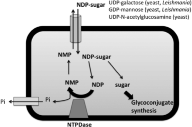
To date, studies of NTPDases in parasites have focused on the role of secreted and surface-located NTPDases. It is worth noting, however, that for the majority of putative NTPDases encoded on parasite genomes the localization is unknown (Table 1). While a number do contain putative N-terminal signal peptides, suggesting secretion from the parasite, a number of others contain predicted transmembrane domains. While these could play a role in anchoring the protein in the membrane on the cell surface, it is equally possible that these proteins are located in organelles such as the Golgi, with the active site located in the lumen of the organelle (as is the case for human NTPDase4 and NTPDase7 (Zimmermann et al. Reference Zimmermann, Beaudoin, Bollen, Goding, Guidotti, Kirley, Robson, Sano, Vanduffel and Lemmens2000). As discussed earlier, in yeast, all NTPDases characterized to date function in the Golgi and deletion results in defective glycosylation. These defects occur because NTPDases are required for nucleotide sugar transport into the Golgi. For example, UDP-galactose is exchanged for UMP, and the galactose molecule used in glycosylation. The Golgi-located NTPDases play crucial roles in regenerating nucleotide monophosphates from nucleotide diphosphates, to allow nucleotide sugar transport to continue efficiently (Figure 3) (Berninsone et al. Reference Berninsone, Miret and Hirschberg1994; Gao et al. Reference Gao, Kaigorodov and Jigami1999).
Schematic of nucleotide-sugar transport into the Golgi of yeast and Leishmania and Trichomonas vaginalis parasites. Different transporters exist for the different nucleotide sugars. The function of an NTPDase in the process is known for some species of yeast, but only postulated for Leishmania and T. vaginalis. In parasites inorganic phosphate may be also incorporated into structures such as lipophosphoglycan, but not all phosphate generated is required for this process, and an NTPDase may still be required. Various enzymes (not shown here) are required to remove the sugar from the nucleotide and incorporate it into the glycoconjugate molecules. Figure adapted from Hirschberg et al. (Reference Hirschberg, Robbins and Abeijon1998).
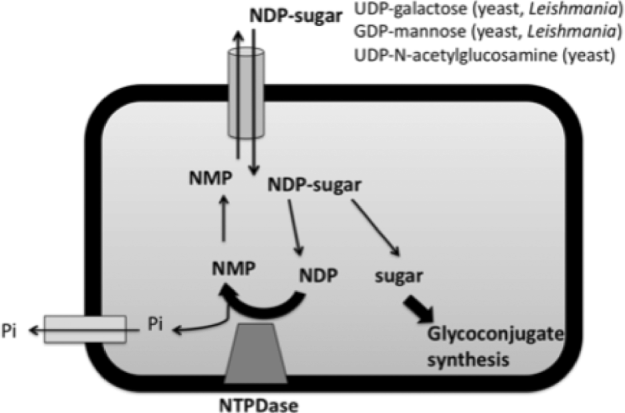
Glycoconjugates of a number of parasites are thought to have key functions in the host-parasite interaction. Depending on the parasite species, important glycoconjugates include lipophosphoglycans (LPG), N-glycans and O-glycans (Guha-Niyogi et al. Reference Guha-Niyogi, Sullivan and Turco2011). The parasites in which the localization of the putative NTPDases is unknown include Trypanosoma, Leishmania and T. vaginalis (Table 1). Of these, both Leishmania and T. vaginalis possess putative NTPDases that contain no signal peptide but do contain predicted transmembrane domains, and these proteins could be present in intracellular organelles. Lending some support to this hypothesis, the Leishmania NTPDases containing predicted transmembrane domains have a closer relationship to the Golgi-located GDA1 of C. albicans than the Leishmania NTPDases containing signal peptides for secretion do (Fig. 2). The T. vaginalis NTPDases are also more closely related to the intracellular organelle-located human NTPDases (NTPDase4 and NTPDase7) than to the cell surface-located NTPDases (Fig. 2). For both parasite species glycoconjugates are important in virulence. In L. major, for example, LPG plays an important role in the ability of the promastigote form of the parasite to resist complement-mediated lysis and oxidative stress, and mutants lacking LPG are significantly delayed in their ability to cause lesions in susceptible mouse strains (Spath et al. Reference Spath, Epstein, Leader, Singer, Avila, Turco and Beverley2000). T. vaginalis parasites require LPG for efficient adherence to host cells, and mutants lacking LPG are also less cytotoxic (Bastida-Corcuera et al. Reference Bastida-Corcuera, Okumura, Colocoussi and Johnson2005). It may be that either or both parasites possess organelle-located NTPDases important in the synthesis of glycoconjugate molecules.
CONCLUSIONS
The diverse roles played by NTPDases in mammals and yeast suggests that a number of functions could exist for this family of enzymes in pathogenic parasites, particularly in the interaction with the host. There is substantial evidence at the genome level for the presence of NTPDases in a number of pathogenic parasites, but varying information in regards to the biological role of these NTPDases. Nonetheless, there is a large body of evidence, although principally indirect, suggesting the importance of these enzymes in parasite biology and pathogenesis. Future work utilizing genetic techniques to more directly address the role(s) of NTPDases would be highly useful in elucidating the function of NTPDases and their suitability as drug targets.
ACKNOWLEDGMENTS
An Australian National Health and Medical Research Council (NH&MRC) Australian Biomedical Fellowship (#567048) supports F.M. Sansom. The support of Professor Malcolm McConville at The University of Melbourne is also gratefully acknowledged.