I. INTRODUCTION
Nanomaterials have continuously attracted worldwide interest because their morphology, dimensionality, and size greatly affect the physical and chemical properties.Reference Hu, Odom and Lieber1–Reference Nakashima and Kimizuka23 Due to their low effective density and high specific surface area, uniform, mondisperse hollow inorganic nanospheres are more important than solid nanospheres in some applications,Reference Peng, Dong and Li24–Reference Son, Hughes, Yin and Alivisatos33 such as catalysis,Reference Ikeda, Ishino, Harada, Okamoto, Sakata, Mori, Kuwabata, Torimoto and Matsumura29 photonic devices,Reference Xu and Asher30 electrochemical cell,Reference Lou, Wong, Yuan, Lee and Archer31 and biotechnology.Reference Dong, Kishimura, Anraku and Kataoka32
Among these inorganic nanomaterials, the lanthanide oxides and lanthanide fluorides are promising host matrices for down- and upconversion (UC) luminescence because of their good chemical and thermal stability.Reference Huang, Liu, Su, Lo, Chen and Yeh34–Reference Li, Zhang, Zhang, Luo, Lu, Hao and Wang40 Monodisperse rare-earth-doped lanthanide oxide and lanthanide fluoride hollow nanospheres are in high demand as both fluorescence and magnetic resonance imaging (MRI) labelsReference Huang, Liu, Su, Lo, Chen and Yeh34 in biomedical field and application in drug-release field because of their low density and high surface area. It is a promising candidate for labeling the location, local environments of the drug carriers in vivo and monitoring the drug delivery conditions instantaneously using the hollow lanthanide oxide and fluoride nanospheres as drug carrier.Reference Zhang, Li, Peng, Chai, Huang, Yang, Cheng and Lin35 When compared with the nanospheres of lanthanide oxides and lanthanide fluorides, the nanospheres of lanthanide oxyfluorides have been more intensively studied due to their unique chemical bonding between metal cations and fluoride anions.Reference Du, Zhang, Yan, Sun and Yan41–Reference Antic-Fidancev, Holsa, Krupa and Lastusarri44 However, to the best of our knowledge, there are few reports on the synthesis of hollow uniform and monodisperse nanospheres of lanthanide oxides and lanthanide fluoridesReference Zhang, Shi, Sun, Zhao and Stucky45–Reference Wang and Liu50 and almost no reports on the synthesis of uniform, monodisperse gadolinium oxyfluoride (GdOF) hollow nanospheres doped with rare earth activators of100-nm diameter, a size needed for biological cell penetration.
In general, two main methodologies had been used to obtain hollow inorganic functional materials: one is template-assisted synthesis and the other is template-free approach. In the first case, hard templates (such as polymer, carbon, silica and metallic core) or soft templates (for instance of surfactant micelles, vesicles, emulsion droplets, gas bubbles, and ionic solvent)Reference Caruso, Caruso and Mohwald51–Reference Wu, Xie, Lei, Hu and OuYang55 have been widely used. In these cases, template cores are coated by controlled surface precipitation of inorganic molecule precursors [titanium dioxide (TiO2), tantalum pentoxide (Ta2O5), silicon dioxide (SiO2), and zinc sulfide (ZnS) etc.] or by surface reaction using specific functional groups on the cores to prepare core-shell composites. Then, the template particles are subsequently removed by calcination at high temperatures in the air or by selective dissolution in an appropriate solvent to give rise to hollow inorganic materials. Template-free synthesis is also used to fabricate hollow nanomaterials via various physicochemical processes, for example, Ostwald ripening,Reference Penn and Banfield56–Reference Liu and Zeng59 oriented attachment,Reference Ostwald60–Reference Yu, Yu, Liu and Mann62 and Kirkendall effect processes.Reference Kirkendall, Thomassen and Upthegrove63–Reference Chiang and Chiang66
The Kirkendall effect is a very facile and inexpensive method to synthesize the hollow nanospheres. Since the first report on the synthesis of hollow cobalt (II) sulfide (CoS) and cobalt (II) oxide (CoO) by sulfidation and oxidation of cobalt (Co) nanoparticles,Reference Yin, Rioux, Erdonmez, Hughes, Somorjai and Alivisatos4 numerous hollow nanospheres have been prepared by this approach. Recently, an alternative ion exchange method has been used to change the composition of materials by replacing the cations (anions) within the precursors with different cations (anions). For example, it is possible to convert cadmium chalcogenides [cadmium sulfide (CdS), cadmium selenide (CdSe), and cadmium telluride (CdTe)] to the corresponding silver chalcogenides with addition of Ag+ to the cadmium chalcogenide nanocrystals.Reference Zhang, Tang, Lee and Ouyang3, Reference Son, Hughes, Yin and Alivisatos33 It is noted that the morphology of these nanostructures was preserved after the ion exchange.
In this work, we present the facile synthesis of uniform and monodisperse GdOF: Yb3+, Er3+ hollow nanospheres of about 100-nm diameter by a controlled ion exchange process. Furthermore, hollow nanospheres of various shell thicknesses were obtained by tuning the reaction condition via the Kirkendall effect. The structure, luminescence, and formation mechanism were investigated in detail. This work opens up new possibilities for synthesizing hollow nanospheres of other lanthanide oxides as also other lanthanide fluorides and extends their applications in biolabel and drug delivery fields.
II. EXPERIMENTAL SECTION
A. Synthesis
1. Synthesis of monodisperse GdOF: Yb3+, Er3+ nanosphere
The synthesis of GdOF: Yb3+, Er3+ hollow nanosphere involved two processes. First, a series of ytterbium ion and erbium ion codoped gadolinium hydroxide [Gd(OH)3: Yb3+, Er3+] nanospheres of about 100-nm diameter were prepared by the solvothermal method. In the synthesis, 0.20 g gadolinium nitrate [Gd(NO3)3∙5H2O], different molar ratios of rare earth nitrate [RE(NO3)3∙5H2O] and 0.031 g iron chloride (FeCl3∙6H2O) were dissolved in 10 mL dimethylformamide (DMF), and then 0.504 g cetyltrimethyl ammonium bromide (CTAB) was added with stirring, and then 0.2 g of polyvinylpyrrolidone (PVP) was added with stirring to form a yellow suspension. The role of Fe3+ ion was to produce minor proton, which could further accelerate the self-assembly process.Reference Wang, Bai, Liu, Dong, Xu, Liu and Song67 The resulting suspension was stirred for 12 h until it became homogeneous, and then sealed in a Teflon-lined stainless steel autoclave. The above steps were performed in ambient atmosphere. The autoclaves were heated at 200 °C for 24 h under static condition and then allowed to cool in ambient atmosphere. The light brown powders were separated by centrifugation and washed three times with ethanol. The light brown powders were redispersed into 6 mL of ultrapure water (18 MΩ, Barnstard). After, forming the colloidal Gd(OH)3: Yb3+, Er3+ solid nanospheres, 2 mL Gd(OH)3: Yb3+, Er3+ colloidal solution, titanium tetrafluoride (TiF4) (1 mL 0.04 M) and deionized water (10 mL) were mixed and stirred for 12 h sequentially. The mixture was then transferred into a Teflon-lined stainless steel autoclave, and the hydrothermal reaction was conducted at 150 °C for 4 h under static condition in an electric oven. The hollow powder was centrifuged and washed three times with ethanol, then air-dried at 60 °C. Experiments for tuning the thickness of shell of GdOF hollow nanospheres were also performed by varying the reaction temperature and reaction time.
2. Thermal conversion of GdOF: Yb3+, Er3+ to crystalline GdOF: Yb3+, Er3+
The as-prepared amorphous hollow GdOF samples were annealed at 400 °C for 4 h in air to obtain the crystalline GdOF products.
B. Characterization
The crystal structure of the GdOF: Yb3+, Er3+ products was inspected with a (XRD; Rigaku model No. D/max 2550, Tokyo, Japan) x-ray diffractometer, using monochromatic Cu radiation (λ = 1.54 Å). The surface morphology of the GdOF: Yb3+, Er3+ nanosphere was observed using field-emission scanning electron microscopy with a (FESEM; JEOL model No. JSM-7500, Tokyo, Japan) field emission scanning electron microscope at an accelerating voltage of 15 kV. The transmission electron micrograph images were recorded on a (TEM; model No. JEM-2010, Tokyo, Japan) transmission electron microscope under a working voltage of 200 kV. The high-resolution transmission electron micrograph images were recorded on a (HRTEM; model No. JEM-2010F, Tokyo, Japan) high-resolution transmission electron microscope under a working voltage of 200 kV. Inductively coupled plasma analysis was performed on a (ICP; model No. Perkin-Elmer Optima 3300DV, Baltimore, MD) spectrometer. The x-ray photoelectron spectra were measured by (XPS; model No. ESCAlab 250 Analytical, Manchester, UK) x-ray photoelectron spectrometer. The zeta potential measurements were carried out at 25 °C using Zetasizer Nano-ZS instrument (Malvern Instruments, Malvern, UK), equipped with a He-Ne ion laser (λ = 633 nm). The emission spectra were recorded on F4500 fluorescence spectrometer (Tokyo, Japan).
III. RESULTS AND DISCUSSION
A. Morphology and structural characterization of GdOF: Yb3+, Er3+ hollow nanospheres
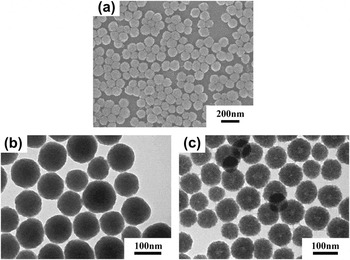
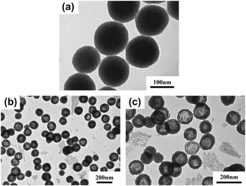
Figure 1(a) shows SEM images of the Gd(OH)3: Yb3+, Er3+ nanospheres. The as- prepared Gd(OH)3: Yb3+, Er3+ powder consists of uniform nanospheres of about 100-nm diameter. Figure 1(b) shows the TEM image of the above-mentioned Gd(OH)3: Yb3+, Er3+ nanospheres. It is worth noting that as-synthesized Gd(OH)3: Yb3+, Er3+ nanospheres are solid. The nanospheres become hollow after they are stored in the water for one month [as shown in Fig. 1(c)] by an Ostwald ripening process.Reference Dong, Ferri, Adalseinsson, Schonhoff, Sukhorukov and Mohwald52
(a) SEM images of Gd(OH)3: Yb3+, Er3+ samples, (b) TEM images of Gd(OH)3: Yb3+, Er3+ samples, (c) TEM images of hollow Gd(OH)3: Yb3+, Er3+ samples.

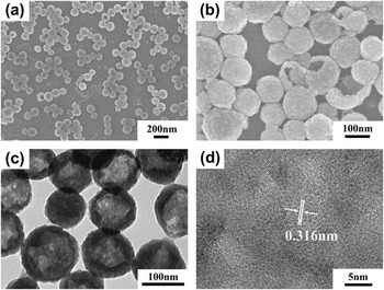
Figure 2(a) shows the SEM image of the GdOF: Yb3+, Er3+ nanosphere precursor. It is noted that uniform, monodisperse GdOF: Yb3+, Er3+ hollow nanospheres inherit the spherical morphology and good size dispersion from the Gd(OH)3: Yb3+, Er3+. The morphology, uniform shape, and hollow structure of nanospheres were not adversely affected by the calcination process [Fig. 2(b)]. The TEM micrograph [Fig. 2(c)] shows strong contrast between the dark edges and the light center confirms that all the nanospheres are hollow. HRTEM image [Fig. 2(d)] of nanocrystalline particles shows lattice planes continuously extending in each nanocrystalline area that shows good nanocrystallinity of the particle. The distance of the lattice planes was calculated to be 0.316 nm using the HRTEM, which is consistent with the (012) planes of rhombohedral phase GdOF: Yb3+, Er3+.
(a) SEM images of GdOF: Yb3+, Er3+ samples, (b) SEM images of GdOF: Yb3+, Er3+ samples after calcination at 400 °C, (c) TEM images of GdOF: Yb3+, Er3+ samples after calcination at 400 °C, (d) HRTEM images of GdOF: Yb3+, Er3+ samples after calcination at 400 °C.

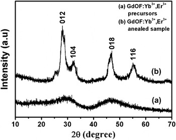
Figure 3 shows the typical x-ray diffraction patterns of the GdOF: Yb3+, Er3+ nanospherical precursor and the crystalline GdOF: Yb3+, Er3+ product obtained by annealing at 400 °C for 4 h. No obvious diffraction peaks appear in the GdOF: Yb3+, Er3+ nanospherical precursor, which indicates that as-synthesized GdOF: Yb3+, Er3+ hollow nanosphere precursor is amorphous before calcination. After calcining at 400 °C for 4 h, all of the diffraction peaks are indexed to the rhombohedral phase of GdOF [Joint Committee on Powder Diffraction Standards (JCPDS) No. 50–0569]. Furthermore, no other impurity peaks could be detected, indicating that the Yb3+ and Er3+ ions have been thoroughly doped into the GdOF host lattice. The peak positions of the GdOF: Yb3+, Er3+ nanospheres do not shift because these doping ions have similar ionic radii as Gd3+ (94 pm). Moreover, no additional peaks of other phases have been found, indicating the formation of pure rhombohedral GdOF: Yb3+, Er3+ phase.
The x-ray powder diffraction patterns of GdOF: Yb3+, Er3+ samples: (a) GdOF: Yb3+, Er3+ precursors, (b) GdOF: Yb3+, Er3+ after calcination at 400 °C.

B. Formation mechanism of GdOF hollow nanospheres
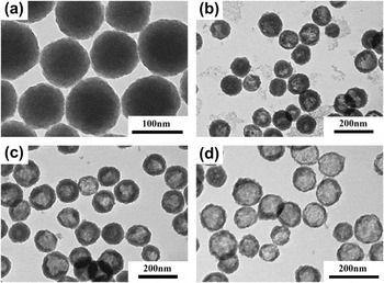
The growth process of the GdOF: Yb3+, Er3+ hollow nanospheres from Gd(OH)3: Yb3+, Er3+ was investigated in detail at different reaction temperatures and times. Figure 4 is a series of TEM images of the samples heated at different reaction temperatures for 4 h. No hollow product was obtained at 120 °C [Fig. 4(a)]. The particles start to become hollow at center and the shell becomes thinner as temperatures rises to 180 °C [Figs. 4(b)–4(d)]. Most of the hollow spheres break down when the reaction temperature reaches 200 °C (as seen in Fig. S1). From the above systematic TEM analysis, it can be understood that the formation of GdOF: Yb3+, Er3+ hollow nanospheres occurs by the Kirkendall mechanism because of competitive atomic diffusions between two phases upon the hydrothermal treatment as related before.Reference Chiang and Chiang66
A series of TEM images of the samples obtained at different reaction temperatures: (a) 120 °C, (b) 130 °C, (c) 150 °C, and (d) 180 °C for 4 h.

Figure 5 shows a series of TEM images of the samples obtained from different reaction time when heated at 150 °C. After 1.0–4.0 h, the products become hollow. However, when the reaction time exceeds 16 h by heated at 150 °C, most of hollow spheres break down (seen in Fig. S2).
A series of TEM images of the samples investigated with different reaction times at 150 °C: (a) 0 h, (b) 1 h, and (c) 4 h.

In general, the starting materials have a great effect on the morphology of the final products. Therefore, a series of experiments with different fluorine sources such as ammonium fluoride (NH4F), and hydrofluoric acid (HF) as starting materials were performed while keeping other parameters constant. The results show that the different fluorine sources determine the morphology and structure of the final products. As shown in Fig. S3(a), when the reaction was performed in the presence of NH4F, no hollow spherical particles were obtained. The XRD pattern of the crystalline sample after calcination shows that the powder is the cubic phase of gadolinium (III) oxide (Gd2O3) (JCPDS No. 86-2477). It is obvious that Kirkendall process does not take place under this condition [Fig. S3(b)]. When HF acid of different amounts such as 20, 40, 60, and 100 μL was used as fluorine source, ytterbium ion and erbium ion codoped gadolinium fluoride (GdF3: Yb3+, Er3+) powders with different morphology were obtained as seen in Fig. S4. It indicates that the uniform and monodisperse Gd(OH)3: Yb3+, Er3+ solid nanospheres reaction to form GdF3: Yb3+, Er3+ when a trace amount of HF acid (20 μL) was introduced into the reaction system. When HF was increased to 100 μL, the hollow microplate product with diameters in the range of 0.5–1.0 μm was observed. Figure S4(e) displays a series of XRD patterns of the as-synthesized samples with 20, 40, 60, and 100 μL HF. It indicates that the uniform and monodisperse Gd(OH)3: Yb3+, Er3+ phase gradually disappears, and GdF3: Yb3+, Er3+ phase gradually appears when HF acid was introduced into the reaction system.
At this point, an interesting question arises, namely, how these different fluorine sources impact the morphology and structure of final products? It can be speculated that the appropriate amount of H+ can play an important role in the reaction process. There was too low H+ (pH = 4.83) when the NH4F was used as fluorine source. So, the Gd(OH)3: Yb3+, Er3+ would not dissolve and no GdOF: Yb3+, Er3+ was formed. However, the dissolution of Gd(OH)3: Yb3+, Er3+ had become dominant compared with the growth of GdOF: Yb3+, Er3+ when the HF acid was used as fluorine source, so that the uniform and monodisperse spherical morphology of Gd(OH)3: Yb3+, Er3+ would not be preserved.Reference Zhang, Shi, Sun, Zhao and Stucky45 The experiments prove that the uniform and monodisperse hollow GdOF: Yb3+, Er3+ nanospheres can be obtained only when the TiF4 was the fluorine source, as described below.
In our reaction system, F− anions can be hydrolyzed to form a small concentration of HF in the hydrothermal environment and Ti4+ ions can react with the OH− to form TiO2 and H+. Once the hydrogen ions (H+) were formed, they would react with F− anions once again to give rise to more HF in this process. Subsequently, the formed appropriate amount of HF would react with the Gd(OH)3: Yb3+, Er3+ to produce the GdOF: Yb3+, Er3+ and hold the former spherical morphology. Previous studies have demonstrated that the Kirkendall processReference Fujihara, Koji and Kimura42–Reference Antic-Fidancev, Holsa, Krupa and Lastusarri44 will take place during this stage because the equilibrium will be set up between the dissolution of Gd(OH)3: Yb3+, Er3+ and growth of GdOF: Yb3+, Er3+. These appropriate amounts of GdOF: Yb3+, Er3+ will form hollow sphere driven by the minimization of the total energy of the system. The titanium element is present as TiO2 clusters with positive static potential. The whole reaction can be described in accordance with following process:
 $${\rm{F}}^{\rm{ - }} \,{\rm{ + }}\,{\rm{H}}_{\rm{2}} {\rm{O}}\, \to \,{\rm{HF}}\,{\rm{ + }}\,{\rm{OH}}^{\rm{ - }} {\rm{\quad,}}$$
 $${\rm{Ti}}^{{\rm{4 + }}} \,{\rm{ + }}\,{\rm{OH}}^{\rm{ - }} \, \to \,{\rm{TiO}}_{\rm{2}} \,{\rm{ + }}\,{\rm{H}}^{\rm{ + }} {\rm{\quad,}}$$
 $${\rm{F}}^{\rm{ - }} \,{\rm{ + }}\,{\rm{H}}^{\rm{ + }} \, \to \,{\rm{HF\quad,}}$$
 $${\rm{Gd(OH)}}_{\rm{3}} \,{\rm{ + }}\,{\rm{HF}}\, \to \,{\rm{GdOF}}\,{\rm{ + }}\,{\rm{2H}}_{\rm{2}} {\rm{O}}{\rm{\quad.}}$$
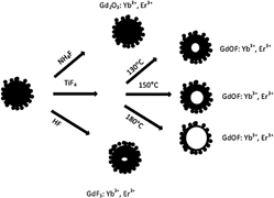
On the other hand, it is well known that the dissolution rate of Gd(OH)3: Yb3+, Er3+ and growth rate of GdOF: Yb3+, Er3+ will be enhanced with increasing reaction temperature. Therefore, the void is enlarged by increasing the reaction temperature, which is in good agreement with the TEM results. The hollow process preferentially takes place in the center of the nanospheres as reported in previous reference literature.Reference Pang, Wang and Zeng68 For validating our conjecture, we measured the zeta potentials of the Gd(OH)3: Yb3+, Er3+ precursor, residual solution, GdOF: Yb3+, Er3+ hollow nanospheres, and the ICP analysis of the residual solution. The results show that the residual solution contains about 0.038M Ti, which is in agreement with the molar concentration of TiF4 (0.04 M). It indicates that the titanium element remains in the residual solution. The measured zeta potentials are +10 mv for Gd(OH)3: Yb3+, Er3+ and +26.5 mv (130 °C), +28.0 mv (150 °C), and +34.2 mv (180 °C) for GdOF: Yb3+, Er3+, respectively. The results show that the Gd(OH)3: Yb3+, Er3+ solid nanospheres and GdOF: Yb3+, Er3+ hollow nanospheres have a positive static potential. It qualitatively suggests that the TiO2 cluster with positive static potential will be excluded from surface of GdOF: Yb3+, Er3+ nanospheres because of the static repulsion and remain in the residual solution. All above-mentioned analyses clearly corroborate that the Kirkendall process is the causal factor in the present system. A schematic illustration for the effect of different fluorine sources for the final product and the formation procession of the GdOF: Yb3+, Er3+ uniform, monodisperse hollow nanosphere at different reaction temperature is presented in Fig. 8.
High-resolution x-ray photoelectron spectra (XPS) were measured to characterize the surface electronic structure and chemical element valence by referencing the C1s line to 284.5 eV, as shown in Fig. 6. The XPS spectra reveal that all the products contain Gd, Yb, and Er as seen in Fig. 6(A).Reference Petoral, Soderlind, Klasson, Suska, Fortin, Abrikossova, Selegard, Kall, Engstrom and Uvdal69, Reference Milanov, Toader, Parala, Barreca, Gasparotto, Bock, Becker, Ngwashi, Cross, Paul, Kunze, Fishcher and Devi70 The bonding energy (BE) at 685.5 eV measured in the samples of GdOF: Yb3+, Er3+ and GdF3: Yb3+, Er3+ can be attributed to the F. However, there is no F in the Gd2O3: Yb3+, Er3+ sample. Figure 6(B) displays the corresponding O1s spectra of Gd2O3: Yb3+, Er3+, GdOF: Yb3+, Er3+, and GdF3: Yb3+, Er3+, respectively. The high BE component at 528.6 and 529.4 eV is due to the presence of lattice O2- on the surface of the samples of Gd2O3: Yb3+, Er3+ and GdOF: Yb3+, Er3+, respectively.Reference Pang, Wang and Zeng68 The second peak at 531 eV can be attributed to the presence of a hydroxyl group and/or carbonate group for the samples of Gd2O3: Yb3+, Er3+ and GdOF: Yb3+, Er3+, respectively.Reference Du, Zhang, Yan, Sun and Yan41 The final peaks at 532.9, 532.4, and 532.6 eV can be assigned to molecular water adsorbed on the surface of Gd2O3: Yb3+, Er3+, GdOF: Yb3+, Er3+, and GdF3: Yb3+, Er3+ products, respectively. These results once again show that different fluorine sources have a great effect on the morphology, component and structure of the final products and match with the above-mentioned SEM and TEM results very well.
The XPS spectrum of as-obtained samples: (A) the survey XPS spectra of F 1s, O 1s, C 1s, Gd 4d, Yb 4d, and Er 4d for different of samples: (a) Gd2O3: Yb3+, Er3+, (b) GdOF: Yb3+, Er3+, and (c) GdF3: Yb3+, Er3+, (B) detailed XPS spectrum of O 1s for the Gd2O3: Yb3+, Er3+ nanosphere, the GdOF: Yb3+, Er3+ hollow nanosphere, the GdF3: Yb3+, Er3+ microplate, respectively.

C. Luminescence properties
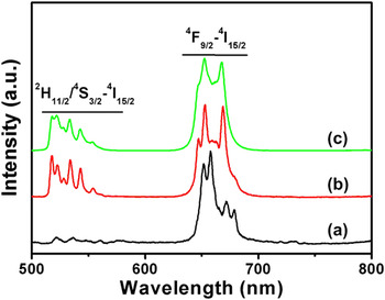
It is well known that GdOF is a good host matrix for UC luminescenceReference Du, Zhang, Yan, Sun and Yan41 due to its chemical durability and lower phonon energy. As reported in reference literature, the rare earth activator ion can be effectively doped into the lanthanide oxides, lanthanide fluoroxides, and lanthanide fluorides host matrices. In this contribution, a stoichiometric amount of rare earth ions (Yb3+ and Er3+) were doped into the Gd2O3, GdOF, and GdF3 host lattices to investigate the UC luminescence properties, respectively. We chose the GdOF:18% Yb3+/2% Er3+ hollow nanospheres that were heated at 150 °C as a typical sample because the normalized UC emission spectra of GdOF:18% Yb3+/2% Er3+ with different shell thicknesses are similar under 980 nm light excitation. Figure 7 shows the normalized UC emission spectra of Gd2O3:18% Yb3+/2% Er3+, GdOF:18% Yb3+/2% Er3+, and GdF3:18% Yb3+/2% Er3+ under 980 nm light excitation. It is well known that codoping not only increases the intensity of luminescence but also induces UC luminescence between the donor and acceptor ions.Reference Milanov, Toader, Parala, Barreca, Gasparotto, Bock, Becker, Ngwashi, Cross, Paul, Kunze, Fishcher and Devi70 In our work, Yb3+ was chosen as the codopant with Er3+ because it has a large absorption cross section at 980 nm and energy transfer (ET) occurs as a result of the large spectral overlap between the Yb3+ emission 2F5/2-2F7/2 and the Er3+ absorption 4I11/2-4I15/2 bands. However, for the samples Gd2O3:18% Yb3+/2% Er3+, GdOF:18% Yb3+/2% Er3+, and GdF3:18% Yb3+/2% Er3+, the emission spectra are quite different. The UC emission color can be adjusted from red to yellow to green when the host matrix was varied from Gd2O3: 18% Yb3+/2% Er3+ to GdOF: 18% Yb3+/2% Er3+ to GdF3: 18% Yb3+/2% Er3+ with all other parameters remaining constant. We can see that the obvious green emissions from 500 to 600 nm regions, which correspond with the 2H11/2-4I15/2 and 4S3/2-4I15/2 transitions of Er3+, and the red lines from 600 to 700 nm can be attributed to the transition of 4F9/2-4I15/2. The enhanced relative intensity of green emissions to red emissions can be attributed to the attenuation of the nonradiative transition rate when the host lattices change from Gd2O3 to GdOF to GdF3.Reference Vetrone, Boyer, Capobianco, Speghini and Bettinelli71–Reference Bai, Song, Pan, Lei, Wang, Ren, Lu, Dong, Dai and Fan73
Normalized emission spectra (λex = 980 nm) in different samples: (a) Gd2O3: Yb3+, Er3+, (b) GdOF: Yb3+, Er3+, and (c) GdF3: Yb3+, Er3+.

Illustration of the effect of different fluorine sources for the final product and the formation procession of the GdOF: Yb3+, Er3+ uniform, monodisperse hollow nanosphere at different reaction temperatures.

IV. CONCLUSIONS
In summary, monodisperse, uniform GdOF: Yb3+, Er3+ hollow nanospheres about 100-nm diameter were successfully synthesized via the simple and facile hydrothermal method. The formation mechanism of the hollow GdOF: Yb3+, Er3+ nanospheres follows the nanoscale Kirkendall effect. The results demonstrate that the TiF4 and the reaction temperature are key parameters for the formation of hollow GdOF: Yb3+, Er3+ nanospheres. The thickness of shell could be tuned from 31 to 13 nm by controlling the reaction temperature. Furthermore, the UC emission color of the final products could be tuned from red to yellow to green by varying the host lattice from Gd2O3 to GdOF to GdF3 using different fluorine sources as the starting materials. The as-prepared GdOF: Yb3+, Er3+ hollow nanospheres might find applications in the fields of molecular imaging, drug delivery and gene therapy due to good chemical stability and luminescence functionality. Moreover, this general and simple synthesis strategy can be extended to prepare other inorganic hollow nanospheres.
ACKNOWLEDGMENTS
The authors are thankful to the National Natural Science Foundation of China (Grant Nos. 20971051 and 10704073). We are very grateful to Prof. Jihong Yu for her helpful discussion and structural characterization. We are also grateful to Prof. Junqi Sun for his helpful discussion.