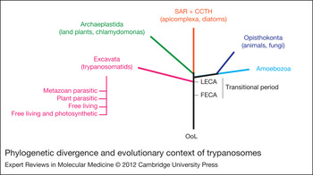
Trypanosomatids constitute a taxonomic grouping of organisms that are highly divergent from animals and fungi (Fig. 1). As a group, trypanosomes have a huge impact on the biota, and representatives include important human disease agents that directly threaten over half-a-billion individuals (Ref. Reference Chatelain and Ioset1). Furthermore, trypanosomatids are increasingly recognised as disease agents of plants, insects and wild animal populations (Refs Reference Catarino2, Reference Lloyd-Smith3, Reference Sturm4), resulting in an unquantified, but probably enormous, impact on agriculture and the flora and fauna of a given region. In Africa, the African trypanosomes (Trypanosoma brucei spp.) have had a major effect on the savannah-like ecology and human history (see Refs Reference Hide5, Reference Rogers and Randolph6 for discussion on this issue). Even more distantly related organisms such as Euglena, which are highly abundant members of aquatic and marine ecosystems, exert great influence over the entire biosphere (Ref. Reference Duttagupta, Gupta and Gupta7). Trypanosomatids are incredibly evolutionarily divergent from animals, fungi and plants, and this is reflected in the multiple unusual and near-unique features of their cell biology, metabolism and gene expression. The eukaryotic root might even lie close to the origin of the trypanosomatid lineage (Fig. 1), suggesting that this divergent biology also reflects some deeper evolutionary origins (Ref. Reference Cavalier-Smith8). Particularly relevant is the dominance of GPI-anchored proteins and glycoconjugates at the cell surface, and polycistronic transcription and trans-splicing of protein-coding genes. The presence of high-abundance surface antigens on several of the better characterised parasitic trypanosomatids, and in many cases, extreme developmental variation resulting in complete remodelling of the surface antigen repertoire during differentiation, suggests that this aspect of trypanosome cell biology particularly relies on strict mechanisms for altering gene expression profiles, which is supported by transcriptome analysis (Refs Reference Jensen9, Reference Koumandou10). Because most studies have focused on pathogenic trypanosomes, and in the absence of well-characterised free-living members of the kinetoplastida lineage, it remains an open question as to how much of the unusual biology of trypanosomes arises from their evolutionary position or selective pressure arising from a parasitic lifestyle.
Phylogenetic divergence and evolutionary context of trypanosomes. Schematic representation of the eukaryotic tree of life. Five supergroups are represented reflecting current thinking on large-scale taxonomic groupings. Trypanosomatids are members of the Excavata (pink) and highly divergent from their most significant hosts, the animals (Opisthokonta, blue), and also land plants (Archaeplastida, green). Positioning of the root reflects recent arguments and is highly speculative. Note the transitional period between the eukaryogenesis event represented by the first eukaryotic common ancestor (FECA, equivalent to the eukaryogenesis event) and radiation of modern lineages represented by the last eukaryotic common ancestor (LECA), which probably corresponds to a period of intense biological innovation. SAR + CCTH, stramenopile, alveolate and Rhizaria plus cryptomonads, centrohelids, telonemids and haptophytes; OoL, origin of life.

T. brucei is the causative agent of African sleeping sickness in humans and a wasting disease, n'agana, in cattle. The parasite is transmitted by the bite of the Tsetse fly. In both host and vector, a complex differentiation pattern is executed that appears balanced to prevent excess damage to the host and to continuously generate highly infective forms for transmission (Refs Reference Field and Carrington11, Reference Gadelha12, Reference MacGregor13). In this regard T. brucei is poorly adapted to modern humans and European cattle (Bos taurus), because the disease is unusually aggressive and invariably fatal unless treated; this is in contrast to many of the endemic mammalian species, including Bos indicus, where infections can persist and are comparatively well tolerated. African trypanosomes are unusual pathogens in that they are exclusively extracellular, and therefore exposed to the immune system in mammalian hosts – a behaviour that is seemingly at odds with the ability of the parasite to survive for prolonged periods within the host. Immune evasion is primarily achieved by antigenic variation (Ref. Reference Gadelha12). Briefly, the surface of the mammalian infective form of the parasite is dominated by the GPI-anchored variant surface glycoprotein (VSG), which contributes an impressive ~90% of cell surface protein. Switching the active VSG gene allows selection of an immunologically distinct VSG coat, which can evade the immune response. The presence of a repertoire of many hundreds of VSG sequences, recombination between them accessing a potentially inexhaustible supply of novel VSG genes, plus monoallelic expression achieved by restricting transcription to a single subtelomeric specialised expression site (ES) allows continued immune evasion for many months or even years.
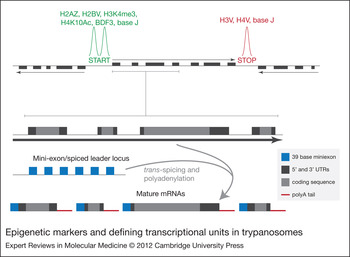
The surface of most stages of T. brucei is dominated by superabundant GPI-anchored antigens, and in addition to VSG in the mammalian infective form, procyclin is present in the insect midgut-dwelling procyclic form, and finally brucei stage alanine-rich protein (BARP) in epimastigote stages that reside within the fly proventricular region (Ref. Reference Urwyler14). Significantly, transcriptional mechanisms in trypanosomes are rather unusual, with the majority of protein-coding genes arranged in long polycistronic transcriptional units (PTUs) initiating from strand-switch regions, at least in the case of divergently oriented PTUs (Fig. 2). These PTUs are transcribed principally by RNA polymerase II (RNA Pol II) and the resulting nascent RNA is resolved by trans-splicing that places an invariant spliced leader at the 5′ end (Ref. Reference Lee and Van der Ploeg15). 5′ cap methylation and polyadenylation of the 3′ end completes mRNA maturation (Fig. 2). This mechanism at first glance appears to function in the absence of clear promoter-mediated control, and transcription of all open reading frames within a given PTU appears equivalent, with the further consequence being that mRNA abundance is principally mediated by post-transcriptional mechanisms (Ref. Reference Haile, Estevez and Clayton16). Although sequences within the 3′ UTR of mRNAs are clearly involved in regulation of mRNA stability, and hence copy number, it is not established whether this accounts for all mRNA copy number control, and it is probable that mRNA transport, which might be affected by the state of the chromatin and other factors, also has a role (Refs Reference Kramer and Carrington17, Reference Serpeloni18). Codon usage bias is also likely to have a large impact on translation control and protein copy number (Ref. Reference Horn19).
Epigenetic markers and defining transcriptional units in trypanosomes. The top schema shows a region of a megabase chromosome, with two strand-switch regions, one marked ‘START’ where the two flanking polycistronic transcriptional units (PTUs) are transcribed in a divergent manner and one marked ‘STOP’, where the PTUs converge. Evidence suggests that transcription initiates at the divergent sites, and terminates at convergent regions; the divergent regions probably resemble RNA-Pol-II-like promoter elements. Recent mapping using ChIP coupled to high-throughput sequencing has found these switch regions to be associated with both variant histones and specific modifications of H3 and H4. The lower schema, connected with a tie bar, shows the resolution of a portion of a polycistronic transcription unit by a combination of trans-splicing, adding a mini-exon (blue) and polyadenylation (red). Arrows indicate nascent mRNAs with the arrowhead to show the direction of transcription.

The principal nuclear architectural features are conserved between trypanosomes and higher eukaryotes, and include the double bilayer nuclear envelope, nuclear pores, a single nucleolus and electron-dense chromatin regions close to the nuclear periphery whose arrangement is under developmental control (Refs Reference Belli20, Reference Elias21, Reference Navarro, Penate and Landeira22, Reference Rout and Field23, Reference Vickerman and Preston24). There is good evidence for positioning of telomeres at the periphery within the nucleus (Ref. Reference Dossin Fde25), and suggestions for variance in the kinetics of the segregation of the different classes of chromosomes (i.e. megabase versus minichromosomes) during mitosis (Refs Reference Ersfeld and Gull26, Reference Landeira27, Reference Ogbadoyi28). The location of the telomeres at the nuclear periphery is highly significant because telomeres are also the loci containing the VSG ESs. Current models suggest that this location serves in some manner to suppress full expression from these sites, a common aspect of epigenetic control (Refs Reference Chaves29, Reference Navarro and Gull30, Reference Perez-Morga31).
Epigenetics
Epigenetics comprises a suite of mechanisms that regulate gene expression but are not directly encoded within the genome, and therefore excluding promotors and other cis-based sequence elements (Refs Reference Figueiredo, Cross and Janzen32, Reference Rando and Chang33). Epigenetic mechanisms appear to exploit the overall organisation of the nucleus and DNA itself, suggesting that they coevolved with the establishment of the nucleus, or soon after (Table 1). Importantly, epigenetic states are heritable and can take many forms (Refs Reference Danchin34, Reference Nowacki, Shetty and Landweber35, Reference Skinner36), and epigenetic control essentially equates with the organisation of chromatin structure in space and time. The major aspects are threefold: (i) direct modification of DNA itself, for example, by methylation or glucosylation of specific bases; (ii) specification of the density and permissiveness of specific DNA sequences to transcription, frequently based around structural organisation of histones and their assembly into nucleosomes, itself based on post-translational modifications of histones; and (iii) three-dimensional positioning of chromatin with, at least in higher eukaryotes, less transcriptionally active chromatin regions (heterochromatin) tending to be positioned at the nuclear periphery and dependent on intermediate filament and nuclear envelope proteins. All these processes are also probably intimately associated with higher-order organisation of the nuclear contents; in the noncondensed interphase state, metazoan chromosomes are compacted and restricted to ‘territories’, whereas even the arrangement of chromosomal territories is ordered (Ref. Reference Geyer, Vitalini and Wallrath37).
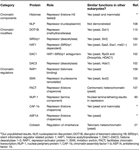
Chromatin-associated proteins that impact transcription at T. brucei telomeres
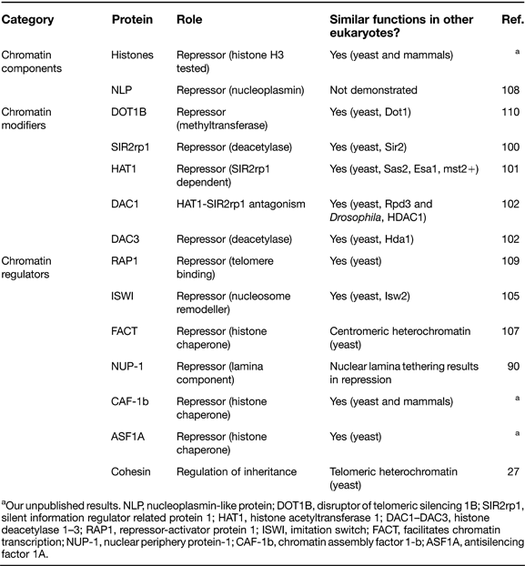
aOur unpublished results. NLP, nucleoplasmin-like protein; DOT1B, disruptor of telomeric silencing 1B; SIR2rp1, silent information regulator related protein 1; HAT1, histone acetyltransferase 1; DAC1–DAC3, histone deacetylase 1–3; RAP1, repressor-activator protein 1; ISWI, imitation switch; FACT, facilitates chromatin transcription; NUP-1, nuclear periphery protein-1; CAF-1b, chromatin assembly factor 1-b; ASF1A, antisilencing factor 1A.
Epigenetic control is used in a great number of contexts, but highly relevant here is developmental regulation of the expression of gene cohorts, reflecting the extremely stable and high dynamic range of relative expression levels of heterochromatin versus euchromatin (active, open chromatin) expression levels. Potentially, such mechanisms ensure against ectopic expression of inappropriate gene products, which is critical in tissue differentiation in metazoa and multicellular plants, and the successful completion of ontological development where abnormal cues at early stages can become amplified into gross morphological defects. In addition, the process can retain environmental information; for example, in Saccharomyces cerevisiae, as a result of DNA-zip-code-dependent positioning of relevant genes, daughter cells retain a memory of the carbon source their parental cell used, facilitating preferential reactivation should the same carbon source be re-encountered (Refs Reference Brickner38, Reference Light39).
In trypanosomes, tight regulation of developmentally expressed surface proteins is a clear requirement. As the parasite progresses through the life cycle, it expresses VSG in the mammalian host, which is replaced by procyclin and then BARP in the insect vector before returning to VSG expression. Although definitive proof is lacking, developmental progression is considered essential for adaptation to specific environments; there is evidence for quality control mechanisms ensuring against ectopic expression, suggesting that trypanosomes are under considerable pressure to fully switch coat composition following differentiation (Ref. Reference Engstler and Boshart40) or to avoid inappropriate surface antigen expression (Ref. Reference Leung41). It is not known whether trypanosomes possess epigenetic memory that persists following progression through the life cycle, but the concept that antigenic variation and selective ES utility has a selective advantage has been discussed many times, although its relevance remains unproven (Refs Reference Gerrits42, Reference Salmon43, Reference Steverding44).
Given probable origins at the earliest stages of eukaryotic evolution and conservation of several features of nuclear biology between trypanosomes and organisms with well-established epigenetic mechanisms, it is important to understand how and to what extent trypanosomes exploit epigenetics. We focus almost exclusively on T. brucei, for the rather pragmatic reason that the vast majority of detailed molecular work has been performed using this organism, and to a first approximation, studies in related trypanosomatids suggest that broadly similar mechanisms probably operate. We will first consider some of the classical work that predates completion of the trypanosome genome sequence, and then more recent studies that have taken candidate-based approaches to analysis of chromatin modifiers. Finally, we present recent data on divergent systems for organisation of heterochromatin and consider recent genome-wide approaches.
The dark ages before the genome sequence
The structure of the nucleus of T. brucei is similar to that of other eukaryotes (Ref. Reference Ersfeld45) and is bound by a membrane punctuated by 200–300 nuclear pores, contains the cell's chromatin and has a single nucleolus for rRNA transcription and ribosome assembly. In contrast, the T. brucei nucleus lacks a metazoan-type nuclear lamina, contains more than 120 chromosomes and extrachromosomal elements, classes of which exhibit different modes of segregation (Ref. Reference Ersfeld and Gull26), hosts extranucleolar RNA Pol I transcription (Ref. Reference Chaves29), and in common with many unicellular taxa, the nuclear membrane and nucleolus maintain integrity throughout mitosis (Ref. Reference Ogbadoyi28).
A suspicion that trypanosomatid DNA was organised in a manner distinct from the higher eukaryotes has been a common refrain, even reaching back to the (with hindsight) bizarre suggestion that trypanosome DNA is not associated with histones (Ref. Reference Beck and Walker46). This refrain of radically divergent mechanisms persisted, being supported by several reports of an absence of histone H1 (Refs Reference Astolfi Filho, Martins de Sá and Gander47, Reference Rubio, Rosado and Castaneda48) and suggestions that the absence of conventional histone complements correlates with greater micrococcal nuclease sensitivity than mammalian chromatin (Ref. Reference Hecker49). The parallel discoveries of trans-splicing and polycistronic transcription also lent credence to conventional wisdom that trypanosome biology was highly divergent from metazoan and fungal systems, with trypanosomes perhaps using completely distinct machinery for gene expression.
This view began to erode when it became clear that H1 was indeed present, initially by biochemical isolation, and a decade later by identification of histone gene orthologues (Refs Reference Burri50, Reference Horn51). Unexpected levels of sequence divergence probably explain the earlier detection issues, whereas differential micrococcal nuclease sensitivity between BSF and PCF DNA suggests life-cycle remodelling of chromatin organisation on a wide scale (Ref. Reference Schlimme52). Furthermore, trans-splicing is performed using essentially the same splicing factors and biochemistry as conventional cis-splicing, indicating that although trypanosomes are indeed unusual, they are essentially exploiting molecular mechanisms with strong commonality to higher eukaryotes.
In the early 1990s, a modified base, β-d-glucopyranosyloxymethyluracil (or J) was reported in trypanosome DNA, and was suggested to associate specifically with telomeric DNA bearing inactive VSG genes (Refs Reference Gommers-Ampt53, Reference van Leeuwen54, see Ref. Reference Borst and Sabatini55 for a discussion on this issue). Evidence that J was essentially absent from the insect form was consistent with a role in VSG repression, and further implied that the mechanism of VSG silencing was distinct between the two life stages. Unfortunately, this concept proved too simplistic because J is present in telomeric and nontelomeric DNA across and beyond the kinetoplastids, with no clear pattern emerging that correlates antigenic variation with J presence and/or location. Recent knockouts of J-binding proteins 1 and 2 failed to relieve VSG repression and the viability of these knockouts demonstrates that base J is nonessential, in vitro at least. This, of course, begs the question of why the biosynthetic pathway for J is retained, although a potential association with strand-switch regions suggests a role in general transcription (see below Ref. Reference Cliffe56).
Biochemical probes revealed over 30 years ago that not all VSG genes are in the same physical environment, a concept that has yielded important and mechanistic insights. Specific interest in telomeres clearly arises from the demonstration that active VSG genes are associated with telomeric regions, and provides an important focus for study. Several VSG genes have enhanced DNAse sensitivity, which extends beyond the telomere repeats and VSG open reading frames to encompass additional protein-coding sequences – a subtelomeric structure now known as an ES (Refs Reference Kooter57, Reference Pays, Lheureux and Steinert58, Reference Pays59). DNAse hypersensitivity implies some opening up of chromatin structure, but not necessarily increased transcriptional activity. Trypanosome telomeres appear to cluster at the nuclear periphery, based on the number of puncta detectable with fluorescent probes being substantially less than the number of telomeres (Ref. Reference Chung60); however, the clusters are dispersed and not concentrated within one or two regions at interphase. There is also potential differential organisation of megabase and minichromosome telomeres, which further adds to the evidence that these two classes of chromosome are distinct in their positioning and behaviour at mitosis (Ref. Reference Ersfeld and Gull26). Furthermore, the arrangement of telomeres varies between life stages, with evidence for increased frequency of peripheral telomere clusters in procyclic forms compared with bloodstream stages, where there is a greater presence within the interior of the nucleus. This relocation was suggested to correlate with repression of VSG ESs in insect forms, and to perhaps reflect a more accessible state for even inactive VSG ESs in the bloodstream stage (Ref. Reference Perez-Morga31). Critically, it is known that transcription from multiple ESs does take place, with only the ‘active’ ES mRNAs progressing to maturity (Ref. Reference Vanhamme61). Taken together, these data suggest a modified, more open chromatin environment surrounding the VSG subtelomeric ES, and which is not restricted to the active ES.
The concept of subnuclear positioning and a relationship to chromatin structure in mediating control of VSG expression is further supported by demonstrations that a metacyclic VSG promoter is more active if placed at a chromosomal internal location compared with the normal subtelomeric location (Ref. Reference Graham, Terry and Barry62). Furthermore, investigations of the location of the active VSG gene using fluorescence in situ hybridisation demonstrated that the active ES was non-nucleolar, which is highly unusual for a Pol-I-transcribed gene. In addition, the procyclin locus also appears to be perinucleolar, but is located closer to the nucleolus than the active VSG gene (Ref. Reference Landeira and Navarro63). Significantly, exclusion of the putative active ES from the nucleolus was independent of the promoter because replacement of the VSG promoter with an rRNA promoter did not lead to incorporation into the nucleolus, suggesting that nonpromoter sequence elements, perhaps DNA zip codes, are required for targeting. Moreover, many VSG ESs, presumed to be inactive, were located at more peripheral sites in the nucleus (Ref. Reference Chaves29).
The extranucleolar spot was subsequently demonstrated to contain an RNA Pol I component shortly afterwards and definitive evidence for the specific inclusion of the active ES within this extranucleolar structure, the expression site body (ESB), was obtained (Ref. Reference Navarro and Gull30). These studies have led to a simple model for monoallelic VSG expression by the inclusion of a single ES within the privileged environment of the ESB. Before the mechanism for targeting of the active ES to the ESB or recruitment of ESB factors to the active ES can be unravelled, detailed characterisation of ESB protein composition and the precise DNA sequences incorporated within the structure need to be determined. The specific telomeric association of a rather unusual variant H3 histone (which is distinct from mammalian H3 variants such as CenH3 that also associate with centromeres) unfortunately also fails to provide a clue here because knockout does not noticeably affect ES silencing (Ref. Reference Lowell and Cross64); because this is the only H3V in the T. brucei genome, we can confidently exclude H3 variants as having prominent roles in control of ES expression, although the role of this histone appears to be rather extensive (see below). Overall, studies prior to completion of genome sequencing indicated that positioning of specific DNA elements was vital for control of VSG gene expression. However, a number of red herrings and perhaps overenthusiastic focus on antigenic variation resulted in a significant level of confusion, and many fundamental questions remained unaddressed.
Genome organisation and segregation
T. brucei has a haploid genome of approximately 32 Mbp distributed across more than 120 chromosomes (Ref. Reference Berriman65). These can be divided into three classes based on size: 11 pairs of megabase chromosomes (MBCs) of at least a megabase in size, 1–5 intermediate-sized chromosomes (ICs) and more than 100 minichromosomes (MCs) of 50–150 kbp (Refs Reference Melville66, Reference Wickstead, Ersfeld and Gull67). The MBCs contain all the known ‘housekeeping’ genes, whereas many of the MCs contain VSG genes, and so contribute to antigenic variation by providing a potential library of distinct variants. The MCs are highly repetitive in nature, being constructed around a palindromic arrangement of a characteristic 177 bp direct repeat (Ref. Reference Wickstead, Ersfeld and Gull67). The intermediate chromosomes also carry VSG genes, although in contrast to the MCs, these are present in ESs (Ref. Reference Ersfeld, Melville and Gull68).
In addition to the chromosomes described above, the T. brucei nucleus also contains an unknown number of circular extrachromosomal DNAs termed as NlaIII repeat (NR)-elements of up to 400 kbp in size; although present in many strains, these appear to be absent or undetectable in the 927 genome strain (Ref. Reference Alsford69). The NR elements contribute approximately 6% of the total DNA content of the nucleus (Ref. Reference Alsford69), but have no known function. In common with the MCs, they are highly repetitive, being made up of short repeats of variable size delineated by NlaIII restriction sites.
The chromosomes of T. brucei apparently do not condense during mitosis to a detectable degree as in metazoa. However, trypanosome chromosomes do appear to have a degree of subnuclear organisation that exhibits some variation through parasite development. As described above, the telomeres are differentially positioned during both the cell and life cycles, and the MCs, which also possess telomeres and therefore make up a significant proportion of the telomere signal, clearly contribute to this positioning and are predominantly peripheral at interphase (Refs Reference Ersfeld and Gull26, Reference Perez-Morga31). In contrast, the circular NR elements are dispersed throughout the interphase nucleus and show no colocalisation with MCs (Ref. Reference Alsford69). In spite of their different structures, the linear MCs and circular NR elements are segregated by a similar mechanism, which is distinct from that of the larger chromosomes. The MBCs appear to be segregated by a spindle–kinetochore interaction through microtubules that are peripheral to the main central spindle (Ref. Reference Ersfeld and Gull26). The MCs and NR elements remain tightly associated with the central spindle and rapidly segregate to the poles of the daughter nuclei, after which the MCs maintain an asymmetrical distribution and the NR elements become dispersed throughout the interphase nucleus (Refs Reference Ersfeld and Gull26, Reference Alsford69).
The lack of a correlation between the small number of kinetochores observable by electron microscopy in the T. brucei nucleus (Ref. Reference Ogbadoyi28) and the much larger number of chromosomes, the manner of MC segregation and the apparent absence of many centrosomal or kinetochore protein-coding genes from T. brucei, led to the proposal of a lateral stacking model (Ref. Reference Gull, Alsford and Ersfeld70). This envisages lateral association between the MCs and the central spindle through kinesin motor proteins, which would not require kinetochores. Recently Kif13-1, a member of the kinesin-13 family, has been identified. It is essential for regulation of spindle assembly in T. brucei (Refs Reference Chan, Matthews and Ersfeld71, Reference Wickstead72). However, motor proteins that could fulfil the role postulated in the lateral stacking model are yet to be identified. The association between the MCs and the spindle might also be dependent upon the 177 bp repeat that makes up a substantial proportion of their sequence and, although unrelated to this repeat, the highly repetitive nature of the NR elements might allow them to use a similar segregation mechanism. Although the details of MC and NR element segregation are still unclear, their segregation occurs with a high degree of fidelity (Refs Reference Alsford69, Reference Alsford73).
Nuclear structure and transcription in trypanosomes
It is well recognised that the nucleus of many taxa possesses internal organisation, with the most prominent features being the nucleolus and differentiation of DNA into euchromatin and heterochromatin based around density in electron micrographs. Furthermore, localisation of specific proteins and DNA sequence elements has led to the suggestion that the metazoan nucleus is organised into regions rich in transcription, splicing and other RNA processing activities. RNA Pol III and tRNA genes cluster and nuclear regions containing highly expressed genes and RNA Pol II have been identified. However, there is uncertainty as to whether these constitute structurally distinct regions of the nucleus that recruit specific loci, that is ‘factories’, or self-assembling transcription zones that form around transcriptionally active loci (Ref. Reference Sutherland and Bickmore74).
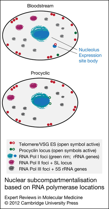
Similar to other eukaryotes, the trypanosome nucleolus is constructed around the RNA-Pol-I-transcribed rDNA arrays (Ref. Reference Berriman65), although this appears to be more complex because of the presence of RNA-Pol-I-transcribed protein-coding genes (Fig. 3). There is also evidence that RNA Pol II and III exhibit a degree of compartmentalisation in trypanosomes. The large subunit of RNA Pol III localises to several foci in the interphase nucleus (Ref. Reference Alsford and Horn75). The large subunit of RNA Pol II is distributed throughout the T. brucei nucleus, but also forms several macrofoci (Ref. Reference Uzureau76), consistent with the constitutive nature of trypanosome transcription where it generally initiates from divergent strand-switch regions (Ref. Reference Siegel77). Trans-splicing adds a common 39 nucleotide 5′-cap containing splice leader (SL) sequence to every mRNA in T. brucei, and the SL genes are organised as an array on chromosome 9 with the only recognisable trypanosome RNA Pol II promoters (Fig. 2) (Ref. Reference Gilinger and Bellofatto78). The macrofoci containing RNA Pol II colocalise with the SL gene array; interestingly, the transcription elongation factors, TFIIS1 and TFIIS2-1, are not enriched at these loci, suggesting that they are primarily involved in the generation of polycistronic RNAs (Ref. Reference Uzureau76). RNA Pol II has a similar distribution in the nucleus of the related kinetoplastid Trypanosoma cruzi, and although dispersed throughout the nucleus as in T. brucei, forms a strong focus that colocalises with the SL gene array in a region that appears to exclude other loci (Refs Reference Dossin Fde and Schenkman79, Reference Schenkman, Pascoalino Bdos and Nardelli80). More recently, the tSNAP complex, which is involved in SL transcript processing (Refs Reference Das81, Reference Schimanski, Nguyen and Gunzl82) has been localised to a distinct region of the T. brucei nucleus (Refs Reference Alsford and Horn75, Reference Lustig83). Taken together, these data indicate that an SL transcription and processing factory might exist in the trypanosome nucleus.
Nuclear subcompartmentalisation based on RNA polymerase locations. Foci for the three RNA polymerases are shown as teal, magenta and grey (RNA Pol I, II and III, respectively. The nuclear envelope is in black, positions of telomeres in red and procyclin loci are in green. Actively transcribed procyclin or telomeric expression sites (ESs) are shown as open symbols whereas transcriptionally repressed elements are closed symbols. Note that RNA Pol I is restricted to the nucleolus and ES body and the rRNA loci are shown as a fuzzy rim at the periphery of the nucleolus–RNA-Pol-I territory, whereas the major sites for RNA Pol II are coincident with the spliced leader loci. In the case of the latter, the localisation is very extensive because of the extensive involvement in transcription of protein-coding genes, and only the most concentrated regions are shown. RNA Pol III is found in numerous small foci throughout the nucleoplasm, but is excluded from the nucleolus. Diagram is based on data from Refs Reference Navarro, Penate and Landeira22, Reference Alsford and Horn75, Reference Uzureau76, Reference Daniels, Gull and Wickstead122. The peripheral localisation of VSG ES telomeres and procyclin loci in bloodstream cells is more speculative.

Nuclear envelope and nucleoskeletal interactions
In metazoa, the nucleus is structurally supported by a family of intermediate filament proteins called lamins (Ref. Reference Shimi84). These proteins, together with many associated factors, are responsible for the construction of a filamentous lamina. The importance of lamins and the lamina in both the structural maintenance of the nucleus and specific roles in the positioning of the chromosomes is well established. Most significant here are observations that the positioning of telomeres is heavily dependent on the correct expression, targeting and assembly of lamins. Inborn defects of lamin targeting and assembly are associated with multiple pathologies in humans, the best known of which is progeria, which results in premature ageing as a result of misregulation of gene expression, telomere shortening and nuclear structural abnormalities. Lamins are restricted to metazoa, with the result that the molecular players controlling nuclear structure and chromosomal positioning in trypanosomes have remained unknown.
A second important element of the nuclear periphery is the nuclear pore complex (NPC), large multisubunit organelles that span the double-membrane nuclear envelope and which act as the gateway for all nucleocytoplasmic transport (Ref. Reference Capelson, Doucet and Hetzer85). NPCs are responsible for export of mRNAs and tRNAs, and for import of DNA replication proteins, transcription factors and the proteins that make and modulate chromatin. Recent work indicates a physical association between NPCs and chromatin, providing a further important player in chromosomal organisation at the nuclear periphery.
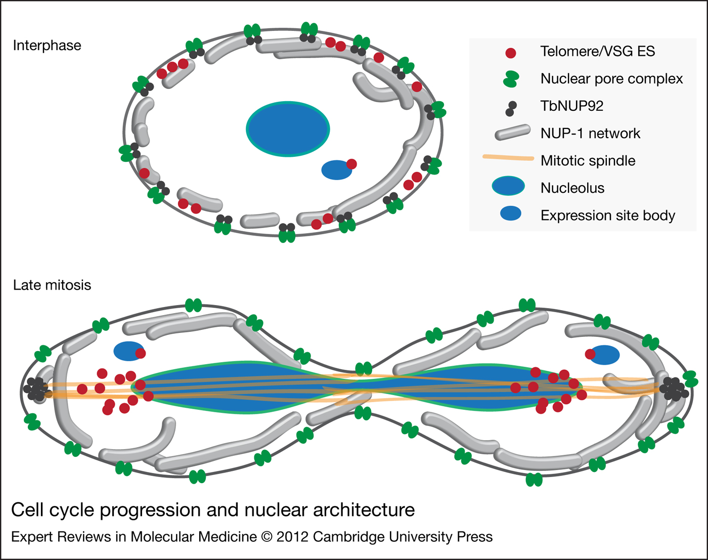
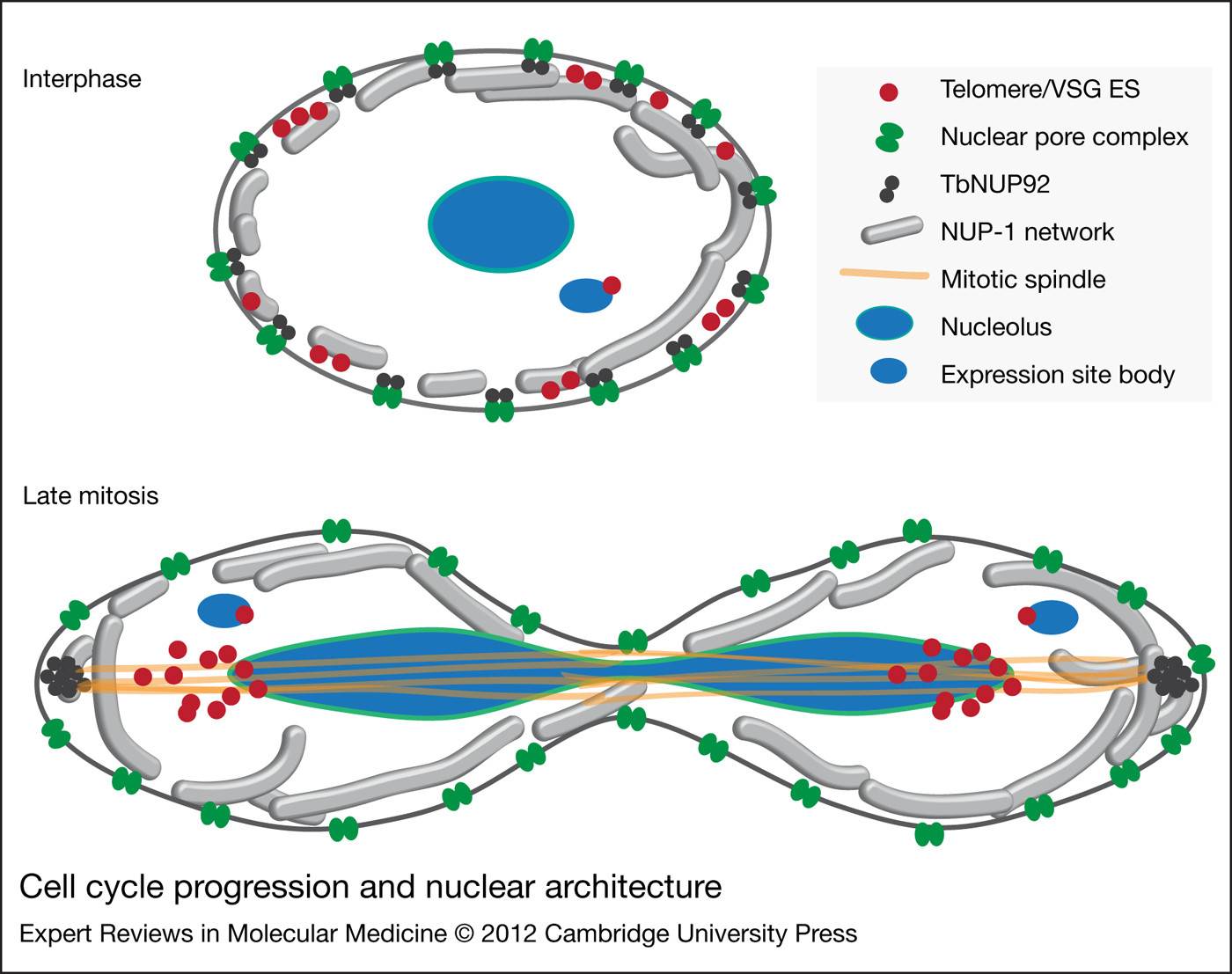
The vast majority of nucleoporins have been identified in trypanosomes by proteomics (Ref. Reference DeGrasse86). Sequence searches had suggested little similarity between trypanosomes and higher eukaryotes, but surprisingly the NUP composition of T. brucei is similar to animals and fungi, reflecting greater conservation of structure rather than sequence, and also suggesting both an ancient common origin for many NUPs and hinting at potential conservation of functions encompassing both mechanisms of nucleocytoplasmic transport and potential interactions with other nuclear processes. An example of the latter is T. brucei NUP92; this protein is a TPR-repeat-containing protein, and uniquely among T. brucei NUPs, has a cell-cycle-dependent location, being exclusively at the NPC during interphase and at a polarised location that is likely to be the spindle-anchoring site during mitosis (Fig. 4). This behaviour is highly reminiscent of Mad-1 and Mad-2 and Tpr1–Tpr2 from S. cerevisiae and these early experiments suggest that a subset of trypanosome NUPs are mobile, and thus participate in the control of mitotic events (Refs Reference Iouk87, Reference Niepel88, Reference Strambio-de-Castillia, Blobel and Rout89). Trypanosome nuclear lamina components have begun to emerge. A giant 450 kDa coiled-coil protein, NUP-1, is a component of fibres at the inner nuclear envelope, and is required to maintain NPC and telomere positioning. Suppression of NUP-1 has highly specific effects on telomeric VSG genes, with clear misregulation of normally silenced VSGs (Ref. Reference DuBois90). Preliminary evidence suggests that at least two additional players interact with NUP-1; one of these, NUP-2, is also a coiled-coil protein that appears to be part of the NUP-1-associated fibrous network. Together, these data indicate that repression by the specific positioning of genes within regions of peripheral heterochromatin is probably a strategy shared with higher eukaryotes. It will be interesting to connect the functions of the NUP-1–NUP-2 networks with factors controlling VSG silencing. Importantly, although the lamins and NUP-1–NUP-2 bear no obvious common ancestry, these studies do indicate that similar, filament-based nucleoskeletons are a basis for much of the higher-order control of nuclear function in animals, fungi and trypanosomes.
Cell cycle progression and nuclear architecture. Top panel shows an interphase bloodstream stage nucleus in schematic, with the focus on the nuclear pore complex (NPC) (green) the putative lamina protein NUP-1 (grey tubes) and positioning of telomeres (red) within small clusters at the nuclear periphery. A single nucleolus and an expression site body (ESB) are shown, both containing RNA Pol I subunits (blue), and in the case of the ESB, an active VSG and associated telomeric region. Note the presence of T. brucei NUP92, a probable nuclear basket component at the NPC (dark grey). Lower panel shows a late mitotic nucleus, where the telomeres have nearly completed migration from the midbody to the opposite poles of the daughter nuclei. The mitotic spindle is shown in orange. Note that NUP92 relocates from the NPC to polarised regions that might correlate with the trypanosome spindle pole body, whereas the remaining NPC subunits remain at the nuclear periphery. The ESB replicates late during mitosis, and only following sister chromatid separation, which is delayed with reference to the remaining chromosomes and the inactive ESs (Ref. Reference Landeira27).
Subtelomeres and the control of VSG ES silencing
Although the precise mechanisms of allelic exclusion itself are only partially uncovered, our understanding of the mechanism(s) underlying VSG silencing has advanced considerably. It appears that two distinct mechanisms operate to repress gene expression at telomeres and VSG ESs (Ref. Reference Horn and McCulloch91). Telomeric silencing was first reported in the budding yeast, S. cerevisiae (Ref. Reference Gottschling92), but subsequent studies showed that genes located within a few kilobase pairs of a telomere are prone to repression in many eukaryotes. The mechanism has been particularly well characterised in S. cerevisiae (see Refs Reference Grunstein93, Reference Tham and Zakian94). Briefly, telomeric silencing depends upon binding of the telomere-binding protein, repressor-activator protein 1 (RAP1) and consequent recruitment of a silencing complex containing a histone deacetylase, silent information regulator 2 (Sir2). Thus, telomeric heterochromatin is defined, at one level, by hypoacetylated chromatin.
Histones both package chromosomes and regulate access to factors involved in a wide range of chromatin transactions, including transcription, DNA replication and repair. Reversible modifications, such as N-terminal acetylation and methylation, extending beyond the nucleosome core, are particularly important for these regulatory functions. Although histone modifications do occur in trypanosomes, they are divergent from higher eukaryote canonical histone modifications (Ref. Reference Figueiredo, Cross and Janzen32). Chip-seq analysis provided the first direct evidence that specific histone modifications have a role in controlling trypanosome gene expression (Ref. Reference Siegel77), and despite their divergence from higher eukaryotes, trypanosomatid histones are subject to reversible modification, in particular for methylation (Refs Reference Alsford and Horn95, Reference Janzen96). Furthermore, several histone acetyltransferases, deacetylases, methyltransferases and demethylases are encoded in the trypanosome genome (Ref. Reference Ivens97). Because histone variants also have roles in transcription regulation and functional specialisation of chromatin domains, similar histone variants were also of interest in trypanosomatids. As noted above, the cenH3 variant is absent, but novel histone variants are present. Trypanosome H2AZ and H2BV are enriched at transcription start sites (TSSs), whereas H3V and H4V are enriched at transcription termination sites (TTSs) (Ref. Reference Siegel77) (Fig. 2). This finding represented a breakthrough in understanding transcriptional control in the apparently promoterless, trypanosome PTUs. The same study also revealed roles for histone acetylation and methylation at initiation sites. Thus, knowledge of trypanosome chromatin biology has advanced considerably in recent years (Ref. Reference Figueiredo, Cross and Janzen32), but many fundamental questions remain, not least of which is how centromeres are defined in the absence of cenH3.
The first direct indication that telomeric silencing operates in T. brucei came from genetic manipulation of the bloodstream form. Using selectable marker genes as reporters inserted adjacent to native telomeres, dramatic repression was observed compared with identical reporters at the active locus (Ref. Reference Horn and Cross98). Notably, this positional effect was developmentally regulated, more consistent with repression by facultative rather than constitutive heterochromatin. It is also important to note a key distinction in the constructs used to interrogate apparent telomere-position effects in T. brucei and in other organisms. Promoters recruiting RNA Pol I were used in T. brucei, but RNA Pol II promoters were used elsewhere. This reflects the unusual properties of transcription units in trypanosomatids and that VSGs are transcribed by RNA Pol I. Because RNA Pol I transcription operates at a much higher rate relative to RNA Pol II transcription, these findings indicated that the positional effect identified at T. brucei telomeres was able to repress even these ‘strong’ promoters.
On the basis of the data at hand, two important questions arise. First, is the telomere itself responsible for silencing, or are additional sequences involved, and second, what factors contribute to this repression? Because it seems that there are two distinct mechanisms operating at these loci, and therefore possibly two distinct sets of factors recruited by distinct sequences, we will consider sequence dependence first. Almost all native telomeres in T. brucei appear to have an adjacent VSG gene or VSG ES. By creating de novo telomeres without VSGs, it was found that the telomere itself can exert negative control on local gene expression (Ref. Reference Glover and Horn99), but significantly, repression was severely diminished at only a short distance from the telomere, immediately suggesting that VSGs or other features of VSG ESs contribute to the more robust and developmentally regulated silencing.
We can also consider the factors known to contribute to this ‘basal’ (VSG and VSG ES independent) telomeric silencing and compare them with factors specifically contributing to VSG ES silencing. At least 14 proteins are linked to the control of transcription at telomeres in T. brucei (Table 1). Telomeric silencing in budding yeast requires Sir2, and consistent with an evolutionarily conserved mechanism, the T. brucei Sir2 orthologue SIR2rp1 also contributes to basal telomeric silencing (Ref. Reference Alsford100). Analysis of several other histone acetyltransferases and deacetylases revealed two additional proteins that modulate SIR2rp1-dependent basal telomeric silencing; the histone acetyltransferase HAT1, thought to constrain SIR2rp1 to telomeric domains through histone H4K10 acetylation (Ref. Reference Kawahara101), and the deacetylase DAC1, which antagonises SIR2rp1 action at telomeres (Ref. Reference Wang, Kawahara and Horn102). Consistent with the paradigm that VSG ES silencing is mechanistically distinct, none of these three factors has a detectable impact on transcription from the VSG ES promoter (Refs Reference Alsford100, Reference Kawahara101, Reference Wang, Kawahara and Horn102).
Basal telomeric silencing is a heterochromatin-based phenomenon and heterochromatin is clearly a likely participant in VSG silencing. Indeed, nucleosomes are specifically depleted from the single active VSG ES (Refs Reference Figueiredo and Cross103, Reference Stanne and Rudenko104). So, what factors repress transcription from the VSG ES promoter, and how do these differ from basal telomeric silencing? The first protein linked to VSG ES silencing was a chromatin-remodelling factor known as Imitation SWI (ISWI) (Ref. Reference Hughes105). ISWI knockdown, however, leads to quite modest derepression, and VSG genes remain repressed to a large degree (Ref. Reference Stanne106). Other proteins showing similar knockdown phenotypes include DAC3, a histone deacetylase (Ref. Reference Wang, Kawahara and Horn102), the chromatin chaperone FACT (Ref. Reference Denninger107), a nucleoplasmin-like protein, NLP1 (Ref. Reference Narayanan108). Knockdown of a telomere-binding protein, RAP1, also generates a similar phenotype (Ref. Reference Yang109), but in this case, multiple telomeric VSGs achieve sufficient expression levels to be detected at the cell surface, which has not been reported for the other proteins; furthermore, the phenotype also includes the production of additional ESBs, which is more consistent with the idea that the ESB associates around an active ES rather than being a stable factory. Our more recent work indicates a role for the nuclear lamina components NUP-1 and NUP2 in regulating telomeric silencing, suggesting that telomere tethering is important for the control of chromatin structure and modification (Ref. Reference DuBois90).
A role for RAP1 in basal telomeric silencing has not been defined, but on the basis of mechanisms in S. cerevisiae, is expected to recruit SIR2rp1, as well as additional factors that spread further along VSG ESs. This rather vague model underlines the fact that mechanisms controlling ‘spreading’ of a repressed domain, probably occurring in cis along the DNA strand, are not particularly well understood in any system. Thus, only modest derepression, which in the case of RAP1 is ~100 times less than full activity (Ref. Reference Yang109), is a common phenotype emerging from knockdowns of candidate regulators for VSG ES repression. It is unclear as to whether this reflects redundancy or the presence of an additional major pathway that remains to be uncovered.
Several factors do have a more direct impact on the monoallelic VSG expression mechanism. First, DOT1B, a nonessential histone lysine methyltranferase, has a role in in situ switching, where transcription is transferred from one VSG ES to another without requiring a DNA rearrangement (Ref. Reference Figueiredo, Janzen and Cross110). Null dot1b cells show very slow in situ switching, suggesting that histone H3K76 methylation is important in inactivating transcription at the original ES, activating transcription at the new ES, or both. Second, chromosomal cohesion appears to have a role in inheritance of the activation status of the VSG ES (Ref. Reference Landeira27). Perturbed sister chromatid cohesion disrupts what is normally very stable inheritance, leading to an increased frequency of in situ switching. Third, an intact nuclear lamina also appears to be important for this stable inheritance, which is disrupted in cells depleted for NUP-1 (Ref. Reference DuBois90).
Overall, much of our understanding here comes from candidate-based approaches, and despite being highly informative, are probably limited in scope, providing only restricted snapshots of the overall processes of telomere repression, mechanisms controlling ES activity and broader issues in terms of epigenetic control. Furthermore, because candidate approaches by necessity target proteins with domain similarity to known participants in epigenetic processes in other systems, the identification of putative trypanosome-specific pathways that exploit truly novel factors is only just beginning.
Genome-wide approaches
Recently, more global approaches have become available for the analysis of post-transcriptional gene expression control. For example, ChIP-Chip has been used to describe the activity of the RNA-binding protein DRBD3 in stabilising T. brucei mRNAs (Ref. Reference Estevez111) and ChIP-qPCR has been used to show that T. brucei ISWI regulates RNA-Pol-I-transcribed loci and is also present at RNA Pol II transcription boundaries (Ref. Reference Stanne106). Furthermore, arginine methyltransferases modify histone tails in higher eukaryotes, among many other probable functions, and thus affect gene regulation. At least five of these enzymes are also present in trypanosomes, and one, T. brucei PRMT6, was found to interact directly with histones by immunoprecipitation, and in vitro assays indicate that PRMT6 can methylate histones (Refs Reference Fisk and Read112, Reference Fisk113). Furthermore, PRMT7 can methylate both histones and RNA-binding proteins, suggesting multiple impacts on the gene expression machinery (Ref. Reference Fisk114). Arginine modification of trypanosome histones has been reported in T. cruzi (Ref. Reference da Cunha115), but the precise contributions of the T. brucei PRMT family and potential roles in chromatin maintenance and modification remain to be elucidated.
Most significantly, high-throughput sequencing is significantly advancing our understanding of epigenetics and allows global analysis. Although these studies frequently remain at the candidate level, the results are more wide ranging. For example, J synthesis and maintenance requires two thymidine hydroxylases, JBP1 and JBP2 (Ref. Reference Cliffe116). Anti-J immunoprecipitation and high-throughput sequencing showed that JBP1 deposits J at chromosome internal sites and there is some association with strand-switch regions, whereas JBP2 acts at telomeric regions (Ref. Reference Borst and Sabatini55). This study suggests that JBP1 induces J synthesis, but maintenance is dependent upon JBP2, which binds DNA through a SWI2–SNF2 domain and hence epigenetically remodels chromatin.
Several recent studies have used Illumina/Solexa high-throughput cDNA sequencing (RNA-seq) to identify splice acceptor sites and polyadenylation signals (Refs Reference Kolev117, Reference Nilsson118, Reference Siegel119, Reference Veitch120, reviewed in Ref. Reference Siegel121). Together, these studies redefined the major splice acceptor site for most genes and identified a surprisingly large number of alternative splice sites. Moreover, a considerable proportion map within the coding region, with clear potential to alter the sequence of the expressed protein. Mechanisms that underpin splice-site selection, and the nature or even presence of signalling pathways that can influence the frequency of splicing at each site are completely unknown. Again, the detailed analysis of these phenomena is only just beginning.
Conclusions and future directions
The relationship between chromatin structure, gene expression and antigenic variation in trypanosomes is becoming clearer, with many proteins now implicated in the silencing and repression of VSG ESs. The genome sequence has done much to refocus the field, with new knowledge and the ability to construct hypotheses based on sound information rather than vague or very incomplete data, as was apparent in earlier studies. There is now excellent evidence placing histone modification as the mediator of telomeric silencing and control of ESs, as well as in the definition of PTUs, which suggests a greater similarity than was previously appreciated between trypanosome TSS regions and conventional eukaryotic promoters, especially in their bidirectional activity. It is also clear that the candidate-based approach still has power because the list is far from exhausted, and further study of orthologues is likely to be highly informative. Falling costs in sequencing mean that genome-wide, rather than target-gene-based, analysis of impacts on transcription is now possible and desirable.
However, several important questions remain: (i) How do the factors operating at telomeres, chromosome internal TSSs and TTSs and at the ESB and other nuclear internal subcompartments interact with each other? Specifically, what are the protein–DNA interaction networks that subtend these functions? (ii) How are these networks incorporated into interactions with the nucleoskeleton, the nuclear envelope and lamina, which in other systems provide much of the assembly framework for high-level organisation of the nucleus, and is also emerging as a participant in transcriptional control and epigenetic memory in higher eukaryotes? (iii) What novel (or taxon-restricted) players are present, and how do they interact and function in the context of the more widely conserved cohorts of gene products? (iv) How are these networks controlled in a spatiotemporal manner, and how does this integrate with life-cycle progression and responses to alterations in the environment? (v) Is the inheritance of VSG transcription status by daughter cells based on the simple retention of all bar one ES at the nuclear periphery through mitosis, or is there a more complex mechanism, perhaps related to the inheritance of nutritional memory in S. cerevisiae?
Addressing these questions will require unbiased sampling of long-range complex interactions with, typically, the application of proteomic and cell biological approaches to identification of new and established players, assessment of post-translational modifications and identification of the DNA elements with which these factors associate and which they modulate in a functional manner. Major advances in our understanding of mammalian and fungal gene regulatory networks and how they impact at the cellular level can provide a framework, but the novel aspects of trypanosome biology should be our priority; technical advances in nucleic acid sequencing, proteomics and immunoisolation methods and RNAi-based screens will offer many opportunities. Indeed, here is where the novel biology and the understanding of genetic mechanisms that kinetoplastids use to express their surface antigens reside with the potential to provide insight to disease and even therapeutic opportunities.
Acknowledgements and funding
Work in the authors' laboratories is mainly supported by the Wellcome Trust, which is gratefully acknowledged. We are grateful to the anonymous reviewers for constructive comments on the early drafts of this article.