Sudden cardiac arrest and sudden cardiac death, although uncommon among children and adolescents, have a significant emotional and social impact over families, physicians, and media. Although many campaigns have been conducted to promote the importance of screening procedures and to improve the awareness and promptness of cardiopulmonary resuscitation in the case of sudden cardiac arrest, a remarkable number of patients continue to die.
In this study, we will review the recent literature, focusing on the epidaemiology and the most common aetiologies of sudden cardiac arrest and sudden cardiac death, also mentioning the primary prevention strategies that might reduce the number of sudden cardiac death, as most of these events occur in the presence of an identifiable structural cardiac abnormality.
Definition
Sudden cardiac arrest is the sudden, unexpected loss of heart function, breathing, and consciousness. Sudden cardiac death is defined as the unexpected non-traumatic death in which the loss of heart function occurs within 1 hour of the onset of collapse symptoms, and postmortem studies identify any pathology of the heart or of the great vessels, and exclude non-cardiac causes of death.Reference Corrado, Basso and Pavei 1
Epidaemiology
Sudden cardiac death is the most frequent non-traumatic cause of death in the paediatric population and in young adults.Reference Meyer, Stubbs and Fahrenbruch 2
It is an uncommon, if not rare phenomenon, but its incidence might be underestimated because most data are collected retrospectively.
The global incidence of sudden cardiac arrest in children and adolescents is estimated between 0.5 and 8 per 100,000 person-yearsReference Triedman and Alexander 3 – Reference Driscoll and Edwards 6 , although higher rates have been reported in the literature. The incidence in young adults, <35 years of age, has been described as high as 20 cases per 100,000 person-years.Reference Donohoe, Innes, Gadd, Whitbread and Moore 7
In Italy, the epidaemiology of sudden cardiac death is more easily traceable starting from 1982, when the screening to obtain eligibility for sports activity became mandatory. 8
The incidence of sudden cardiac death in Italy, in a population 12–35 years of age in the period 1979–2004, has been estimated between 1 and 1.9 per 100,000 person-years.Reference Corrado, Basso and Pavei 1 , Reference Corrado, Basso, Rizzoli, Schiavon and Thiene 9
A retrospective study from the State of Washington reports an overall incidence of 2.28 per 100,000 person-years, and underlines some differences between the various age groups. Sudden cardiac death incidence is 2.1 cases per 100,000 person-years for patients between 0 and 2 years; 0.61 per 100,000 person-years between 3 and 13 years; and 1.44 per 100,000 person-years between 14 and 25 years.Reference Meyer, Stubbs and Fahrenbruch 2
The trend of sudden cardiac death incidence is not clear. Some reports describe an increase in sudden cardiac death cases in the past few yearsReference Spurgeon 10 , Reference SoRelle 11 , whereas Corrado et al described a reduction in sudden cardiac death incidence among italian young athletes and only a modest increase in non-athletic population.Reference Corrado, Basso and Pavei 1 , Reference Corrado, Basso, Rizzoli, Schiavon and Thiene 9
Many studies suggested a correlation between sudden cardiac death and sports. Approximately 20–25% of sudden cardiac arrest occur during physical activity, and it has been shown that adolescent athletes have a 2.5 times relative risk compared with an age-matched non-athletic population. Moreover, sudden cardiac death occurs more frequently in the male population.
Many factors are implicated in the correlation between physical activity and sudden cardiac death. The main link might be the triggering of life-threatening arrhythmias in a heart with structural abnormalities during sports.Reference Corrado, Basso and Pavei 1 , Reference Corrado, Basso, Rizzoli, Schiavon and Thiene 9 , Reference Gajewski and Saul 12 – Reference Maron 16
Survival rates of sudden cardiac arrest are low. The percentage of “aborted sudden death” varies between 27% and 40% depending on the age, but has increased in the past 40 years thanks to the improvement of cardiopulmonary resuscitation ability and of availability of automatic external defibrillators.Reference Meyer, Stubbs and Fahrenbruch 2 , Reference Atkins, Everson-Stewart and Sears 5
Aetiology and pathophisiology
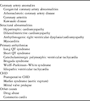
The main final pathway of sudden cardiac arrest and sudden cardiac death is ventricular tachycardia/ventricular fibrillation occurring on a pathological substrate. There are several different causes responsible for the predisposition to these arrhythmias in children and young adults (Table 1). Epidaemiology and relative incidence of each aetiology are not clearly defined, mostly because of geographic and age differences, but also because of lack of extensive data. Cardiac causes can be divided into five categories: (1) coronary artery abnormalities, (2) myocardial structural diseases, (3) primary arrhythmic diseases, (4) congenital cardiac diseases, and (5) other cardiac causes (Table 1).
Causes of sudden cardiac death.

Before discussing the cardiac causes of sudden cardiac death, we briefly present the sudden infant death syndrome, which is another important cause of death in the young.
Sudden infant death syndrome
Sudden infant death syndrome is a condition in which an infant, usually in the early postnatal period and nearly always before 6 months of age, dies during sleep for unexplained reasons and the standard autopsy fails to disclose an aetiology.Reference Rubens and Sarnat 17
Around 10–20% of sudden infant death syndrome cases are due to primary electrical heart diseases caused by genetic variants in either ion channel or ion channel-associated proteins, including long QT syndrome, short QT syndrome, and Wolff–Parkinson–White syndrome. Most sudden infant deaths, however, have non-cardiac aetiologies, which include respiratory infections, disorders of ventilation, metabolic derangements, hyperthermia, and asphyxia because of environmental factors.Reference Tfelt-Hansen, Winkel, Grunnet and Jespersen 18 , Reference Berkowitz 19
Coronary artery diseases
Coronary artery disease, caused either by premature atherosclerosis or by congenital coronary artery anomalies, is a frequent cause of sudden cardiac death.Reference Meyer, Stubbs and Fahrenbruch 2 , Reference Corrado, Basso and Poletti 20
Atherosclerotic coronary artery disease
Atherosclerotic coronary artery disease has been identified in individuals between 25 and 35 years of age in several studies, whereas congenital anatomical anomalies can be found in cases of sudden cardiac death also in younger patients.Reference Meyer, Stubbs and Fahrenbruch 2 , Reference Corrado, Basso, Rizzoli, Schiavon and Thiene 9 , Reference Corrado, Basso and Poletti 20
Congenital anatomic anomalies
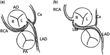
The most frequent anomaly is the ectopic origin of one coronary artery from the opposite aortic sinus with a course between the aorta and the pulmonary artery (Fig 1). Although a right coronary artery arising from the left aortic sinus is more commonly observed (Fig 1a), a left main coronary artery arising from the right aortic sinus appears to be more commonly associated with sudden cardiac death (Fig 1b). There are four main mechanisms postulated to be responsible for myocardial ischaemia: (1) reduction in myocardial blood flow owing to the presence of an acute angle as the coronary artery arises from a hypoplastic ostium; (2) compression of the anomalous artery between the pulmonary artery and aorta, especially during exercise, when the calibre of the two large vessels increases; (3) coronary spasm because of endothelial dysfunction; and (4) increased wall tension in the aorta during exercise causing compression of the intramural portion of the anomalous coronary artery.
Ectopic coronary artery origins. Abnormal coronary artery origins associated with sudden cardiac death. ( a ): Right coronary artery (RCA) ostium situated in the coronary cusp (L) of the aorta (AO) and RCA course between the aorta and the pulmonary artery (PA). ( b ): Left main (LM) ostium situated in the right coronary cusp (R) and LM course between the aorta and the pulmonary artery. Cx=circumfliex; LAD=left anterior descending artery.

In the presence of sudden cardiac death with coronary anomalies, premature atherosclerosis is usually absent.Reference Gajewski and Saul 12 , Reference Chandra, Bastiaenen, Papadakis and Sharma 15 , Reference Maron 16 , Reference Liberthson 21 – Reference Taylor, Byers, Cheitlin and Virmani 23
The preventive diagnosis of anatomical coronary artery anomalies is clinically difficult. Basal electrocardiogram and exercise testing are usually negative, and in most cases the first symptom is sudden cardiac death.
Although coronary angiography, cardiac computed tomography, and MRI are more accurate, some of these abnormalities can be also diagnosed through transthoracic echocardiogram with a small rate of false-negative results. The recognition of coronary anomalies during transthoracic echocardiogram, however, needs a focused examination and often an experienced operator.Reference Davis, Cecchin, Jones and Portman 22 , Reference Labombarda, Coutance and Pellissier 25
Coronary anomalies can be suspected from prodromal symptoms, such as chest pain, syncope, or pre-syncope, particularly during exercise.Reference Chandra, Bastiaenen, Papadakis and Sharma 15 , Reference Angelini 26 , Reference Attili, Hensley, Jones, Grabham and DiSessa 27
Kawasaki disease
A rare cause of sudden cardiac arrest is Kawasaki disease, an acute vasculitis that primarily affects the coronary arteries. Endothelial inflammation causes the development of a combination of coronary aneurysms and thrombotic occlusions. The disease is usually self-limiting, but sequels are frequent: coronary artery complications develop in up to 25% of affected children if the disease is left untreated.
Coronary abnormalities – aneurysms, stenosis, or occlusions – can persist and are responsible for ischaemic sudden death, even at an advanced age.Reference Wood and Tulloh 28 – Reference Suzuki, Kamiya, Arakaki, Kinoshita and Kimura 31
Structural abnormalities
Several structural cardiac abnormalities can cause sudden cardiac death in the young, and many studies suggest that hypertrophic cardiomyopathy accounts for most.Reference Meyer, Stubbs and Fahrenbruch 2 , Reference Maron 16 , Reference Liberthson 21
In the United States, structural myocardial anomalies account for most cases of sudden cardiac death.Reference Maron 16
Geographical differences are, however, well recognised. Most deaths in athletes in Italy are caused by coronary artery abnormalities followed by arrhythmogenic right ventricular dysplasia, whereas hypertrophic cardiomyopathy is responsible for only 5.5% of sudden cardiac deaths.Reference Berger and Campbell 32 , Reference Basso, Corrado and Thiene 33 This finding might be explained by the use of electrocardiography in pre-participation screening for athletes in Italy, thus reducing the frequency of hypertrophic cardiomyopathy deaths by early identification of the disease.
Hypertrophic cardiomyopathy
Hypertrophic cardiomyopathy is a primary disease of the myocardium, defined as left ventricular wall thickening occurring in the absence of increased after-load or other secondary causes.
Prevalence is rather high. It is the second most common form of heart muscle disease, affecting about 1 in 500 adults. Most forms of hypertrophic cardiomyopathy are genetic and are due to mutations of different genes involved in the myocite structure or metabolism, usually inherited with autosomal dominant pattern. The mechanisms of sudden death are ischaemic, arrhythmic, and haemodynamic. Myocite hypertrophy and disarray of muscle fibres cause microvascular abnormalities and microcirculation ischaemia. Moreover, hypertrophic cardiomyopathy can lead to myocardial bridging and ischaemia due to epicardial vessel compression. When hypertrophic cardiomyopathy is associated with left ventricular outflow tract obstruction, increased end-diastolic pressure can cause subendocardial ischaemia through increased wall compression. Ischaemic damage can induce fibrosis and scarring, thus creating an arrhythmic substrate, in which re-entry ventricular tachyarrhythmias are frequent. Despite being a less frequent cause of sudden cardiac death, haemodynamic collapse because of reduced systolic stroke volume and left ventricular outflow tract obstruction can occur. Sudden cardiac death in hypertrophic cardiomyopathy is usually exercise-induced, and thus restriction from sports is mandatory in these patients.
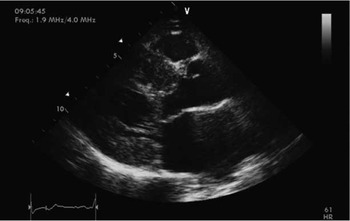
Diagnosis can be presumed by electrocardiogram, and usually confirmed by echocardiography (Fig 2). Family screening after a diagnosis of hypertrophic cardiomyopathy should be performed to assess the risk of relatives.Reference Maron 16 , Reference Liberthson 21 , Reference Maron and Maron 34 – Reference Keren, Syrris and McKenna 38
Hypertrophic cardiomyopathy. Parasternal long-axis view of a hypertrophic cardiomyopathy patient with a prominent septal hypertrophy and a moderate posterior wall hypertrophy. A systolic anterior displacement of mitral valve apparatus is observed. Myocardial texture is patchy because of myocardial fibres disarray.

The literature recognises several risk factors for sudden cardiac death in patients with hypertrophic cardiomyopathy: family history of sudden cardiac death, unexplained recent syncope, multiple repetitive non-sustained ventricular tachycardia on ambulatory electrocardiogram, exercise-induced hypotension, septal wall thickness>30 mm, extensive and diffuse late gadolinium enhancement at cardiac magnetic resonance, end-stage phase (ejection fraction<50%), left ventricular apical aneurysm and scarring, and substantial left-ventricular outflow gradient at rest. When one or more of these risk factors is present, the implantation of an implantable cardioverter–defibrillator in primary prevention should be evaluated.Reference Maron and Maron 34
Arrhythmogenic right ventricular dysplasia
Arrhythmogenic right ventricular dysplasia is a rare hereditary disorder, with estimated incidence between 1:1000 and 1:5000 cases.
It primarily affects right ventricular myocardium, although progression of the disease can lead to the involvement of the left ventricle in 50% of the cases. Even less common is primary left ventricular involvement. Arrhythmogenic right ventricular dysplasia is characterised by progressive myocite death and replacement with the fibrous and fatty tissue.Reference Basso, Bauce, Corrado and Thiene 39 Pathogenesis of arrhythmogenic right ventricular dysplasia is not completely understood, but it is likely caused by mutations in genes involved in myocite intercellular junctions, the desmosome, and is usually transmitted with autosomal dominant pattern.
A total of 12 genes have been linked to arrhythmogenic right ventricular dysplasia, but it is assumed that there are other genes involved that still need to be identified.Reference Azaouagh, Churzidse, Konorza and Erbel 40 It is not uncommon for these patients to have multiple genetic defects in the same gene (compound heterozygosity) or in a second complementary gene (digenic heterozygosity). The penetration of the genetic defects is largely incomplete. Genetic testing is available and it is advised in first-degree family members when the affected individual carries a known mutation.Reference Rigato, Bauce and Rampazzo 41 , Reference Marcus, Edson and Towbin 42
Myocardial dysplasia subsequently induces wall thinning and dilation of the right ventricle, and predispose to ventricular arrhythmias with left bundle branch block morphology, arising from the right ventricle.
The disease can be clinically silent in children and young adults, but its first symptoms, other than palpitations and syncope, can be sudden cardiac arrest and sudden cardiac death. Sudden death is mostly caused by ventricular tachiarrhythmias originating from the right ventricle.Reference Azaouagh, Churzidse, Konorza and Erbel 40 , Reference John, Tedrow and Koplan 43 , Reference Basso, Corrado, Marcus, Nava and Thiene 44
Arrhythmogenic right ventricular dysplasia can be suspected by some resting electrocardiographic features: (1) inverted T-waves in leads V1–V3; (2) esilon Wave following QRS in lead V1; (3) right bundle branch block; and (4) frequent premature ventricular complexes and/or ventricular tachycardia with left bundle branch block morphology.Reference Marcus, McKenna and Sherrill 45
T-wave inversion in leads V1–V3, however, can be found in children and even in around 3% of apparently healthy young adults, and thus this finding alone is usually of benign significance.Reference Marcus 46
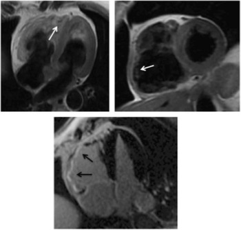
In some cases, electrophysiological abnormalities develop later in the course of the disease, thus making early diagnosis difficult. In addition, echocardiographic diagnosis can be difficult, but usually MRI and/or right ventriculography are conclusive, at least in later stages of the disease (Fig 3).Reference Gomes, Finlay and Ahmed 47 The diagnosis of arrhythmogenic right ventricular cardiomyopathy in the paediatric population remains challenging because of incomplete phenotype. In the early stages of arrhythmogenic right ventricular cardiomyopathy, neither magnetic resonance nor angiography are typically conclusive.
Arrhythmogenic right ventricular dysplasia. Cardiac magnetic resonance of a patient affected by arrhythmogenic right ventricular dysplasia. The upper panels show inversion recovery sequences with fat suppression. White arrows show microaneurysms of the right ventricle with fatty infiltration. The bottom panel shows diffuse late qadolinium enhancement of the right ventricle (black arrows).

As previously discussed, arrhythmogenic right ventricular dysplasia is more frequent in Italy, especially in the region of Veneto, probably as a consequence of a particular genetic substrate.Reference Corrado, Basso and Pavei 1 , Reference Corrado, Schmied and Basso 13
Dilated cardiomyopathy
Dilated cardiomyopathy is another myocardial-related cause of sudden cardiac arrest and sudden cardiac death; however, in most cases, dilated cardiomyopathy-related deaths are not sudden and they are not the first presentation of the disease.Reference Wren 36 , Reference Borjesson and Pelliccia 48
Dilated cardiomyopathy is characterised by progressive dilation of the left ventricle, with a concomitant reduction of the ejection fraction. The pathogenesis of dilated cardiomyopathy is various: it can be primary – most likely genetic – or secondary to inflammation, drugs, toxin, metabolic, and infectious diseases. However, aetiology can be determined only in 50% of the cases.Reference Hershberger and Siegfried 49 , Reference Hamilton and Azevedo 50 Despite a lack of agreement in the literature, we do not classify post-ischaemic ventricular dysfunction in the context of dilated cardiomyopathy.
Dilated cardiomyopathy is the second most frequent cardiomyopathy in children. The prevalence of dilated cardiomyopathy in the paediatric population ranges between 2.6 and 7 cases per 100,000 persons. The prevalence of dilated cardiomyopathy in the adult population is much higher.Reference Hamilton and Azevedo 50 , Reference Grimm and Maisch 51
Sudden cardiac death can occur in up to 10% of children with dilated cardiomyopathy, and it is usually caused by ventricular tachyarrhythmia secondary to left ventricular remodelling. Sudden cardiac death can also occur before the impairment of the ejection fraction. Diagnosis of dilated cardiomyopathy can be achieved through imaging studies, particularly echocardiography.Reference Borjesson and Pelliccia 48 , Reference Gavazzi, De Maria and Renosto 52 , Reference Burch, Siddiqi and Celermajer 53
Interestingly, some genetic mutations associated with dilated cardiomyopathy, such as Lamin A/C, carry a higher risk for sudden cardiac death. Some authors suggest the preventive implantation of implantable cardioverter–defibrillator in patients with such mutations.Reference Meune, Van Berlo and Anselme 54
Myocarditis
Myocarditis accounts for 5–20% of sudden cardiac deaths in the young. In a population of young victims of sudden death, a viral infection of the myocardium has been found in 12.5% of postmortem studies.Reference Borjesson and Pelliccia 48 , Reference Puranik, Chow, Duflou, Kilborn and McGuire 55 , Reference Gaaloul, Riabi and Harrath 56
The main cause of sudden cardiac death in acute and hyper-acute myocarditis is arrhythmic death. This can be related to the inflammatory micro-environment in the myocardium. Myocarditis can be presumed by clinical symptoms, such as shortness of breath, palpitations, and chest pain, but fatal arrhythmias can represent the first manifestation of the disease.
Electrocardiogram and echocardiographic findings can suggest the presence of myocarditis, but MRI is usually needed for the differential diagnosis, and sometimes myocardial biopsy is necessary.Reference Feldman and McNamara 57 , Reference Kyto, Saukko and Lignitz 58 Chronic myocardial dysfunction related to a previous myocarditis, with subsequent dilated cardiomyopathy, can also result in sudden cardiac death.
Primary arrhythmias
Primary arrhythmic causes are responsible for a high percentage of sudden cardiac arrest in young patients, which can be even higher if we consider all deaths without a clear diagnosis at autopsy. The relative frequency varies among different studies and ranges between 7% and 23%.Reference Corrado, Basso and Pavei 1 , Reference Meyer, Stubbs and Fahrenbruch 2
Long QT syndrome
Long QT Syndrome is a hereditary disorder with a prevalence varying between 1:2500 and 1:7000. It is caused by mutations in different genes encoding for ion channels involved in the heart conduction system.Reference Gajewski and Saul 12 , Reference Wren 36 , Reference Berul 59 , Reference Schwartz, Stramba-Badiale and Crotti 60 The risk for sudden cardiac death is considered greater when corrected QT is longer than 500 ms.Reference Schwartz, Spazzolini and Priori 61
This ion channel disorder is clinically associated with syncope, both during exercise and at rest, polymorphic ventricular tachycardia and sudden death. Its main feature is the alteration – delay – of ventricular repolarisation, which results in prolongation of corrected QT interval on the electrocardiogram. Up to one quarter of patients, however, can have a normal corrected QT interval.Reference Vincent, Timothy, Leppert and Keating 62 , Reference Lu and Kass 63
Nowadays 12 genes have been involved in the pathogenesis of long QT syndrome, with different clinical presentations. The most frequent mutations are the loss of function of one of the two potassium channels KCNQ1, causing LQT1 syndrome, and KCNH2, causing LQT2 syndrome, or the gain of function of sodium channel SCN5A, causing LQT3. All these mutations are inherited with autosomal dominant pattern.
LQT1 is characterised by malignant arrhythmias during exercise or conditions that increase adrenergic tone, thus making β-blocker therapy useful for the reduction of events. LQT1 is also frequently associated with deafness.Reference Ocal, Imamoglu, Atalay and Ercan Tutar 64 However, risk for sudden death with LQT1 phenotype appears lower.Reference Ellinor, Milan and MacRae 65
Jervell Lange–Nielsen Syndrome is a rare, but highly lethal disorder, characterised by congenital profound bilateral sensorineural hearing loss, long QTc, usually greater than 500 ms and specific mutations in potassium channels (KCNQ1 or KCNE1).Reference Tranebjaerg, Samson and GE 66
LQT2 is characterised by arrhythmias that usually occur at arousal or with surprise. In LQT3, the risk of cardiac death is higher when sympathetic tone is low, such as at rest.Reference Schwartz, Priori and Spazzolini 67 , Reference Vincent 68
Comprehensive genetic testing is an important component of the family evaluation, although the presence of many genetic variants of unknown significance ensures that ascribing true pathogenicity remains challenging.Reference Abrams and Macrae 69
The first symptom of long QT syndrome can be sudden cardiac death, although prodromes such as pre-syncope, syncope, and palpitations usually occur.
Long QT syndrome diagnosis can be difficult and several different criteria have been developed. Electrocardiogram can be normal, and therefore family history of sudden death is very important in suggesting the need for a screening in relatives of affected people. In addition, clinical history of pre-syncope or syncope can be useful, but usually stress testing or electrocardiogram analysis after infusion of epinephrine are needed for a definite diagnosis, as tachycardia can cause corrected QTc prolongation in affected patients and unmask the syndrome. Genetic testing can be needed for the final diagnosis, however.Reference Hofman, Wilde and Kaab 70 In the cases of diagnostic electrocardiogram (prolonged QT interval), the pattern is usually different among the various mutations. LQT1 is characterised by a broad T wave, LQT2 by low-amplitude T wave with notching, and LQT3 by long isoelectric ST segment.
β-blockers are used to reduce the incidence of sudden cardiac arrest, but usually patients with long QT syndrome require an implantable cardioverter–defibrillator as a prevention strategy.Reference Barsheshet, Dotsenko and Goldenberg 71
Sympathetic denervation is an additional therapeutic tool, which has recently been introduced in the clinical practice. Current indications for sympathetic denervation in long QT syndrome include: patients with appropriate ventricular fibrillation-terminating defibrillator shocks; patients with long QT syndrome-triggered breakthrough cardiac events when receiving adequate drug therapy; patients who fail to tolerate β-blocker therapy because of unacceptable adverse effects or asthma; and high-risk young children in whom primary drug therapy might not be sufficiently protective, with an intention for denervation to serve as a “bridge” to an implantable cardioverter–defibrillator.Reference Schwartz 72
Short QT syndrome
Although less common than long QT syndrome, short QT syndrome is another cause of sudden cardiac death in the young.Reference Algra, Tijssen, Roelandt, Pool and Lubsen 73 Short QT syndrome is a hereditary, mostly autosomal dominant, syndrome, usually associated with gain of function mutations in genes encoding for K+ channels. These mutations cause an increased outward K+ current, thus reducing the duration of ventricular repolarisation and shortening of the QT interval on the electrocardiogram (typically QTc<320 ms).Reference Gaita, Giustetto and Bianchi 74 , Reference Crotti, Taravelli, Girardengo and Schwartz 75
Short QT syndrome is frequently symptomatic. Patients can have a wide range of clinical manifestations, ranging from cardiac arrest to syncope to palpitations, usually due to atrial fibrillation, which can be associated with short QT syndrome. The mechanism of cardiac arrest and sudden cardiac death is ventricular fibrillation.Reference Crotti, Taravelli, Girardengo and Schwartz 75
Diagnostic criteria for short QT syndrome are not currently available. Familial history of sudden cardiac death, an electrocardiogram finding of a shortened QTc interval, sometimes associated with atrial fibrillation, and/or tall, peaked, and symmetrical T waves, however, can raise the suspicion.Reference Lu, Zhou and Zhang 76
Today, implantable cardioverter–defibrillator implantation is the only effective therapy in preventing sudden cardiac death. However, in very young patients at higher risk for peri-implantation complications and in patients who refuse the implantable cardioverter–defibrillator, hydroquinidine can lengthen ventricular repolarisation duration, thus increasing the QTc interval and reducing the risk for sudden cardiac death.Reference Giustetto, Di Monte and Wolpert 77 , Reference Boriani, Biffi, Valzania, Bronzetti and Martignani 78
Catecholaminergic polymorphic ventricular tachycardia
Catecholaminergic polymorphic ventricular tachycardia is a rare congenital arrhythmia characterised by bidirectional and polymorphic ventricular tachycardia that can lead to sudden cardiac arrest in paediatric age. Arrhythmias usually occur during exercise, as a consequence of increased adrenergic tone.Reference van der Werf and Wilde 79 , Reference Arias, Fernandez-Guerrero, Herrador and Pagola 80
Prevalence of catecholaminergic polymorphic ventricular tachycardia is estimated to be 1:10000 in Europe, and it carries an extremely high mortality, reaching 31% under 30 years of age when left untreated.
Catecholaminergic polymorphic ventricular tachycardia is generally inherited with autosomal dominant pattern, and it is caused by type 2 ryanodine receptors (RyR2) mutations. When catecholaminergic polymorphic ventricular tachycardia is due to the mutation of calsequestrin 2 (CASQ2) gene, it is inherited by autosomic recessive way. The mechanism involved is the alteration in myocite calcium homoeostasis.Reference Uchinoumi, Yano and Suetomi 81 , Reference Kontula, Laitinen and Lehtonen 82
Catecholaminergic polymorphic ventricular tachycardia is characterised by episodic syncope during exercise or acute emotion in individuals without structural cardiac abnormalities, and caused by reproducible, exercise-induced ventricular polymorphic arrhythmia. It is usually rare before the age of 2 years, but some RyR2 mutations have been identified postmortem in cases of sudden infant death syndrome.Reference Kontula, Laitinen and Lehtonen 82 , Reference Tester, Dura and Carturan 83
Resting electrocardiogram is normal, thus making catecholaminergic polymorphic ventricular tachycardia difficult to identify in asymptomatic patients.Reference Sumitomo, Harada and Nagashima 84 Family history is of main importance, as about 30% of patients with catecholaminergic polymorphic ventricular tachycardia have a family history of exercise-related syncope or sudden death. This makes family screening mandatory in case of catecholaminergic polymorphic ventricular tachycardia diagnosis.Reference Napolitano and Priori 85 , Reference Pflaumer and Davis 86
Treatment includes the use of β-blockers to reduce adrenergic tone that can elicit arrhythmias.Reference Sumitomo, Harada and Nagashima 84 , Reference Pflaumer and Davis 86
In addition, different studies showed that flecainide is useful in patients in which β-blockage is not enough to reduce ventricular tachycardia.Reference Watanabe, Chopra and Laver 87 , Reference van der Werf, Kannankeril and Sacher 88
A promising approach is left cervical sympathetic denervation. However, implantable cardioverter–defibrillator implantation is sometimes necessary to reduce the risk for sudden cardiac death.Reference Wilde, Bhuiyan and Crotti 89
Several studies are testing new drugs such as RyR2 channel inhibitors, but these approaches are not yet applied in the clinical practice.Reference Liu, Ruan and Denegri 90 , Reference Napolitano, Liu and Priori 91
Brugada syndrome
First described in 1992, Brugada syndrome has been associated with a high risk of sudden cardiac death predominately in young male patients.Reference Brugada and Brugada 92 Its prevalence is around 1:2000 individuals. It accounts for about 20% of deaths in patients without structural cardiac defects, and 4% of all sudden deaths.Reference Nademanee, Veerakul and Nimmannit 93 – Reference Viskin and Rosso 96
It is usually caused by the mutation of a sodium channel gene, SCN5A, but several different mutations have been reported.Reference Crotti, Marcou and Tester 97 , Reference Skinner and Love 98
Brugada syndrome can manifest itself with different arrhythmias such as atrial fibrillation, atrioventricular blocks, slowed atrial, or sinus node conduction. Reference Morita, Kusano-Fukushima and Nagase 99 – Reference Bordachar, Reuter and Garrigue 101
Wolff–Parkinson–White syndrome and atrioventricular nodal re-entry tachycardia also sometimes associate with Brugada Syndrome.Reference Fazio, Augugliaro and Lunetta 102
Nevertheless, the most common presentations of Brugada syndrome are ventricular arrhythmias – polymorphic ventricular tachycardia or ventricular fibrillation – which constitute the usual cause of sudden cardiac death.Reference Priori, Napolitano, Giordano, Collisani and Memmi 103 , Reference Wilde and Priori 104
Clinical manifestations include pre-syncope, syncope, palpitations, and sudden cardiac death, although most patients can be asymptomatic lifelong. Sudden cardiac death can occur during exercise or at rest.Reference Sidik, Quay, Loh and Chen 105
Diagnosis can be often suspected by resting electrocardiogram. Brugada syndrome is electrocardiographically characterised by a coved ST-segment elevation of at least 2 mm in the right precordial leads (V1–V3) followed by negative T waves. However, Richter et alReference Richter, Sarkozy and Paparella 106 suggested that lead V3 does not yield diagnostic informations in Brugada syndrome, as in their cohort of patients lead V3 alone was not diagnostic in any patient.
The electrocardiographic pattern can be evident at rest or can only be elicited by sodium channel blockers such as flecainide, which can be given in case of clinical suspicion, fever, vagotonic or adrenergic drugs, tricyclic antidepressants, combination of glucose and insulin, electrolyte alterations, alcohol, or cocaine abuse.Reference Richter, Sarkozy and Paparella 106 This electrocardiogram appearance is usually described as Brugada pattern type 1, which is the one with the higher risk for sudden cardiac death. The risk for sudden cardiac death is even higher when this pattern is manifest on rest electrocardiogram, whereas it is lower when it becomes evident only after provocative manoeuvres. In contrast, pattern type 2 and 3, characterised by a linear ST segment elevation <2 mm with positive or negative T waves, carry a lower risk for sudden cardiac death. As for other genetic diseases, family history is fundamental.Reference Yokokawa, Noda and Okamura 108 , Reference Smits, Eckardt and Probst 109
Currently, no pharmacologic treatment has been proven to prevent sudden cardiac death in Brugada syndrome, and the mainstay of preventive therapy is implantable cardioverter–defibrillator implantation. As a matter of fact, amiodarone and β-blockers are not effective, whereas class IC agents such as flecainide and propafenone can be. Some specific class IA agents, such as high-dose quinidine, and isoproterenol proved to be more effective compared with other anti-arrhythmic agents, but are not commonly used in the clinical practice.Reference Antzelevitch, Brugada and Borggrefe 107 , Reference Mizusawa, Sakurada, Nishizaki and Hiraoka 110 , Reference Belhassen 111
It is important to underline that, as fever is frequently associated with the elicitation of electrocardiographic Brugada pattern and increased risk for cardiac arrest, prompt antipyretic treatment must be provided in the presence of pyrexia.Reference Arbelo and Brugada 112
Risk stratification of Brugada syndrome is fundamental to identify patients with higher risk for sudden cardiac death: despite conflicting data, the presence of inducibile ventricular arrhythmias on electrophysiological testing have been advocated by some authors to be related with a worse outcome. Moreover, male gender, typical electrocardiogram features at rest, and previous symptoms associate with higher risk for sudden arrhythmic death.Reference Priori, Napolitano, Giordano, Collisani and Memmi 103 , Reference Priori, Gasparini and Napolitano 113
Nowadays, the only effective preventive strategy in high-risk patients or in secondary prevention is implantable cardioverter–defibrillator implantation.Reference Brugada, Brugada and Brugada 114 , Reference Brugada, Brugada and Brugada 115
Wolff–Parkinson–White syndrome
Wolff–Parkinson–White syndrome was first described in 1930. It is characterised by a ventricular pre-excitation associated with symptomatic paroxysms of tachycardia.Reference Wolff, Parkinson and White 116
The prevalence of Wolff–Parkinson–White syndrome is 1–3:1000 individuals according to different studies.Reference Guize, Soria and Chaouat 117 , Reference Cohen, Triedman and Cannon 118 Diagnosis is usually achieved during childhood. Most patients are symptomatic, and thus they are diagnosed with Wolff–Parkinson–White syndrome. Up to 25% of patients, however, undergo an incidental diagnosis of ventricular pre-excitation – Wolff–Parkinson–White syndrome pattern. Clinical presentations include supraventricular tachycardia, palpitations, chest pain, syncope, and less frequently sudden cardiac death. A not so negligible percentage of patients, ranging between 7% and 20%, have a concomitant structural heart disease.Reference Cain, Irving, Webber, Beerman and Arora 119
Typical electrocardiographic findings include a shortened PR interval and a delta wave, which originates from the small amount of ventricular myocardium that prematurely depolarises, resulting in a mildly enlarged QRS complex. The mechanism of arrhythmia is usually a re-entrant circuit involving the atrio-ventricular node and the accessory pathway, resulting in an atrioventricular reciprocating tachycardia. Sudden cardiac death is usually caused in Wolff–Parkinson–White syndrome patients by atrial fibrillation with rapid conduction to the ventricles through an accessory pathway with a short refractory period, resulting in ventricular fibrillation.Reference Cohen, Triedman and Cannon 118 , Reference Klein, Bashore and Sellers 120
The risk for sudden cardiac death in Wolff–Parkinson–White syndrome is estimated between 1.25 and 2.8 per 1000 person-years.Reference Cain, Irving, Webber, Beerman and Arora 119 , Reference Obeyesekere, Leong-Sit and Massel 121
Percutaneous transcatheter ablation of the accessory pathway in patients with Wolff–Parkinson–White syndrome with normal left ventricular ejection fraction, and no other electrocardiographic anomaly resolves the condition. Recently, the zero fluoroscopy approach has been demonstrated to be safe and effective to ablate supraventricular arrhythmias and accessory pathways.Reference Kerst, Parade, Weig, Hofbeck, Gawaz and Schreieck 122 – Reference Anselmino, Sillano, Casolati, Ferraris, Scaglione and Gaita 124 The zero fluoroscopy ablation is particularly attractive for children and adolescents owing to the absence of risk for radiation-related damages.
In patients resuscitated from a previous sudden cardiac arrest, ablation prevents recurrences. Thus, placement of an implantable cardioverter–defibrillator is usually not necessary in Wolff–Parkinson–White syndrome patients.Reference Antz, Weiss and Volkmer 125
On the other hand, in patients with asymptomatic Wolff–Parkinson–White syndrome pattern, ablation is advised only in the case of persistent pre-excitation confirmed at exercise stress testing, with specific electrophysiological findings, including shortest pre-excited RR interval in atrial fibrillation <250 ms or inducible sustained ventricular tachycardia.Reference Cohen, Triedman and Cannon 118
Idiopathic ventricular tachycardia
Idiopathic ventricular tachycardia refers to ventricular tachycardia occurring in structurally normal hearts, in the absence of myocardial scarring.
Classification of monomorphic idiopathic ventricular tachycardia is made on the site from which the arrhythmia originates and includes: outflow tract ventricular tachycardia, fascicular ventricular tachycardia, papillary muscle ventricular tachycardia, annular ventricular tachycardia, and miscellaneous – ventricular tachycardia from the body of the right ventricle and crux of the heart.
However, the most common subtype is right ventricular outflow tract ventricular tachycardia, which thus has a left bundle branch block morphology.Reference Hoffmayer and Gerstenfeld 126
Its precise aetiology is not known, but it was demonstrated that the mechanism of tachycardia is triggered activity due to catecholamine-mediated delayed after depolarisations.Reference Lerman 127
Ventricular tachycardia is typically exacerbated by exercise or stress, and may also occur during hormonal cycles in women.
Although idiopathic monomorphic ventricular tachycardia can cause syncope, sudden death is rare, expecially in right ventricular outflow tract ventricular tachycardia, the most common form. Two exceptions exist, however: patients who have a more malignant form of short-coupled premature ventricular complex-induced polymorphic ventricular tachycardia, or patients who develop left ventricular dysfunction secondary to tachycardia-induced cardiomyopathy.Reference Yarlagadda, Iwai and Stein 128 , Reference Viskin, Rosso, Rogowski and Belhassen 129
Diagnosis is made through 24-hour electrocardiographic monitoring, and echocardiogram must be subsequently performed to exclude structural causes, including arrhythmogenic right ventricular dysplasia, and secondary ventricular tachycardia.Reference Antz, Weiss and Volkmer 125
Treatment is not always necessary in asymptomatic patients. In patients with frequent symptoms first-line therapy includes a β-blocker or calcium-channel blocker, effective in about half of the patients. Patients with persistence of symptoms, despite pharmacological therapy are best treated with radiofrequency catheter ablation, a safe and low-risk procedure with high overall success rate (>90–95%). Implantation of a cardioverter–defibrillator is not usually necessary.Reference Altemose and Buxton 130
CHD
Postoperative CHD
Patients who have undergone cardiac surgery during neonatal or infant life have a postoperative risk for sudden cardiac death, which is considered around 1:1000 patient-years.Reference Mondesert and Khairy 134 The risk for sudden cardiac death mostly depends on the congenital repaired defect. It is higher for the transposition of the great arteries, intermediate for the tetralogy of Fallot, and low for other malformations such as aortic stenosis or coarctation.Reference Liberthson 21 , Reference Wren 36 , Reference Silka, Hardy, Menashe and Morris 131
Sudden cardiac death in patients with CHD is mostly due to malignant arrhythmias. It is important, however, to remind that up to 20% of sudden cardiac deaths may be due to non-arrhythmic causes such as cerebral or pulmonary embolism, myocardial infarction, heart failure, and aortic or aneurysmal rupture.Reference Wren, Irving and Griffiths 132
Arrhythmias may be the consequence of displaced or disfunctionant nodal tissue or atrioventricular conduction systems, coexisting primary myocardial disease, hypoxic tissue injury, residual or postoperative sequelae such as scarring, and genetic influences.Reference Silka, Hardy, Menashe and Morris 131
All kinds of arrhythmias may develop in patients with CHD, and often several different arrhythmias coexist in these patients. For example, it has been estimated that ∼50% of 20-year-old patients will develop an atrial tachyarrhythmia during their lifetime. Although less frequent, ventricular arrhythmias are the leading cause of sudden death.
The approach to patients with asymptomatic arrhythmias is controversial: evidence regarding which patients should be screened and which screening tests should be conducted is limited but growing. On the other hand, treatment in symptomatic patients is mandatory, as they are at a high risk for sudden death. Several therapeutic options are available, including pharmacological treatment and catheter ablation, but cardioverter–defibrillator implantation is the only effective option to reduce the risk of sudden death significantly.Reference Khairy, Van Hare and Balaji 133
Cardioverter–defibrillator implantation is indicated as secondary prevention in the case of resuscitated sudden cardiac arrest or in patient with symptomatic sustained ventricular tachycardia.
On the other hand, selecting appropriate candidates for primary prevention defibrillator remains a major challenge. Implantation should be considered in patients with ejection fraction <35%, in patients with recurrent syncope of undetermined origin in the presence of ventricular dysfunction or inducible ventricular arrhythmias, in the setting of recurrent syncope associated with complex CHD and advanced systemic ventricular dysfunction, and in specific cases of complex congenital heart defects such as tetralogy of Fallot with additional risk factors.Reference Mondesert and Khairy 134
Marfan syndrome
Marfan syndrome is an autosomal dominant collagen disorder caused by mutations in the fibrillin gene and characterised by alterations in the skeletal, ocular, and vascular tissue. It affects around 1:5000 newborns and is a frequent cause of sudden cardiac death, accounting for around 1–3% of sudden deaths, especially among athletes.Reference Chandra, Bastiaenen, Papadakis and Sharma 15
Sudden death in Marfan syndrome is usually associated with aortic dissection and rupture, occurring mostly during exercise as a result of increased aortic pressure. Ventricular arrhythmias can sometimes be responsible for cardiac arrest. These are the consequences of an arrhythmogenic substrate caused both by fibrillin mutation and left ventricular dilation.Reference Savolainen, Kupari, Toivonen, Kaitila and Viitasalo 135 – Reference Devereux and Roman 137
Mitral valve prolapse
Despite being very common, affecting up to 5% of the general population, mitral valve prolapse has been associated with sudden cardiac death occurring in structurally normal hearts in different studies. The mechanism of death is not clear, however, and this finding might be only incidental.Reference Chandra, Bastiaenen, Papadakis and Sharma 15 , Reference Anders, Said, Schulz and Puschel 138 , Reference Kligfield, Levy, Devereux and Savage 139
Athletes with mitral valve prolapse are not excluded from sport activity, including agonistics. The rate of complications of mitral valve prolapse is low, unless there is a concomitant heart disease, history of syncope and/or chest pain, arrhythmias, significant mitral regurgitation, or family history of sudden cardiac death.Reference Chandra, Bastiaenen, Papadakis and Sharma 15 , Reference Jeresaty 140
Other causes
Primary pulmonary hypertension
Primary pulmonary hypertension is a rare (1–2 cases per million people) disorder characterised by pulmonary arterial hypertension (mean pulmonary-artery pressure of more than 25 mmHg at rest, or 30 mmHg with exertion) in the absence of heart disease, chronic thromboembolic disease, underlying pulmonary disorder, or other secondary causes.Reference Rich, Dantzker and Ayres 141 , Reference Gaine and Rubin 142 It can be familial or sporadic. In the familial form, BMPR2 has been identified as the causal gene. BMPR2 mutations induce morphological changes in the pulmonary vasculature: the precapillary pulmonary arteries are affected by medial hypertrophy, intimal fibrosis, microthrombosis, and plexiform lesions.
Most individuals present with dyspnoea or evidence of right heart failure, but sudden cardiac death can be the first manifestation of the disease.Reference Kawato, Hitosugi and Kido 143
Sudden death is usually due to exacerbation of right heart failure or ventricular arrythmias and only less frequently to respiratory failure.Reference Runo and Loyd 144
Physical examination can detect right ventricular failure. Electrocardiography commonly reveals right-axis deviation, prominent P waves in inferior leads, R waves greater than S waves in lead V1, and right ventricular strain pattern. Echocardiography is the best non-invasive test for screening and follow-up of primary pulmonary hypertension. After exclusion of secondary causes, right heart catheterisation with vasodilator testing must be performed to confirm diagnosis.
Current therapy includes epoprostenol – that is, prostacyclin – which slows progression of the disease. Many promising new therapeutic options, including prostacyclin analogues, endothelin-1-receptor antagonists, and phosphodiesterase inhibitors, improve clinical function and haemodynamic measures and may prolong survival, but are not yet used in the clinical practice.Reference Tonelli, Arelli and Minai 145
Drug abuse
Drug abuse is a cause of sudden cardiac death. Several performance-enhancing drugs such as steroids, ephedrine, and human recombinant erythropoietin can cause sudden cardiac death.Reference Hausmann, Hammer and Betz 146 , Reference Dhar, Stout and Link 147
In addition, recreational drugs can be the cause of sudden cardiac arrest and sudden cardiac death both in patients with normal hearts, and, even more frequently and with lower doses, in patients with previously unknown heart disease. Agents that are most commonly associated with sudden cardiac death include cocaine and amphetamines.Reference Isner, Estes and Thompson 148 , Reference Raymond, van den Berg and Knapp 149
Psychiatric medications – also for therapeutic use and at therapeutic doses – such as antidepressants, antipsychotic agents, and psychotropic drugs, lengthening QTc interval, can also be a cause of sudden cardiac death.Reference Taylor 150 , Reference Chong 151 These drugs together with macrolides, quinolones, and H1 hystamine antagonists should be avoided in known LQTs.
Commotio cordis
Commotio cordis is defined as a phenomenon in which a sudden blunt impact to the chest, especially in the sternum area, causes sudden death without inducing structural cardiac injury, cardiac contusion.Reference Maron, Gohman, Kyle, Estes and Link 152
Commotio cordis primarily affects young athletes who participate in sports that involve a small, solid ball, such as baseball and lacrosse, or in sports that involve body contact (martial arts).Reference Maron, Doerer and Haas 153 , Reference Maron, Boren and Estes 154
Risk for sudden cardiac death increases with male sex, particular conformation of the chest, and genetic susceptibility.Reference Alsheikh-Ali, Madias, Supran and Link 155
The mechanism of sudden cardiac death is ventricular fibrillation that develops through an R on T phenomenon induced by mechanic depolarisation occurring during the vulnerable phase of ventricular repolarisation, which is between 10 and 30 ms before the peak of the T wave on the surface electrocardiogram.Reference Link, Wang and Pandian 156
Primary prevention strategies include the presence of automatic external defibrillator on sports fields and the use of chest barriers in sports carrying a higher risk.Reference Doerer, Haas, Estes, Link and Maron 157
Symptoms and prodromes
Although sudden cardiac arrest and sudden cardiac death are often the presentation event, many retrospective studies have demonstrated that prodromal or antecedent symptoms occur in up to 70% of patients. Prodromic symptoms, however, can be often overlooked by the patients themselves or the medical personnel. 14 , Reference Wisten and Messner 158
The most common prodromic symptom are presyncope, syncope, or the development of fatigue. Syncope is the manifestation of cerebral hypoperfusion, which can occur as a consequence of a self-limiting arrhythmia. Less frequently, patients with sudden cardiac death have reported chest pain, palpitations, or dyspnoea before the event.Reference Jabbari, Risgaard and Holst 159 , Reference Drezner, Fudge and Harmon 160
Despite these symptoms being usually benign in the young, a detailed personal and familiar anamnesis is of main importance to avoid missing the diagnosis of the prodromes of sudden cardiac death.
Preventive strategies
As the incidence of sudden cardiac death in the young is not negligible, we believe that the implementation of preventive strategies is required.
However, the cost–benefit effectiveness of screening in healthy population has been investigated by few studies, and results have been controversial.Reference Ino, Yabuta and Yamauchi 161 , Reference Vetter, Dugan and Guo 162
Screening among athletes could be more beneficial, as up to one-third of sudden cardiac death occurs in this high-risk population.
Whether young athletes should be screened and how the screening should be performed are still controversial, and pre-participation screening programmes differ around the world.
Although some authors argue that screening has not been scientifically proven to improve sudden cardiac mortality, both the American Heart Association and the European Society of Cardiology state that screening before participation in sports is an effective method to reduce cardiovascular death and increase safe exercise.Reference Asif, Rao and Drezner 163
Thus, most of the current debate is on the methods to identify asymptomatic patients with conditions predisposing to sudden cardiac death, and most of all, whether electrocardiogram should be performed as a form of pre-participation screening in addition to history and physical examination.
As a matter of fact, electrocardiogram sensitivity is variable among different conditions: it can reach 95% in some conditions such as long QT syndrome, Wolff–Parkinson–White syndrome and hypertrophic cardiomyopathy, but is much lower in other cardiac abnormalities.Reference Rodday, Triedman and Alexander 164 , Reference Haneda, Mori and Nishio 165
A 25-year observational study in Italy showed a 90% reduction in the rate of sudden cardiac death in athletes after the implementation of a screening programme characterised by the acquisition of a yearly 12-lead electrocardiogram. Although similar studies in the United States and Israel showed conflicting results, pre-participation electrocardiogram screening in Italy nowadays includes electrocardiogram, as opposed to the United States.Reference Corrado, Basso and Pavei 1 , Reference Fuller, McNulty and Spring 166 – Reference Maron, Thompson and Ackerman 169
On the other hand, a pooled analysis by Maron et al showed similar rate of mortality, despite electrocardiogram screening. Thus, the American Heart Association states that electrocardiographic screening does not have a positive cost–benefit ratio, as sensitivity and specificity are low.Reference Maron, Haas, Doerer, Thompson and Hodges 170 – Reference Schoenbaum, Denchev, Vitiello and Kaltman 172
Most authors against electrocardiographic screening raise concerns about: (1) high number of false positives; (2) unfavourable cost–benefit ratio, given the low prevalence of sudden cardiac death among young athletes; (3) insufficient ability of the physicians to interpret such a high number of electrocardiograms; and (4) few therapeutic options for young athletes with electrocardiographic abnormalities, increasing the risk of a sedentary life and its negative consequences.
Thus, owing to the lack of global consensus, the National Institute of Health in the United States has recently launched a national registry of sudden cardiac death to collect comprehensive data and improve the knowledge about this condition.Reference Mitka 173
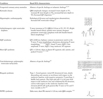
Typical resting electrocardiogram findings of the most frequent causes of sudden cardiac death are shown in Table 2.
Electrocardiographic findings in frequent conditions predisposing to sudden cardiac death.

ECG=electrocardiogram; LQT=long QT; WPW=Wolff–Parkinson–White
Conclusions
The incidence of sudden cardiac death in the young population is rather low ranging between 0.5 and 8 patients per 100.000 persons a year. There are many different aetiologies, but the most frequent include coronary artery abnormalities, hypertrophic cardiomyopathy, and primary arrhythmic diseases, and most deaths occur during physical exercise.
Most conditions predisposing to sudden cardiac death are congenital or have a genetic/familiar basis, and sudden cardiac death is frequently preceded by symptoms, thus making personal history and family history fundamental for its prevention.
In more than 50% of cases, however, sudden cardiac death is the first manifestation of the cardiac disease. Despite the lack of consensus on its use owing to the low benefit/cost ratio, a 12-lead screening electrocardiogram might be particularly useful in identifying asymptomatic subjects carrying higher risk of sudden mortality.
Acknowledgement
None.
Financial Support
This research received no specific grant from any funding agency, commercial, or not-for-profit sectors.
Conflicts of Interest
None.