Introduction
Air-containing cysts in the paratracheal region were first described in 1838 by Rokitansky as retention cysts of the mucous glands.Reference Rokitansky1 These cysts are usually found incidentally on chest imaging and are managed conservatively. Their incidence has been reported as 1 per cent on autopsyReference MacKinnon and Tracheal2 and 0.75–2 per cent on computed tomography (CT) imaging.Reference Mineshita, Tajima and Kondo3, Reference Goo, Im, Ahn, Moon, Chung and Park4 They have also been reported on fibre-optic bronchoscopy in 0.3 per cent of children aged over 10 years.Reference Abramovic and Zoric5
As with many rare clinical entities, numerous terms have been used to describe these lesions over the years. These include paratracheal air cyst, tracheal diverticulum, tracheal diverticulosis, intratracheal diverticulum, bronchogenic cyst, aerocoele, bronchocoele and aerial goitre (air-goitre).
By recent convention, the term tracheocoele has become synonymous with acquired tracheal diverticulum.
We present the case of a 72-year-old man with an acquired tracheal diverticulum.
Case report
A 72-year-old Indian man presented with an eight-week history of right lower neck pain and cough productive of yellow sputum. He also complained of night sweats, low-grade fever, weight loss (6 kg in two months) and loss of appetite. His past medical history included chronic obstructive pulmonary disease (COPD), myocardial infarction, a cerebrovascular event and gastroesophageal reflux disease. He had been an ex-smoker for 40 years but had a previous 120 pack-year history.
On examination, there was an expansile mass arising from the right supraclavicular fossa, which increased in size when the patient performed the Valsava manoeuvre. This sign has been previously described in a case of air-goitre.Reference Gronner and Trevino6
Five years previously, during a hospital admission for gastroenteritis, a routine chest radiograph and subsequent CT scan had demonstrated an air cyst in the right upper mediastinum (3.6 × 2.7 × 2.5 cm). The cyst had been seen to cause some displacement of surrounding structures (trachea, right subclavian vessels and common carotid arteries) but otherwise had not displayed any adverse features. The remaining lung fields had been mildly hyper-inflated, with appearances consistent with COPD, but had no other significant abnormalities. Subsequent investigation with flexible nasendoscopy and formal bronchoscopy had failed to reveal any abnormalities. The air cyst had been considered an incidental finding and thus ignored.
During the current presentation, a repeated chest CT scan (Figure 1) demonstrated that the lesion had increased in size, to 4.1 × 3.4 × 4.8 cm. An air–fluid level was now apparent within the cyst. In the lung fields, non-specific pulmonary nodules were seen in the right upper lobe and left lower lobe, together with scarring of the right middle lobe. Other investigations included negative sputum microscopy and microbiological culture (including mycobacterial culture). Arterial blood gases were normal.
Typical axial computed tomography appearance of a tracheocoele (A) as a thin-walled cyst in the right paratracheal region (trachea is shown as B). The absence of cartilage within the wall differentiates it from a congenital diverticulum. A = anterior; R = right; L = left; P = posterior

The patient's clinical symptoms had initially responded to a short course of antibiotics.
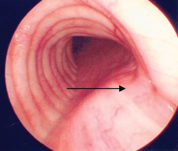
Microlaryngobronchoscopy demonstrated one tract and two further indentations in the posterolateral aspect of the upper trachea at the junction of the trachealis muscle and tracheal cartilages (Figure 2). Pharyngoesophagoscopy was normal.
Endoscopic view showing indentations (black arrow) representing an unusually narrow connection between trachea and tracheocoele.

Two days after endoscopy, the patient re-presented with a further significant episode of fever, this time with odynophagia. A repeated CT excluded any post-operative complication. In light of his persistent and recurrent symptoms, a decision was made to undertake surgical resection.
Surgery was performed under general anaesthesia, via a right collar neck incision. The inferior thyroid vessels were ligated and the right lobe of the thyroid mobilised to expose the paratracheal region. The cyst was identified (Figure 3) and a capsular dissection performed. Two fibrous connections to the posterolateral aspect of the trachea were identified and ligated. The recurrent laryngeal nerve was identified, intimately related to the deep surface of the cyst, and was preserved.
Operative view showing a thin-walled cyst identified during surgical resection.

The post-operative period was complicated by a temporary ipsilateral recurrent laryngeal nerve palsy, which resolved over nine months.
Histologically, the specimen consisted of a simple cyst lined by ciliated columnar epithelium, with a fibrous tissue wall up to 2 mm thick containing small foci of smooth muscle. The lumen was filled with frothy mucus.
Discussion
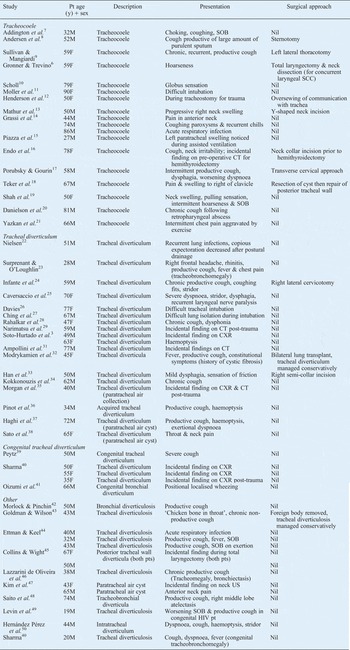
We performed a literature review of all English language reports describing paratracheal air-filled abnormalities in adults (Table I). Changing trends in nomenclature made accurate comparison of cases difficult. We therefore separated cases into four groups according to the original description: tracheocoele, tracheal diverticulum, congenital tracheal diverticulum and others (predominantly tracheal diverticulosis). We identified 18 cases described as tracheocoele, 18 as tracheal diverticulum and five as congenital tracheal diverticulum; a further 14 we grouped together as ‘other’.
Previous english-language reports of paratracheal air-filled abnormalities in adults*

* Older than 18 years. Pt = patient; y = years; M = male; F = female; SOB = shortness of breath; SCC = squamous cell carcinoma; CT = computed tomography; CXR = chest X-ray; US = ultrasound; HIV = human immunodeficiency virus
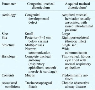
By recent convention,Reference Soto-Hurtado, Penuela-Ruiz, Rivera-Sánchez and Torres-Jimenez30, Reference Sharma40 those lesions are now defined as tracheal diverticula and sub-divided into congenital or acquired lesions (See Table II).
Distinguishing features of congenital and acquired tracheal diverticula

*Also known as tracheocoele
Acquired tracheal diverticula are true diverticula resulting from mucosal herniation through weak points, most commonly in the right posterolateral trachea (98 per cent) and usually at the level of the thoracic inlet. They are thin-walled and consist of normal respiratory epithelium on a narrow layer of fibrous stroma. These lesions were previously commonly referred to as tracheocoeles.
A congenital tracheal diverticulum represents a malformed, vestigial, supernumerary budding of the trachea.Reference Boyden51 The wall of a congenital diverticulum is similar to the wall of the trachea, containing smooth muscle fibres, cartilage and respiratory epithelium. While congenital tracheal diverticula may occur in isolation, their embryological origins mean they are more likely to occur in association with other upper aerodigestive tract anomalies. These include Mournier–Kuhn disease (congenital dilatation of the trachea and bronchi),Reference Lazzarini de Oliveira, de Barros Franco, de Salles and de Oliveira46 tracheoesophageal fistulaReference Dinner, Ward and Yun52 and tracheobronchomegaly.Reference Sharma40
Tracheal diverticula have also been reported in association with: trachiectasis (dilatation of the trachea with multiple small herniations of the membranous portion of the trachea),Reference Andersen, Clagett and Good8 congenital human immunodeficiency virus infection,Reference Levin, Weingart, Adam and Vicencio49 cystic fibrosisReference Modrykamien, Abdallah, Kanne and Mehta32 and Duchenne muscular dystrophy.Reference Piazza, Bolzoni, Cavaliere and Peretti15
Other air-filled abnormalities can occur at the thoracic inlet. The differential diagnosis includes lymphoepithelial cyst, bronchogenic cyst, laryngocoele, pharyngocoele, Zenker's diverticulum, pneumomediastinum, apical hernia of the lung, and apical paraseptal blebs or bullae.Reference Goo, Im, Ahn, Moon, Chung and Park4, Reference Teker, Lorenz, Lee and Murthy18, Reference Soto-Hurtado, Penuela-Ruiz, Rivera-Sánchez and Torres-Jimenez30
Acquired tracheal diverticula are thought to occur as a result of a persistently raised intratracheal pressure associated with chronic respiratory conditions. They occur in pre-existing areas of potential weakness in the posterolateral tracheal wall between the tracheal cartilage and trachealis muscle. They occur almost exclusively on the right side, probably due to the relative positions of the trachea and oesophagus on the left. Previous reports suggest that they should have relatively wide connections to the trachea, compared with congenital diverticula; however, this was not the case in our patient.
It is likely that the majority of cases are asymptomatic, being discovered incidentally on routine radiological investigation, including X-ray,Reference Soto-Hurtado, Penuela-Ruiz, Rivera-Sánchez and Torres-Jimenez30, Reference Sharma40 CTReference Endo, Saito, Hasegawa, Sato and Sohara16, Reference Ampollini, Bobbio, Carbognani and Rusca31 and ultrasound.Reference Kim, Kim, Kim and Park47 Most reported cases are symptomatic, usually due to a local mass effect and direct compression, resulting in cough, dyspnoea, stridor, dysphagia, and chest, neck and/or right clavicular pain. Symptoms can also arise due to vagal irritation (resulting in cough, dysphonia or vocal fold paralysis) or alternatively due to retained secretions (serving as a reservoir of chronic infection) or recurrent irritation of the upper airways. Tracheal diverticula have also been found incidentally during tracheostomy,Reference Henderson, Harrington, Izenberg, Dyess and Silver12 laryngectomy,Reference Collins and Wight45 difficult intubation,Reference Möller, Ten Berge and Stassen11, Reference Davies26, Reference Ching, Chow and Ng27 and following trauma.Reference Sharma40, Reference Narimatsu, Yama, Danjoh, Kurimoto and Asai29, Reference Morgan, Perone and Yeghiayan35
CT scanning has been shown to be the most effective method for evaluating the presence and features of tracheal diverticula.Reference Polverosi, Carloni and Poletti53 In most cases, a CT scan and barium swallow will exclude the differential diagnoses listed above.
Previous authors have suggested that surgery is only indicated in young, symptomatic patients in whom conservative measures (e.g. antibiotics, mucolytics and physiotherapy) have been unsuccessful. The current case demonstrates that asymptomatic lesions can become problematic over time. It also demonstrates that age and other pre-existing co-morbidities should not be considered as absolute contraindications to surgery.
• Tracheal diverticulum is a rare anomaly of the tracheobronchial tree
• The nomenclature describing paratracheal air cysts is confusing
• Such lesions are now divided into congenital and acquired tracheal diverticula (the latter also known as tracheocoeles); these subtypes have different aetiologies and characteristics
• This case report illustrates the necessity of surgical resection in cases of symptomatic tracheal diverticulum
Where surgery is considered, the most commonly described approach is resection via a transverse or lateral neck incision.Reference Porubsky and Gourin17, Reference Infante, Mattavelli, Valente, Alloisio, Preda and Ravasi24 Other reported options have included fulguration,Reference Early and Bothwell54 endoscopic cauterisation with laser or electrocoagulation,Reference Soto-Hurtado, Penuela-Ruiz, Rivera-Sánchez and Torres-Jimenez30, Reference Modrykamien, Abdallah, Kanne and Mehta32 and endoscopic division with biopsy forceps.Reference Johnson, Cotton and Rutter55
Conclusion
Most of the cases identified in our literature review, other than those reported as congenital tracheal diverticula, probably represent similar clinical entities. However, an inconsistent approach to the naming of these lesions makes accurate comparison of previously reported cases difficult. With regards to the clinical management, it is important to identify congenital lesions so that associated abnormalities can be investigated and managed appropriately. For the remaining acquired lesions, any further sub-classification has little bearing on clinical decision-making. We would therefore advocate the simple classification of cases into congenital or acquired lesions. For acquired lesions, surgery should be considered in symptomatic cases in which simple medical treatment options have been unsuccessful.