Introduction
Porcine circovirus (PCV), a small, non-enveloped, circular, single-stranded DNA virus, was first discovered as a picornavirus-like contaminant of porcine kidney-15 cells (PK-15 cells) in 1974 (Tischer et al., Reference Tischer, Rasch and Tochtermann1974). Although further studies on the virus indicated that it was non-pathogenic (Tischer et al., Reference Tischer, Mields, Wolff, Vagt and Griem1986), the discovery of the virus later designated PCV type 1 (PCV1) initiated worldwide investigations into the prevalence of PCV in the swine population and led to the discovery of PCV type 2 (PCV2). PCV2 shares 68–76% nucleotide sequence identity with PCV1 (Hamel et al., Reference Hamel, Lin and Nayar1998; Morozov et al., Reference Morozov, Sirinarumitr, Sorden, Halbur, Morgan, Yoon and Paul1998). Unlike PCV1, PCV2 was initially associated with clinical disease in the 1990s (Allan et al., Reference Allan, McNeilly, Kennedy, Daft, Clarke, Ellis, Haines, Meehan and Adair1998; Harding et al., Reference Harding, Clark, Strokappe, Willson and Ellis1998) and today it is of economic importance in the swine industry worldwide. Based on sequence analyses of PCV2 isolates, they can be divided into five subtypes (PCV2a–e). PCV2a and PCV2b have been documented worldwide (Segalés et al., Reference Segalés, Olvera, Grau-Roma, Charreyre, Nauwynck, Larsen, Dupont, McCullough, Ellis, Krakowka, Mankertz, Fredholm, Fossum, Timmusk, Stockhofe-Zurwieden, Beattie, Armstrong, Grassland, Bækbo and Allan2008), PCV2c has been documented in archived samples from Denmark (Dupont et al., Reference Dupont, Nielsen, Bækbo and Larsen2008), and PCV2d and PCV2e have only been identified in China (Wang et al., Reference Wang, Guo, Ge, Wang, Chen, Cha and Yang2009). In 2010, a new genotype, PCV1/2a, whose name was based on nucleotide identities in the open reading frame 1 (ORF1) of PCV1 and the ORF2 from PCV2a, was identified in tissue homogenates from swine in Canada (Gagnon et al., Reference Gagnon, Music, Fontain, Tremblay and Harel2010).
Clinical signs of PCV2 infection manifest in a variety of ways including multisystemic disease with wasting, reproductive failure, enteritis, respiratory disease, porcine dermatitis and nephropathy syndrome (PDNS), or a combination of these (Opriessnig et al., Reference Opriessnig, Meng and Halbur2007) summarized as porcine circovirus-associated disease (PCVAD). In cases of PCVAD, a morbidity rate of 4–30% with a mortality rate of 70–80% in affected animals has been reported (Segalés et al., Reference Segalés, Allan and Domingo2005a). It is clear that PCV2 is ubiquitous and was present many years prior to the onset of clinical PCVAD. To date, the question of how a highly prevalent virus such as PCV2 was able to cause epidemics of disease in populations of swine worldwide remains unanswered. The presence of PCV2 prior to the recognition of clinical PCVAD outbreaks in multiple countries indicates that the presence of co-factors contributes to the development of clinical disease. Multiple factors including co-infections, immune stimulation, immune suppression, management practices and many others have recently been reviewed (Opriessnig et al., Reference Opriessnig, Meng and Halbur2007; Madec et al., Reference Madec, Rose, Grasland, Cariolet and Jestin2008; Gillespie et al., Reference Gillespie, Opriessnig, Meng, Pelzer and Buechner-Maxwell2009; Ramamoorthy and Meng, Reference Ramamoorthy and Meng2009). To the authors' knowledge, an in-depth review of the epidemiology and horizontal transmission of PCV2 has not been conducted to date.
Epidemiology
PCV2 is prevalent worldwide
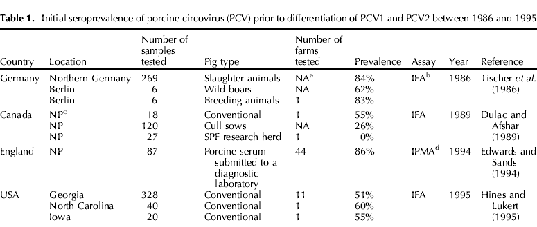
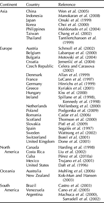
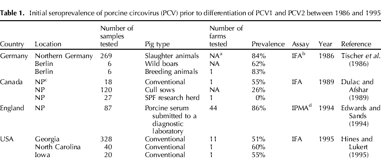
Following the initial detection of PCV in 1974 (Tischer et al., Reference Tischer, Rasch and Tochtermann1974), the seroprevalence of this virus was determined in Germany (Tischer et al., Reference Tischer, Mields, Wolff, Vagt and Griem1986), Canada (Dulac and Afshar, Reference Done, Gresham, Potter and Chennells1989), England (Edwards and Sands, Reference Edwards and Sands1994) and the USA (Hines and Lukert, Reference Hines and Lukert1995). As these studies were published in the early 1990s, the exact PCV-type represented in the reports is unknown. Specifically, persistently infected porcine kidney-15 cells (PK-15 cells) were used for an immunofluorescent assay (IFA) making the differentiation of PCV1 and PCV2 difficult. These initial studies determined that PCV was widespread in swine populations in the investigated countries (Table 1). By the late 1990s, however, diagnostic tests that differentiated non-pathogenic PCV1 from pathogenic PCV2 became available, allowing further characterization of PCV isolates. PCV2 has since been identified in countries worldwide (Table 2).
Initial seroprevalence of porcine circovirus (PCV) prior to differentiation of PCV1 and PCV2 between 1986 and 1995
a NA, not applicable.
b IFA, immunofluorescence assay.
c NP, not provided.
d IPMA, immunoperoxidase monolayer assay.
Porcine circovirus type 2 (PCV2) detection separated by countries/continents by serological, molecular or antigen-based assays

PCV2 was identified in archived tissue samples prior to the onset of clinical disease
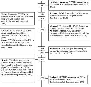
Retrospective studies have indicated that PCV2 had been present years prior to the onset of clinical cases of PCVAD (Fig. 1). However, based on the limited sequencing information from these studies, little to no changes were detected in the viral genome between isolates recovered prior to and following clinical outbreaks of PCVAD (Grierson et al., Reference Grierson, King, Sandvik, Hicks, Spencer, Drew and Banks2004; Jacobsen et al., Reference Jacobsen, Krueger, Seeliger, Bruegmann, Segalés and Baumgaertner2009). Interestingly, there has been a shift in the predominant PCV2 subtype from PCV2a to PCV2b which might have contributed to the increased severity and frequency of PCVAD.
Timeline of the first identification of PCV2 in conventional pigs in various countries as part of retrospective analyses by immunoperoxidase monolayer assay (IPMA), immunofluorescence assay (IFA), immunohistochemistry (IHC), polymerase chain reaction (PCR) or in-situ hybridization (ISH).

A global shift in the predominant PCV2 subtype
Several studies have evaluated the temporal differences in the identification of PCV2a and PCV2b and concluded that a global shift in the predominant subtype has occurred. In a study published in 2007, 218 PCV2 genome sequences were analyzed and the authors concluded that a major shift from PCV2a to PCV2b occurred on a global scale in or prior to 2003 (Dupont et al., Reference Dupont, Nielsen, Bækbo and Larsen2008). Interestingly, based on the analysis in the study which included sequences from 1997 to 2006, the shift in subtypes did not affect Korea, Japan or Australia (Dupont et al., Reference Dupont, Nielsen, Bækbo and Larsen2008). Since then, PCV2b has been demonstrated as the predominant subtype in Korea based on the analysis of isolates from 2005 to 2007 (Kim et al., Reference Kim, Park, Hyun, Park, Jeong, Shin, Chun, Hosmillo, Lee, Kang and Cho2009b) and in Japan where both PCV2a and PCV2b were detected in isolates collected between 2006 and 2007 (Takahagi et al., Reference Takahagi, Nishiyama, Toki, Yonekita, Morimatsu and Murakami2008). Additionally in a study by Olvera et al. (Reference Olvera, Cortey and Segalés2007) 148 PCV2 isolates were analyzed. Of these, 63.5% were PCV2b sequences of which 92.5% were published after 2003. Numerically fewer (62.2%; 33/53) PCV2a sequences) were published after 2003 (Olvera et al., Reference Olvera, Cortey and Segalés2007). However, the authors cautioned that the time of sequence publication in relation to the time the virus was first detected can be variable (Olvera et al., Reference Olvera, Cortey and Segalés2007). In the following section, summaries of selected reports which document the predominance of the PCV2b subtype are provided for each continent.
Asia
In China, 49 isolates from various geographic regions were sequenced (Wang et al., Reference Wang, Guo, Ge, Wang, Chen, Cha and Yang2009). Results revealed that only PCV2a isolates were recovered in 2001 (n=3). In isolates recovered between 2002 and 2006 (n=34), a mixture of PCV2a, PCV2b, PCV2d and PCV2e was detected, with the majority of samples (n=18) clustering with PCV2b. Of the isolates recovered in 2007 and 2008 (n=12), all clustered with PCV2b (Wang et al., Reference Wang, Guo, Ge, Wang, Chen, Cha and Yang2009). Similarly, in a 2010 publication evaluating 136 isolates from Eastern China collected between 2001 and 2009, 127 of the isolates clustered with PCV2b (Li et al., Reference Li, Wang, Ma, Feng, Li and Jiang2010), while only nine clustered with PCV2a. Of these nine, eight were collected between 2001 and 2004.
In Korea, PCV2 isolates detected by PCR in tissue samples collected from PCVAD-affected herds (n=29) and non-PCVAD-affected herds (n=9) in 2007 were sequenced and categorized as PCV2a and PCV2b. All 29 sequences from PCVAD-affected herds and four of the sequences from non-PCVAD-affected herds clustered into the PCV2b group (Chae and Choi, Reference Chae and Choi2010). Similarly, in a study by An et al. (Reference An, Roh, Song, Park and Park2007) 36 Korean PCV2 sequences from pigs with PDNS or PCVAD collected from 1999 to 2006 were compared to other PCV2 sequences available in GenBank®. The phylogenetic analysis indicated that PCV2b was detected in 66.7% (24/36) of the samples. Although the specific year when these 24 isolates were collected is not provided, the authors suggest that more severe PCVAD cases in Korea in 2004 were related to the increased incidence of PCV2b (An et al., Reference An, Roh, Song, Park and Park2007).
In Japan, 150 serum samples were collected from 2- to 4-month-old pigs on 10 farms (15 samples/farm). PCV2 DNA positive samples were analyzed by the restriction fragment length polymorphism (RFLP) analysis. In two farms, RFLP-type 311 (which corresponds to PCV2a) was identified in 2006. Six months later in the two farms, RFLP-type 111 (which corresponds to PCV2b) was identified, indicating a shift in the PCV2 subtype over a short temporal span (Takahagi et al., Reference Takahagi, Nishiyama, Toki, Yonekita, Morimatsu and Murakami2008).
Europe
In Ireland, PCV2a was recovered from five of six farms classified as PCVAD-affected based on samples collected from 1997 to 2003 (Allan et al., Reference Allan, McNeilly, McMenamy, McNair, Krakowka, Timmusk, Walls, Donnelly, Minahin, Ellis, Wallgren and Fossum2007). However, PCV2b was isolated from the remaining farm which had increased mortality (peak mortality of 35%) in comparison to the other five farms. In all samples after 2003 from both PCVAD-affected and non-PCVAD-affected farms, only PCV2b was isolated (Allan et al., Reference Allan, McNeilly, McMenamy, McNair, Krakowka, Timmusk, Walls, Donnelly, Minahin, Ellis, Wallgren and Fossum2007). As severe PCVAD outbreaks occurred between 2003 and 2006 in Ireland, this is suggestive of a temporal link between the identification of PCV2b and severe outbreaks (Allan et al., Reference Allan, McNeilly, McMenamy, McNair, Krakowka, Timmusk, Walls, Donnelly, Minahin, Ellis, Wallgren and Fossum2007).
In Brittany, de Boisséson et al. (Reference de Boisséson, Béven, Bigarré, Thiéry, Rose, Eveno, Madec and Jestin2004) sequenced and compared isolates from PCVAD (n=25) and non-PCVAD (n=31) affected farms collected from 2000 to 2002. Results indicated that two main clusters were present, with the majority of the PCV2 sequences clustering into the PCV2b group (de Boisséson et al., Reference de Boisséson, Béven, Bigarré, Thiéry, Rose, Eveno, Madec and Jestin2004). While the authors concluded that a new subtype of PCV2 was not responsible for the increased outbreaks of PCVAD in Brittany as PCV2b was detected in both PCVAD- and non-PCVAD-affected herds, this is consistent with PCV2b being the predominant strain during this time.
In Denmark, Dupont et al. (Reference Dupont, Nielsen, Bækbo and Larsen2008) sequenced and compared a total of 45 isolates from PCVAD-affected and non-PCVAD-affected herds collected between 2003 and 2004. Similar to the study by de Boisséson et al., a distinctive clustering of isolates by disease status was not noted; however, all isolates clustered within the PCV2b group.
North America
In North America, the shift to increased prevalence of PCV2b was documented by several recent studies. The first identified the presence of PCV2b in Canada in samples from 13 PCVAD cases collected in 2005 in the province of Quebec (Gagnon et al., Reference Gagnon, Tremblay, Tijssen, Venne, Houde and Elahi2007). Investigation of 83 sequences collected from both PCVAD-affected and non-PCVAD-affected animals from four provinces in Canada between 2005 and 2006 revealed that 79.5% (66/83) of sequences clustered with PCV2b (Gagnon et al., Reference Gagnon, Tremblay, Tijssen, Venne, Houde and Elahi2007). In a separate study, 121 cases from animals with and without clinical signs of PCVAD submitted to the Institut National de Santé Animale between October 2006 and January 2007 were analyzed by a PCV2a/PCV2b differential PCR (Gagnon et al., Reference Gagnon, del Castillo, Music, Fontaine, Harel and Tremblay2008). Of the 121 cases, PCV2b was detected in 92.6%, PCV2a in 4.1% and both subtypes were detected in 3.3% of cases (Gagnon et al., Reference Gagnon, del Castillo, Music, Fontaine, Harel and Tremblay2008). Another publication identified 12 PCV2b isolates in four finishing facilities in Kansas that experienced severe PCVAD (Horlen et al., Reference Horlen, Schneider, Anderson, Nietfeld, Henry, Tokach and Rowland2007). PCV2b was also identified in tissue homogenates from pigs manifesting severe PCVAD in Kansas, North Carolina and Iowa in 2005 (Cheung et al., Reference Cheung, Lager, Kohutyuk, Vincent, Henry, Baker, Rowland and Dunham2007) and in Cuba from porcine tissues collected between 2007 and 2008 (Pérez et al., Reference Pérez, Díaz de Arce and Frías2010b). Prior to these reports, PCV2a had been the only strain detected in North America (Fenaux et al., Reference Fenaux, Halbur, Gill, Toth and Meng2000; Larochelle et al., Reference Larochelle, Magar and D'Allaire2002; Olvera et al., Reference Olvera, Cortey and Segalés2007).
Oceania
To the authors' knowledge, there are only two studies evaluating isolates from this region. In the first, Muhling et al. (Reference Muhling, Raye, Buddle and Wilcox2006) evaluated seven PCV2 isolates collected between 1999 and 2002. Six of the isolates clustered with previous isolates from Japan and Canada and had an overall 94–99% nucleotide identity with previously reported strains from other countries. Specific designations of the isolates into PCV2a and PCV2b were not made. The second study evaluating isolates from New Zealand found that isolates from the 2006 PCVAD outbreak share 94–100% nucleotide identity with other reported PCV2 isolates (Neumann et al., Reference Neumann, Dobbinson, Welch and Morris2007).
South America
In tissues from pigs with respiratory signs collected between 2000 and 2004, eight PCV2 sequences were identified (Castro et al., Reference Castro, Cortez, Heinemann, Brandão and Richtzenhain2008). In a later study by Chiarelli-Neto et al. (Reference Chiarelli-Neto, Yotoko, Vidigal, Silva, Castro, Fietto, Silva and Almeida2009) 11 Brazilian isolates from PCVAD-affected animals collected in 2005 were sequenced. The majority of Brazilian isolates clustered with the PCV2b group and the authors concluded that Brazilian strains emerged from a common ancestor identical or descendent from strains that were common in Europe and Asia. While this provides information that PCV2b is currently likely the predominant subtype, it does not determine when or whether a shift occurred. The information for other South American countries is limited (Cano et al., Reference Cano, Rodríguez-Cariño, Sogbe, Utrera, Díaz, Segalés, Olvera and Calsamiglia2005).
The available data suggest that the PCV2b subtype currently predominates worldwide. The shift appears to have originated in Europe prior to or around 2003 followed by shifts in North America and Asia in or around 2005 and potentially in Oceania in or around 2006. The underlying reason for the shift in subtypes outside Europe has not been confirmed, but may be related to importation of subclinically infected animals; especially in South and Latin America (Chiarelli-Neto et al., Reference Chiarelli-Neto, Yotoko, Vidigal, Silva, Castro, Fietto, Silva and Almeida2009; Pérez et al., Reference Pérez, Díaz de Arce and Frías2010b). Additional sequences, especially from isolates collected prior to 2005, would aid in getting a more precise estimate of the time when the shift in subtypes occurred. In addition, correlation of these sequences with epidemiological data on the presence of PCVAD and pig movement between countries would be beneficial in determining how the shift occurred. Interestingly, the trend toward the discovery of PCV2b has often correlated with PCVAD outbreaks of increased severity within the geographical region. However, PCV2b isolates were often detected in both PCVAD-affected and non-affected herds. This suggests that an unknown viral factor, host factor, environmental conditions or a co-infecting pathogen is complicating PCV2b infection in animals with severe PCVAD. Finally, as new subtypes of PCV2 emerge, including potential recombinations of subtypes (Hesse et al., Reference Hesse, Kerrigan and Rowland2008) and genotypes such as PCV1/2a (Gagnon et al., Reference Gagnon, Music, Fontain, Tremblay and Harel2010), the prevalence of these subtypes and association with clinical disease should be monitored.
Horizontal transmission
Effective transmission depends on factors falling under three main categories: (1) characteristics of the host, (2) characteristics of the pathogens and (3) the type of exposure (Thrusfield, Reference Thrusfield2005). Specifically, the host must be susceptible; the pathogen must be virulent, infective and stable in the environment; and the interaction between the pathogen and host must be favorable for infection (correct infectious dose, adequate contact time, etc.) (Thrusfield, Reference Thrusfield2005). In general, horizontal transmission can occur by one of two routes, direct contact with an infected host or the infected host's secretions or through indirect contact of a virus with a living vector, contaminated inanimate object or aerosol droplets (Thrusfield, Reference Thrusfield2005). The following focuses on routes of PCV2 shedding, environmental stability and evaluation of the transmission of PCV2 through direct and indirect contact.
PCV2 is shed in large quantities through multiple routes
Following the experimental infection of naïve animals with PCV2, PCV2 DNA was detected in multiple sample types. Caprioli et al. (Reference Caprioli, McNeilly, McNair, Lagan-Tregaskis, Ellis, Krakowka, McKillen, Ostanello and Allan2006) oronasally inoculated 4-week-old colostrum-deprived (CD) specific pathogen-free (SPF) pigs with a PCV2 isolate recovered from a clinically affected Canadian pig. Blood, fecal swabs and tonsillar swabs were collected at 1, 3, 6, 9, 12, 15 and 21 days post inoculation (dpi). Results indicated that 10/12 and 11/12 pigs had PCV2 DNA-positive tonsillar and fecal swabs, respectively, at 1 dpi. Interestingly, at dpi 6, only 1/12 and 0/12 animals were PCV2 DNA positive on tonsillar and fecal swabs, respectively. However, by the last day of the trial (dpi 21), 11/12 and 11/11 animals were PCV2 positive by tonsillar and fecal swabs, respectively. PCV2 DNA was not detected in serum, plasma or whole blood until 9 dpi. Based on these findings, the authors concluded that polymerase chain reaction (PCR) had detected inoculum in the nasal and fecal swabs prior to dpi 6. In a separate study, Bolin et al. (Reference Bolin, Stoffregen, Nayar and Hamel2001) inoculated mixed breed, cesarean-derived, colostrum-deprived (CDCD) pigs intranasally and subcutaneously at 20 or 25 days of age with a PCV2 isolate obtained from a clinically affected Canadian pig for determining the tissue distribution of PCV2 DNA and the duration of viral persistence. Virus was recovered by isolation from nasal, rectal or tonsil swabs between 12 and 19 dpi. Analysis of shedding in CDCD pigs was also evaluated by Shibata et al. in 2003. Pigs were intranasally inoculated with PCV2 at 2 weeks of age and oropharyngeal and nasal swabs and feces were collected among other samples at the time of necropsy (necropsy occurred on dpi 7, 14, 21, 35, 49 or 70). There was early and consistent shedding in the majority of animals for an extended period of time. All oropharyngeal and nasal swabs and fecal samples contained PCV2 DNA on dpi 1 (excluding 1 nasal swab). Between dpi 1 and dpi 70, all fecal samples were PCV2 PCR positive and 83 out of 86 nasal and oropharyngeal swabs were PCV2 DNA positive (Shibata et al., Reference Shibata, Okuda, Yazawa, Ono, Sasaki, Itagaki, Nakajima, Okabe and Hidejima2003). In a similar study in which CDCD pigs were inoculated with PCV2, PCV2 DNA was detected in nasal and rectal swabs in a high percentage of sampled animals (67–100% excluding 1 nasal sampling at dpi 35 in which only 1 of 3 animals had detectable PCV2 DNA) between 7 and 49 dpi (Harms et al., Reference Harms, Sorden, Halbur, Bolin, Lager, Morozov and Paul2001). Several other studies detected PCV2 DNA or viable virus (by virus isolation) from nasal swabs, feces, ocular swabs or a combination of these samples at various time points in low numbers of animals following experimental infection (Krakowka et al., Reference Krakowka, Ellis, Meehan, Kennedy, McNeilly and Allan2000; Magar et al., Reference Magar, Larochelle, Thibault and Lamontagne2000a; Resendes et al., Reference Resendes, Balasch and Calsamiglia2002).
Data on the amount of PCV2 DNA present in various secretions, excretions and serum are also available. Chung et al. (Reference Chung, Chan, Chaung, Lien, Wu and Huang2005) intranasally challenged 14 cross-bred commercial pigs that were serologically naïve to porcine reproductive and respiratory syndrome virus (PRRSV) and PCV2 at 10 weeks of age with a tissue homogenate containing PRRSV and PCV2 (n=12); two animals were left as negative controls. As part of the study, six of the animals challenged with PCV2 and PRRSV were injected intramuscularly with inactivated Salmonella choleraesuis and complete Freund's adjuvant prior to infection. In experimentally infected pigs PCV2 concentrations at 14 dpi were similar to those in naturally infected animals. Samples were collected from nine dyspneic pigs naturally infected with PRRSV and PCV2 (based on PCR detection in serum), and nasal fluid and saliva of these pigs contained between 3.7±0.2 to 4.4±0.2 log10 PCV2 genomic copies/ml (Chung et al., Reference Chung, Chan, Chaung, Lien, Wu and Huang2005). In a study in which five gnotobiotic pigs were experimentally infected with PCV2, between 1×10⁶ to 1×10¹² copies/ml in serum and liver were detected (McIntosh et al., Reference McIntosh, Tumber, Harding, Krakowka, Ellis and Hill2009).
While the above studies involved relatively low numbers of pigs with varying immune status (CDCD, CD or commercial SPF pigs) and pigs were inoculated with different PCV2 isolates, PCV2 DNA was consistently detected in the secretions and excretions evaluated. In addition, PCV2 DNA was detected early in the infection process (dpi 1) and generally persisted in a high number of animals through the end of the evaluation period indicating persistent infection. Based on the available quantitative data, PCV2 DNA is present in high amounts in various secretions, serum and hepatic tissues; however, data on the amount of PCV2 DNA present in various sample types throughout the infection process are not available to the authors' knowledge.
Additional studies have described PCV2 shedding following natural infection of pigs and PCV2 was detected and quantified in multiple sample types. The PCV2 shedding patterns in three naturally PCV2-infected farms were investigated between 1999 and 2000 (Shibata et al., Reference Shibata, Okuda, Yazawa, Ono, Sasaki, Itagaki, Nakajima, Okabe and Hidejima2003). Nasal swabs and fecal samples were collected from 313 pigs in five age groups. Results suggested that infection occurred post-weaning as an increased frequency of PCV2 detection occurred in both sample types after the nursery phase (n=64), peaked in 3- to 4-month-old animals (n=81) and declined in the sampled adult animals (n=47). Overall, PCV2 was detected in 20.4% (64/313) of fecal samples and 19.2% (60/313) of nasal samples (Shibata et al., Reference Shibata, Okuda, Yazawa, Ono, Sasaki, Itagaki, Nakajima, Okabe and Hidejima2003).
In 2009, a longitudinal case-control study of two PCVAD farms located in Spain and Denmark was designed to describe the dynamics of a natural PCV2 infection (Grau-Roma et al., Reference Grau-Roma, Hjulsager, Sibila, Kristensen, López-Soria, Enøe, Casal, Bøtner, Nofrarías, Bille-Hansen, Fraile, Bækbo, Segalés and Larsen2009). In contrast to the previously described cross-sectional study, this study provided data on a specific cohort of animals over time. Serum, nasal swabs and fecal swabs were collected from selected animals at four time points from 1 to 9 (Denmark) or 11 (Spain) weeks of age and at the time of necropsy (between 9 and 21 weeks of age). Of the 108 Spanish animals evaluated, 78 animals were defined as ‘case’ and 30 as ‘controls’ based on clinical evaluation. A similar clinical evaluation resulted in 91 cases and 27 control animals from farms in Denmark. On Spanish farms, PCV2 was detected by PCR sporadically in all samples between week 1 (first sampling point) and week 11. At the time of necropsy, PCV2 was detected in 87.0% of nasal and 50.5% of fecal swabs. There was a significantly higher percentage of positives in animals classified as ‘case’ animals. The amount of PCV2 DNA in nasal and fecal samples ranged from approximately 4–6 log10 genomic copies of PCV2 per swab throughout the study period with significantly higher amounts in ‘case’ animals. In Danish farms at 1 week of age, 69.3% of nasal swabs and 22.8% of rectal swabs were PCV2 DNA positive. By the time of necropsy, the percentage of PCV2 positive PCR samples had risen to 100% in nasal samples and to 98.5% in rectal swabs. Throughout the study period, the amount of PCV2 DNA ranged from approximately 4 to 8 log10 genomic copies PCV2 per swab in fecal samples and from 4 to 10 log10 genomic copies PCV2 per swab in nasal samples. By the time of necropsy, ‘case’ animals had significantly larger amounts of virus in both nasal and fecal swabs in comparison with healthy controls (Grau-Roma et al., Reference Grau-Roma, Hjulsager, Sibila, Kristensen, López-Soria, Enøe, Casal, Bøtner, Nofrarías, Bille-Hansen, Fraile, Bækbo, Segalés and Larsen2009).
Quantitative data on the amount of PCV2 DNA in various samples collected from 146 pigs submitted to the Pathological Diagnostic Service at the Veterinary School of Barcelona between 2000 and 2001 were reported in 2005 (Segalés et al., Reference Segalés, Calsamiglia, Olvera, Sibila, Badiella and Domingo2005b). Serum samples (n=57), nasal swabs (n=99), tonsillar swabs (n=108), tracheobronchial swabs (n=72), urinary swabs (n=91) and fecal swabs (n=42) were tested quantitatively for PCV2 DNA. The results were divided into three categories: PCVAD-affected pigs, PCV2 subclinically infected pigs and non-PCVAD pigs, based on the presence of histopathological lesions and the amount of PCV2 antigen detected by in-situ-hybridization and divided into two categories based on age (≥1.5 and >1.5 months of age). PCV2 was found in 100% of all swabs and serum samples from animals classified as PCVAD-affected pigs, with significantly more PCV2 DNA present in comparison with PCV2 subclinically infected pigs and non-PCVAD pigs. Additionally, significantly more PCV2 DNA was present in older animals (≥1.5 months of age) except for tonsillar swabs. Quantitative results for PCVAD pigs ranged from approximately 5 to 8 log10 PCV2 genomic copies/ng for all sample types with the highest PCV2 load detected in trachea-bronchial swabs followed by serum, tonsillar, nasal, fecal and urinary swabs. In all sample types, the amount of PCV2 was higher in PCVAD-affected pigs compared to non-PCVAD-affected pigs. The authors concluded that the more severe the PCV2 lesions, the more PCV2 is present within serum and various swabs (Segalés et al., Reference Segalés, Calsamiglia, Olvera, Sibila, Badiella and Domingo2005b). McIntosh et al. (Reference McIntosh, Harding, Parker, Krakowka, Allan and Ellis2008) compared the amount of PCV2 DNA present in pooled fecal samples from various age groups from both a PCVAD-affected and a non-PCVAD-affected farm. Results of this study indicated that the highest amount of PCV2 DNA detected in pooled fecal samples occurred in nursery-aged pigs on a PCVAD-affected herd, with approximately 1×10⁷ copies of PCV2 per gram of pooled fecal material detected. This was significantly different from the mean amount of PCV2 detected in nursery-aged pigs from PCVAD non-affected herds (1×10² copies of PCV2 per gram of pooled fecal material). Of note, shedding of PCV2 in feces was reported in all age groups from recently weaned pigs to boars and lactating sows (McIntosh et al., Reference McIntosh, Harding, Parker, Krakowka, Allan and Ellis2008). The amount of PCV2 DNA present in serum and feces from three naturally infected conventional pigs exhibiting clinical signs of wasting has also been reported, with amounts ranging from 3.30×10⁸ to 2.91×10¹⁰ copies/ml in serum and between 1.57×10⁹ and 7.04×10¹⁰ copies/g in feces (McIntosh et al., Reference McIntosh, Tumber, Harding, Krakowka, Ellis and Hill2009).
The above studies suggest that PCV2 is present in high concentration in nasal and fecal excretions and serum for extended periods of time following natural PCV2 infection of a population. Also, they support the hypothesis that increased shedding is present in animals with clinical signs associated with PCV2 in comparison to healthy controls. Finally, they demonstrate the relationship between maternal antibodies and piglet viremia. Specifically, in populations where maternal antibodies are present, viremia is delayed and increases with age (Grau-Roma et al., Reference Grau-Roma, Hjulsager, Sibila, Kristensen, López-Soria, Enøe, Casal, Bøtner, Nofrarías, Bille-Hansen, Fraile, Bækbo, Segalés and Larsen2009). This is in contrast to a study by McIntosh et al. (Reference McIntosh, Harding, Parker, Krakowka, Allan and Ellis2008) in which the highest shedding was noted in nursery aged pigs. Although the antibody status of dams in the publication by McIntosh et al. (Reference McIntosh, Harding, Parker, Krakowka, Allan and Ellis2008) is unknown, it is likely that there were lower levels of maternal antibodies present in the population based on previous work (McKeown et al., Reference McKeown, Opriessnig, Thomas, Guenette, Elvinger, Fenaux, Halbur and Meng2005). While these studies which evaluated naturally PCV2-infected pigs provide valuable information on PCV2 shedding, the impact of management, co-infections and other co-factors on shedding limits the external usefulness of the results.
In summary, the above information suggests that PCV2 DNA can be detected in fecal, nasal, oral and tonsillar swabs as well as in urine and feces from both naturally and experimentally infected pigs. In addition, PCV2 DNA can be detected early in the infectious process and persists for extended periods of time. However, in the majority of the studies, the infectivity of the PCV2 DNA was not investigated.
PCV2 is stable in the environment and resistant to disinfectants
In studies, which have evaluated the stability of PCV2, results indicate that the virus is very resistant under high temperatures and a wide range of pH conditions. Welch et al. (Reference Welch, Bienek, Gomperts and Simmonds2006) subjected PCV2 virus stock to two treatments. One treatment was pasteurization at 60°C for 0, 2, 5, 10 and 24 h and the other treatment was dry heat applied to freeze-dried virus (selected vials were partially rehydrated) at 80°C for 8, 24, 48 or 72 h. Following the treatments, an infectivity reduction factor was calculated. For pasteurization, a 1.33 log reduction (corresponding to 5% of the starting material) was calculated at 24 h. Following dry-heat treatment, a 0.75 log reduction (corresponding to 18% of the starting material) was calculated at 72 h. Due to the lack of efficacy of the temperatures utilized, additional temperatures were evaluated. Results indicated pasteurization at a temperature of 65, 70 and 75°C for 30 min lead to a 0.25, 1.59 and 1.92 log reduction, respectively. Interestingly, only a 1 log reduction in infectivity was calculated following dry-heat treatment at 120°C for 30 min (Welch et al., Reference Welch, Bienek, Gomperts and Simmonds2006). Resistance of PCV2 to heat was also shown in a study in which PCV2 virus stock in cell culture media was incubated in a water bath at temperatures of 56, 65, 70, 75, 80 and 85°C (O'Dea et al., Reference O'Dea, Hughes, Davies, Muhling, Buddle and Wilcox2008). The presence of PCV2 antigen following treatments was determined by immunohistochemistry (IHC) and the presence of replicating PCV2 was determined by reverse transcription (RT)–PCR for replicative intermediates. Results indicated that PCV2 antigen was detected at temperatures up to 70°C and PCV2 RNA was detected up to 75°C but not at 80°C. The authors suggested that PCV2 was more labile when heated in liquid culture than when freeze-dried explaining the differences between this study and previous work by Welch et al. (Reference Welch, Bienek, Gomperts and Simmonds2006) and O'Dea et al. (Reference O'Dea, Hughes, Davies, Muhling, Buddle and Wilcox2008) .
In 2009, the in vitro stability of PCV2 was again investigated by incubating PCV2 at various temperatures and ranges of pH. Results confirmed extreme environmental resistance as evidenced by an unchanged virus titer following incubation at 56°C for 1 h, a reduction from 105.5 to 102.5 50% tissue culture infective dose (TCID50) per ml following incubation at 70°C for 1 h, and a reduction from 105.5 to 102.7 TCID50/ml following incubation at a pH of 2 for 30 min (Kim et al., Reference Kim, Lyoo and Joo2009a).
In addition to showing environmental stability, PCV2 has been shown to withstand several types of disinfectants. Kim et al. (Reference Kim, Lyoo and Joo2009a, Reference Kim, Park, Hyun, Park, Jeong, Shin, Chun, Hosmillo, Lee, Kang and Chob) evaluated various disinfectants for efficacy against PCV2 in an in vitro model. Incubation of a potassium peroxomonosulphate, a sodium hypochlorite product, or 3% sodium hydroxide with 1×104.5 to 1×105.5 TCID50/ml for 10 min resulted in complete reduction of PCV2 (Kim et al., Reference Kim, Lyoo and Joo2009a). When the incubation time was increased to 30 min with a quaternary ammonium compound or 12 h with a quaternary ammonium/gludaraldehyde mixturethere was also a complete reduction of PCV2 infectivity (Kim et al., Reference Kim, Lyoo and Joo2009a). When PCV2 was incubated with 0.05 or 0.1% formalin, PCV2 was detected; however, a specific mean TCID50/ml was not provided (Kim et al., Reference Kim, Lyoo and Joo2009a).
In an earlier study, an in vitro suspension assay was performed to determine the virucidal efficacy of nine disinfectants against a French PCV2 isolate (Martin et al., Reference Martin, Le Potier and Maris2008). Equal volumes of double-strength disinfectant were mixed with PCV2 virus stock for 30 min at 20°C. There was a reduction of the mean PCV2 titer of greater than 1.61 log10 at the authorized concentration for the two disinfectants which contained potassium monopersulfacte as the active ingredient and the oxidizing agent with a combination of peracetic acid and hydrogen peroxide as active ingredients. Products containing a combination of aldehydes and quaternary ammonium active ingredients resulted in a reduction of the PCV2 titer greater than 1.74 log10. Products containing iodine and phenolic compounds as active ingredients did not reduce the infectivity titer of PCV2. While results indicate several of the products evaluated reduced the infectivity titer of PCV2, none of the products resulted in a 3 log10 TCID50/ml reduction in infectivity titer (French regulation for the determination of virucidal efficacy) because of the low initial titer of the PCV2 used in the suspension (Martin et al., Reference Martin, Le Potier and Maris2008).
In a similar study by Royer et al. (Reference Royer, Nawagitgul, Halbur and Paul2001), PCV2 virus stock was incubated at room temperature for 10 min with the recommended dilution of various disinfections, passed through a detoxification column and assessed for infectivity by cell culture. Results indicated several disinfects significantly reduced the mean infectivity titers of PCV2 following exposure including two phenol products (30.6–40.3% mean reduction titer), two quaternary ammonium compounds (34.7–50.0%), two oxidizing agents (45.8–73.6%) and an alkali agent (61.1%). However, products containing alcohol, iodine, formaldehyde or chlorhexidine compounds did not result in a significant reduction in mean infectivity titer. While several products were efficacious, none completely eliminated infectious PCV2 under optimal, laboratory conditions (i.e. no organic material present, optimal contact time and optimal physical surface) (Royer et al., Reference Royer, Nawagitgul, Halbur and Paul2001). Yilmaz and Kaleta (Reference Yilmaz and Kaleta2004) evaluated the efficacy of four disinfectants (two glutaraldehyde and formaldehyde combination products, a formic acid product and a potassium peroxomonosulphate product) against PCV2. Virucidal activity was assessed by incubating a virus carrier (autoclaved poplar pieces) coated with 0.1 ml of virus suspension with 40% fetal calf serum for various lengths of time (15, 30, 60, 120 and 180 min) in 4 ml of diluted disinfectant. Infectivity was determined by an indirect IFA test following the incubation of filtered supernatant with PK-15 cells. Virus was detectable following incubation with all four disinfectants at concentrations of 1 and 2% for the 15 and 30 min exposure periods. Disinfection (lack of detection of virus by IFA) was achieved by a product containing 21% glutaraldehyde and 17% formaldehyde when applied at 20°C, at 1% concentration for 60 min. At higher concentrations (2%) or for longer exposure times (120 or 180 min), the other three disinfectants were also able to inhibit the detection of PCV2 (Yilmaz and Kaleta, Reference Yilmaz and Kaleta2004).
Taken together, these studies provide evidence that reduction of PCV2 titers under in vitro conditions is most likely to occur when an oxidizing, halogen, or sodium hydroxide containing product is used, while products containing iodine, alcohol, phenol or formaldehyde are likely to be less effective. Complete inactivation of PCV2 under in vitro conditions is difficult (only accomplished in two of the four studies) and results are difficult to interpret as neither a cytotoxicity control nor a virus control (virus plus media alone) were used in one of the studies (Kim et al., Reference Kim, Lyoo and Joo2009a) and extended period of exposure time were required in the other study (Yilmaz and Kaleta, Reference Yilmaz and Kaleta2004). Finally, all published work with disinfectants has been done under in vitro conditions and it is unknown if these results are repeatable under in vivo conditions.
Documentation of horizontal transmission of PCV2 by direct contact with infected animals
Most of the evidence on the transmission of PCV2 by direct or nose-to-nose contact comes from investigations where infected pigs were commingled with naïve pigs following experimental infection. Bolin et al. (Reference Bolin, Stoffregen, Nayar and Hamel2001) commingled naïve pigs with mixed-breed, CDCD pigs that were intranasally and subcutaneously inoculated at 20 or 25 days of age with a Canadian PCV2 isolate. The pigs were commingled at 42 dpi and all 3 naïve pigs seroconverted within 3 weeks of contact exposure (Bolin et al., Reference Bolin, Stoffregen, Nayar and Hamel2001). In a similar study, six 10- to 13-week-old pigs from a PCVAD-affected conventional farm were commingled with 10-week-old SPF pigs (Albina et al., Reference Albina, Truong, Hutet, Blanchard, Cariolet, L'Hospitalier, Mahé, Allée, Morvan, Amenna, Le Dimna, Madec and Jestin2001). Following co-mingling, 5/5 contact pigs developed clinical signs including pyrexia and diarrhea. Additionally, separate groups of 5–9-week-old SPF or conventional pigs were inoculated with a tissue homogenate from pigs showing clinical signs of PCVAD. PCV2-naïve contact animals were commingled with these animals immediately following experimental inoculation. Ninety percent of contact animals (10 of 11 pigs) developed pyrexia and growth retardation. These clinical signs were also seen in 82% (45/55) of the inoculated animals. Severe wasting was noted in 7.2% (4/45) of the inoculated animals and was not noted in contact animals. The onset of disease in contact animals was approximately 16 days following the onset of clinical disease in inoculated animals (Albina et al., Reference Albina, Truong, Hutet, Blanchard, Cariolet, L'Hospitalier, Mahé, Allée, Morvan, Amenna, Le Dimna, Madec and Jestin2001).
While the above studies documented that naïve animals can be infected with PCV2 following exposure to infected animals, low numbers of animals were used and information on specific transmission parameters including latency, basic transmission rate and mean disease generation time were not determined. The incubation period was found to be approximately 16 days in one study (Albina et al., Reference Albina, Truong, Hutet, Blanchard, Cariolet, L'Hospitalier, Mahé, Allée, Morvan, Amenna, Le Dimna, Madec and Jestin2001); however, the information was based on only four animals.
Several aspects of transmission have been evaluated by Andraud et al. (Reference Andraud, Grasland, Durand, Cariolet, Jestin, Madec, Pierre and Rose2009) using a time-dependent transmission model. Using experimentally infected pigs commingled with naïve contact pigs in six successive transmission trials, the latency period for PCV2 was estimated to be 8 days, the basic reproduction ratio was estimated to be 5.9 and the mean disease generation time was estimated to be 18.4 days. Interestingly, the transmission rate decreased between 15 and 55 dpi even though pigs remained viremic with viral loads between 103 and 106 genomic copies/ml. This suggested a correlation between the presence of neutralizing antibodies and decreased transmission secondary to reduced viremia. Alternatively, the infectious period did not correspond to the period of viremia (Andraud et al., Reference Andraud, Grasland, Durand, Cariolet, Jestin, Madec, Pierre and Rose2009).
In a second study by the same research group, further modeling was done to determine specific transmission parameters including the basic reproduction ratio (R o), within pen transmission rate (βwithin) and the transmission rate between pens (βbetween) by commingling experimentally infected and naïve animals (Andraud et al., Reference Andraud, Grasland, Durand, Cariolet, Jestin, Madec and Rose2008). A maximum likelihood model including a constant 6-day latent period (based on previous in vitro infectivity titer data) and an assumption of the duration of infection were used. Under the assumption that pigs were ‘recovered’ following seroconversion and decreased PCV2-viremia, the within pen R 0 was 8.9 (5.1–15.4) and βwithin was 0.28 (0.18–0.42). Using the same assumptions, the R 0 between-pen was 1.2 (0.5–2.9) and the βbetween was 0.04 (0.02–0.08). In the between pens or indirect contact group, the first viremic contact pig was detected at 17 days post commingling; by the first necropsy date (35 days), 16/16 pigs were viremic. In this group, seroconversion was first noted at 35 days; 3/16 pigs had seroconverted at the time of either the first or second necropsy (35 and 42 days, respectively). In the within pen or direct contact group, viremia was first detected at 21 days and all 16 pigs were viremic by 35 days post exposure. In this group, seroconversion was first detected at 35 days and 7/16 pigs were positive at either the first or second necropsy date. These results suggest that the movement of PCV2 between pens is expected to be low, and hence the risk of movement of PCV2 between farms without direct contact with PCV2-infected pigs or fomites is low. Combined with information on age susceptibility to PCV2, the authors suggested that segregation of pigs from different sources until the risk of PCV2 infection decreases (more than 7 weeks; Rose et al., Reference Rose, Eveno, Grasland, Nignol, Oger, Jestin and Madec2009) would likely result in decreased incidence of clinical disease associated with PCV2 (Andraud et al., Reference Andraud, Grasland, Durand, Cariolet, Jestin, Madec and Rose2008).
While these studies provide the only available information on transmission parameters for PCV2, they are based on low numbers of animals using a single replicate. For example, the information for the direct contact group is based on a single replicate of four experimentally infected PCV2 animals which were commingled with four PCV2 naïve animals for a 2-day contact period. Assumptions in the model included homogenous infectious potential of experimentally infected animals and random mixing of the experimentally infected pigs and naïve contact animals. These assumptions may or may not be valid under field conditions. In addition, as noted by the authors, these results were generated from subclinically infected animals, whereas results may differ in animals experiencing systemic clinical manifestations of PCVAD (Andraud et al., Reference Andraud, Grasland, Durand, Cariolet, Jestin, Madec, Pierre and Rose2009).
In addition to providing evidence that PCV2 can be transmitted from pig to pig, the ability of PCVAD to ‘spread’ between groups of pigs has been evaluated. One such investigation commingled 108, 6- to 11-week-old pigs from four herds defined as PCVAD-affected with 216, 4- to 5-week-old pigs from two non-affected herds for 46–48 days. Pigs were divided into four compartments to evaluate direct contact, nose-to-nose contact between pens and no contact (separation by an aisle). In each compartment, there were two pens containing commingled pigs from PCVAD-affected and non-affected herds (direct contact), one pen of non-affected pigs with nose-to-nose contact with the commingled pigs, one pen of pigs from PCVAD-affected pigs in nose-to-nose contact with commingled pigs and one pen of non-affected animals with no contact (Dupont et al., Reference Dupont, Hjulsager, Kristensen, Bækbo and Larsen2009). Compartments had concrete floors with straw bedding and partitions between pens allowed nose-to-nose contact and movement of feces between pens (Kristensen et al., Reference Kristensen, Bækbo, Bille-Hansen, Bøtner, Vigre, Enøe and Larsen2009). Disinfection was done with formaldehyde prior to initiation of the study with 2-week downtime without pigs. PCVAD was diagnosed by histopathology and immunofluorescence in 44 of the 324 pigs in the study. Of the 44 PCVAD animals, 30 pigs were from PCVAD-affected herds and 14 were from non-affected herds. Transmission was confirmed by comparing PCV2 sequence data within individual animals (prior to the trial and following exposure) and among all animals. Sequencing results indicated that of the 14 animals from non-affected herds, eight were infected through direct contact, four from nose-to-nose contact and two were infected without direct contact with infected animals. On-site control animals housed in a separate compartment did not develop clinical disease; however, PCV2 genomic copies per ml serum increased from 104 to 109 during the study in two animals, indicating exposure by an indirect means (Dupont et al., Reference Dupont, Hjulsager, Kristensen, Bækbo and Larsen2009). During the study, personnel changed gloves, clothes and boots between pens (Kristensen et al., Reference Kristensen, Bækbo, Bille-Hansen, Bøtner, Vigre, Enøe and Larsen2009). Based on this work, the transmission of PCV2 with the subsequent development of clinical PCVAD can occur following commingling or nose-to-nose contact of animals from PCVAD-affected and non-PCVAD-affected farms.
Overall, information on the horizontal transmission of PCV2 comes from studies in which PCV2-naïve animals were exposed to experimentally or naturally PCV2-infected animals for a minimal contact period. However, it is clear that PCV2 can be transmitted to naïve animals by direct or close contact. For further evaluating transmission parameters for PCV2, multiple replicates documenting the transmission of PCV2 between a single experimentally infected and a single naïve SPF animal would be desirable. Information on the latent period, length of infectivity and required exposure period would be gained by varying the length of exposure time and by performing replicates of exposure at different time points post experimental infection.
Possible transmission of PCV2 by the oral route, aerosol route and contact with other species
Transmission of PCV2 by the oral route
Published information suggests that PCV2 may be infectious via the oral route as PCV2-naïve animals became infected following the ingestion of uncooked meat from an experimentally infected pig (Opriessnig et al., Reference Opriessnig, Patterson, Meng and Halbur2009b). In this study, skeletal muscle, bone marrow and lymphoid tissues from experimentally PCV2-inoculated pigs transmitted PCV2 by the oral route to naïve animals, evidenced by seroconversion and viremia. Tissues were collected from animals at the time of peak infection and fed raw to naïve animals for three consecutive days. PCV2 genomic copies in the inoculum were 107.8 and 105.8 in lymphoid tissues and bone marrow, respectively. Interestingly, skeletal muscle, which is not a commonly demonstrated source of PCV2, contained 104.0 PCV2 genomic copies/ml and transmitted PCV2 by the oral route to naïve animals. It was not determined whether the low levels of PCV2 were due to residual blood within the muscle or the presence of the virus within endothelial cells. It was noted that seroconversion (both anti-IgM and anti-IgG responses) was delayed in pigs inoculated with skeletal muscle and bone marrow, suggesting that a lower infectious dose may result in delayed infection (Opriessnig et al., Reference Opriessnig, Patterson, Meng and Halbur2009b). Based on this study, it is known that PCV2 can be transmitted through the ingestion of uncooked meat; however, delayed seroconversion suggests that this route is not as efficient as direct contact with infected animals. The significance of this route of infection in animals with passive or active immunity to PCV2 remains unknown.
Two studies have been done on indirect transmission of PCV2 through contaminated feed additives. Fenaux et al. (Reference Fenaux, Opriessnig, Halbur, Xu, Potts and Meng2004b) found that a porcine-derived commercial pepsin product contained PCV2 DNA (Fenaux et al., Reference Fenaux, Opriessnig, Halbur, Xu, Potts and Meng2004b). However, both in vitro analysis and a bioassay indicated the PCV2 detected by PCR was non-infectious. The bioassay portion of the study included 16, 5-week-old SPF pigs of which six were intranasally and intramuscularly inoculated with the reconstituted commercial pepsin product. Evaluation of animals for clinical signs, seroconversion and viremia was done for 42 dpi (Fenaux et al., Reference Fenaux, Opriessnig, Halbur, Xu, Potts and Meng2004b).
In the second study, oral transmission of PCV2 via spray-dried plasma was evaluated. Numerous studies have documented the value of incorporating spray-dried plasma protein into the diet of weanling pigs to improve feed intake and growth performance, especially following disease challenge (Coffey and Cromwell, Reference Coffey and Cromwell2001; Torrallardona, Reference Torrallardona2010). However, owing to the inherent stability of PCV2, withstanding the spray-drying process is possible. To date, one study has evaluated the presence of PCV2 in spray-dried plasma. In this study, pigs with low levels of maternal PCV2 antibody were fed a diet containing 8% of a commercial spray-dried plasma product which contained 2.47×10⁵ PCV2 DNA copies/ml for 45 days (Pujols et al., Reference Pujols, López-Soria, Segalés, Fort, Sibila, Rosell, Solanes, Russell, Campbell, Crenshaw, Weaver and Polo2008). Results of weekly PCV2 PCR and ELISA analysis on serum samples indicated that neither treated nor control pigs became viremic or seroconverted to PCV2 during the trial period. Based on the results, the authors suggested that spray-dried plasma containing PCV2 DNA fed to pigs with low levels of maternal antibodies represents a low risk of infection (Pujols et al., Reference Pujols, López-Soria, Segalés, Fort, Sibila, Rosell, Solanes, Russell, Campbell, Crenshaw, Weaver and Polo2008). However, it is unknown whether the animals were susceptible to infection as a PCV2 challenge control was not included in the study. Previously, in animals with high levels of passively acquired antibodies, PCV2 viremia and seroconversion were decreased following experimental challenge with PCV2; however, low levels of passively acquired antibody titers were not found to be generally protective (McKeown et al., Reference McKeown, Opriessnig, Thomas, Guenette, Elvinger, Fenaux, Halbur and Meng2005). As anti-PCV2 antibodies can mask or delay infection, PCV2 seroconversion, viremia or both might have been detected if the study by Pujols et al. (Reference Pujols, López-Soria, Segalés, Fort, Sibila, Rosell, Solanes, Russell, Campbell, Crenshaw, Weaver and Polo2008) had been carried out for a longer period. As commercial products are known to contain PCV2 DNA, the use of spray-dried plasma on farms does represent a potential risk for PCV2 infection but the risk is likely low in comparison with direct exposure to infected animals or their secretions.
PCV2 transmission via the aerosol route
There is a lack of conclusive evidence that exposure by the airborne route results in PCV2 transmission to naïve animals. Hermann et al. (Reference Hermann, Brockmeier, Yoon and Zimmerman2008) inoculated four 63-day-old pigs with PCV2 at a dose of 1×104.8 TCID50/ml. Following infection, oral and nasal swabs, air samples from the pigs and samples of ambient room air were collected at various intervals for 28 dpi and tested by PCR. Air samples from pigs were collected for 5 min using an impinger and a surgical mask which covered the snout of the pig. PCV2 DNA was not detected in nasal swabs until 14 dpi and in only 3 of 20 samples taken. PCV2 DNA was also not detected from either the air samples or directly from the pig. The limit of detection for PCV2 using the impinger detection system is unknown; however, 1×101.1 TCID50 and 1×101.4 TCID50 of PRRSV and swine influenza virus were previously detected in a 5 min sampling period (Hermann et al., Reference Hermann, Brockmeier, Yoon and Zimmerman2008).
No studies have been conducted to evaluate the spread of PCV2 by aerosol contaminates under field conditions. However, PCV2 DNA has been detected in air samples obtained from swine facilities. Verreault et al. (Reference Verreault, Létourneau, Gendron, Massé, Gagnon and Duchaine2010) took air samples from 23 commercial swine buildings including multiple finisher facilities, one nursery and one farrowing building. Each farm was sampled 2 or 3 times, the air samples were evaluated by quantitative real-time PCV2 PCR, and correlated with total dust and bacterial concentrations. While airborne concentrations of PCV2 ranged from 2×10³ to 107 genomes per m3 of air, only 40.5% of the virus concentration was explained by the variation in dust concentration and no correlation was found between PCV2 concentrations and airborne bacterial concentrations (Verreault et al., Reference Verreault, Létourneau, Gendron, Massé, Gagnon and Duchaine2010). There was no correlation between PCV2 concentration and relative humidity, temperature, CO2 concentration, or animal density, which were evaluated by questionnaire or monitoring devices in the buildings. Interestingly, on three sampling occasions, PCV2 was not detected from the farrowing site indicating the presence of less than 1389 to 2778 PCV2 genomic copies per m3 of air (the limit of detection for the sampling apparatus) (Verreault et al., Reference Verreault, Létourneau, Gendron, Massé, Gagnon and Duchaine2010). Because only one farrowing site was sampled and individual animals were not tested for the presence of virus or antibodies, it is unclear whether passive immune protection of piglets resulted in reduced shedding or if this concentration of airborne PCV2 on this site was within normal farm-to-farm variation.
Results of this study indicate that PCV2 DNA can be detected in high concentrations in air samples from commercial swine buildings. However, it is unknown whether the high concentrations of airborne PCV2 DNA were correlated to PCV2 prevalence in the herds. Additionally, health factors including PCV2 vaccination and the presence of concurrent disease challenges were not analyzed in the study (Verreault et al., Reference Verreault, Létourneau, Gendron, Massé, Gagnon and Duchaine2010). To the authors' knowledge there are only two studies on aerosol transmission of PCV2 and as only one of the studies evaluated PCV2 infection of animals in conjunction with air samples, much more information is needed before it can be determined whether aerosol transmission is a significant risk factor in horizontal transmission of PCV2.
Potential for the transmission of PCV2 through contact of pigs with non-porcine species
Previous studies have shown that PCV2 can replicate in mice (Kiupel et al., Reference Kiupel, Stevenson, Choi, Latimer, Kanitz and Mittal2001; Cságola et al., Reference Cságola, Cadar and Tuboly2008; Opriessnig et al., Reference Opriessnig, Patterson, Jones, Juhan, Meng and Halbur2009a) and can be transmitted between mice (Cságola et al., Reference Cságola, Cadar and Tuboly2008) indicating a potential role for mice in the transmission of PCV2. Clinical signs were not observed in mice and microscopic lesions in PCV2-experimentally infected mice were minimal and not similar to those in PCVAD-affected pigs (Kiupel et al., Reference Kiupel, Stevenson, Galbreath, North, HogenEsch and Mittal2005; Opriessnig et al., Reference Opriessnig, Patterson, Jones, Juhan, Meng and Halbur2009a). In a recent study by Lőrincz et al. (Reference Lőrincz, Cságola, Biksi, Szeredi, Dán and Tuboly2010), PCV2 DNA was detected in tissues from 13/20 (65%) dead mice and 5/21 (23.8%) of dead rats collected from two PCV2-infected swine farms. PCV2 DNA was not detected in tissues from 25 rodents (yellow-necked mice, house mice and common voles) collected in areas not directly connected to PCV2-infected farms. Limitations of this short communication included a lack of information on the source of control rodents and determining the basis of PCV2-infection status of the herd on the detection of PCV2 DNA in 8 or 10 sampled porcine tissues without correlation to data on mortality or clinical PCVAD cases. However, based on the information available, indirect transmission of PCV2 secondary to contact with PCV2-infected rodent species is a potential risk factor. Increased information on the presence of PCV2-infected rodents in association with the PCVAD status of multiple herds is necessary for determining the magnitude of the risk.
Experimental infection of cattle (Ellis et al., Reference Ellis, Konoby, West, Allan, Krakowka, McNeilly, Meehan and Walker2001), sheep (Allan et al., Reference Allan, McNeilly, McNair, Curran, Walker, Ellis, Konoby, Kennedy and Meehan2000) and rabbits (Quintana et al., Reference Quintana, Balasch, Segalés, Calsamiglia, Rodríguez-Arrioja, Plana-Durán and Domingo2002) with PCV2 isolates from pigs with PCVAD failed to induce clinical disease, microscopic lesions, PCV2 viremia or seroconversion. Based on the apparent failure of PCV2 to replicate in these species, it is unlikely that these species pose a risk for the transmission of PCV2 to swine. No information exists to the authors' knowledge on experimental infection of avian species with PCV2. However, due to the lack of detection of PCV2 in species other than swine (Segalés et al., Reference Segalés, Allan and Domingo2005a) and the host specificity of circoviruses within avian species (Todd, Reference Todd2004), transmission by this route seems unlikely.
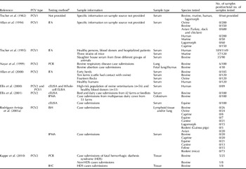
To date, there has been no serological evidence of PCV2 infection in human, avian, bovine, ovine, caprine and equine species (Table 3). These data support the hypothesis that transmission of PCV2 to swine from other species is unlikely. However, PCV2 has been detected by PCR from bovine respiratory and abortion cases (Nayar et al., Reference Nayar, Hamel, Lin, Sachvie, Grudeski and Spearman1999) and from cases of hemorrhagic diathesis syndrome (Kappe et al., Reference Kappe, Halami, Schade, Alex, Hoffmann, Gangl, Meyer, Dekant, Schwarz, Johne, Buikamp, Böttcher and Müller2010) (Table 3) indicating there remains some risk of cross-species infection. Serological evidence of PCV1 is inconclusive as two studies did not find antibodies against PCV1 in human, bovine, ovine, caprine, murine, avian and lagomorph species (Tischer et al., Reference Tischer, Gelderblom, Vettermann and Koch1982; Allan et al., Reference Allan, Phenix, Todd and McNulty1994), while one study reported the evidence of exposure to PCV1 in humans, murine and bovine species (Tischer et al., Reference Tischer, Bode, Apodaca, Timm, Peters, Rasch, Pociuli and Gerike1995) (Table 3).
Prevalence of porcine circovirus (PCV) in species other than porcine

a Testing methods: IFA, immunofluorescent assay; PCR, polymerase chain reaction; cELISA, competitive enzyme-linked immunosorbent assay; ISH, in-situ hybridization; IPMA, immunoperoxidase monolayer assay; IHC, immunohistochemistry.
Potential for the transmission of PCV2 through vaccines
The increased use of next-generation sequencing methods including pyrosequencing has improved the ability to detect viruses in various samples without a priori knowledge of viral sequences. Using this technology, PCV1 was identified in an oral rotavirus vaccine intended for human usage (Victoria et al., Reference Victoria, Wang, Jones, Jaing, McLoughlin, Gardner and Delwart2010). Further testing by the manufacturer revealed the presence of PCV1 DNA in the working cell bank and viral seed stock (Victoria et al., Reference Victoria, Wang, Jones, Jaing, McLoughlin, Gardner and Delwart2010). While this vaccine is manufactured for human applications, this work highlights the potential for vaccines, including swine products, to be contaminated with viruses even following the stringent testing associated with product development. Therefore, vaccines may be a potential source of the transmission of viruses including PCV2 especially if the products are not evaluated for the presence of virus. However, as extensive safety and efficacy studies are done prior to product release, the finding of PCV2 in a product would need to be correlated with epidemiological and pathological data suggesting that the presence of a virus outweighs the benefits of vaccination. The discovery of a PCV1/2a virus that displays high-nucleotide identity with the ORF1 of PCV1 and the ORF2 of PCV2a in three tissue homogenates (2.5% of the samples tested) submitted to a Canadian diagnostic laboratory between September 2008 and March 2009 has also been reported (Gagnon et al., Reference Gagnon, Music, Fontain, Tremblay and Harel2010). The transmission of this virus is likely not associated with the use of the inactivated chimeric PCV2 product (Fenaux et al., Reference Fenaux, Opriessnig, Halbur, Elvinger and Meng2004a, Reference Fenaux, Opriessnig, Halbur, Xu, Potts and Mengb), although the viruses share 99.6–99.7% nucleotide identity, due to the nature of the vaccine (an inactivated product), inability to isolate the virus from the vaccine and the low observed prevalence (2.5%).
In summary, there is a potential for the transmission of PCV2 through indirect routes including oral, aerosol and vaccine routes. However, the relative risk of each of these routes must be weighed with the cost of prevention of transmission by these routes. For example, if the operation is a research or genetic multiplier herd and the entry of PCV2 has to be prevented, oral, aerosol and vaccine routes should be considered in the risk assessment. In other populations, such as a commercial finisher barn, the risk of transmission of PCV2 by indirect routes is less of a concern compared to the risk of movement of contaminated pigs and personnel between buildings. Also, in these situations, PCV2 DNA is likely already present in the population. To date, little (bovine, ovine, lagomorphs, murine species) or no work (avian) has been done to directly evaluate the transmission of PCV2 between porcine and other species. Of the available information, only the murine species seems to support PCV2 replication making this species a potential for horizontal transmission of PCV2.
Conclusion
It is well documented that PCV2 is a ubiquitous virus based on serological and viremia data from countries worldwide. Furthermore, PCV2 DNA has been discovered in archived samples prior to the first reports of clinical disease and a shift in PCV2 subtype has occurred worldwide. Following both natural and experimental infection of naïve animals with PCV2, PCV2 DNA was consistently detected in secretions and excretions. Under experimental conditions, PCV2 DNA was detected early in the infection process (dpi 1) and generally persisted in a high number of animals through the end of the evaluation period indicating persistent infection. PCV2 is very resistant to high temperatures and a wide range of pH and complete inactivation of PCV2 under in vitro conditions is difficult. Reduction of PCV2 titers under in vitro conditions is most likely to occur when an oxidizing, halogen, or sodium hydroxide containing product is used; products containing iodine, alcohol, phenol or formaldehyde are likely to be less effective. Direct transmission of PCV2 between experimentally or naturally infected animals and naïve animals has been documented and the introduction of clinically or subclinically infected animals into a population represents a risk to the herd. Indirect transmission through the oral, aerosol or vaccine routes is likely a lesser risk for transmission in most swine populations but may be worth evaluating in high-health herds.