Anomalous left coronary artery form the pulmonary artery is usually a solitary cardiac lesion; however, it has been reported to occur in association with a variety of other cardiac malformations including patent ductus arteriosus, ventricular septal defect, coarctation of the aorta, and tetralogy of Fallot.Reference Awasthy, Marwah, Sharma and Dalvi 1 – Reference Laux, Bertail, Bajolle, Houyel, Boudjemline and Bonnet 6 We report a case of anomalous left coronary artery from the pulmonary artery that was diagnosed by echocardiography after repair of the total anomalous pulmonary venous return.
Case report
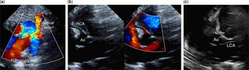
A 6-week-old, full-term infant presented to the emergency department with irritability, tachycardia, and cyanosis. Vital signs and physical examination were significant for a heart rate of 179 beats/minute, respiratory rate of 48 breaths/minute, oxygen saturation of 90% in room air, gallop, and hepatomegaly. Chest radiography showed cardiomegaly with pulmonary oedema. The electrocardiogram demonstrated sinus rhythm with right atrial enlargement and right ventricular hypertrophy. There was no evidence of myocardial ischaemia. The echocardiogram revealed levocardia with normal cardiac segments, supracardiac total anomalous pulmonary venous return draining to the innominate vein via a vertical vein (Fig 1a), secundum atrial septal defect, severe dilation of the right-sided chambers, and a tricuspid regurgitation velocity of 3.4 m/second. The right coronary artery had a normal origin and course by both two-dimensional imaging and color Doppler (Fig 1b). The left coronary artery appeared to have a normal origin by two-dimensional imaging but was not clearly seen by color Doppler (Fig 1c).
Echocardiography images demonstrating ( a ) pulmonary venous confluence draining to a vertical vein, ( b ) normal origin of the right coronary artery (RCA) from the aorta (Ao) seen by two-dimensional (2D) imaging and color Doppler, and ( c ) possible origin of the left coronary artery (LCA) from the Ao seen only by 2D imaging. MPA=main pulmonary artery.

The patient underwent surgical repair with anastomosis of the pulmonary venous confluence to the left atrium and closure of the atrial septal defect. He was preemptively placed on inhaled nitric oxide, which was discontinued on postoperative day 1. He was weaned from inotropic support by postoperative day 5. He was noted to have some fussiness, but was discharged home on postoperative day 11.
The patient was admitted 10 days after discharge for inconsolable crying, poor feeding, diaphoresis, and perioral cyanosis. His echocardiogram demonstrated good biventricular function, mild right ventricular dilation, and unobstructed pulmonary venous drainage. The coronary arteries were not evaluated. There was no evidence of myocardial ischaemia on electrocardiogram. The patient’s symptoms were attributed to colic and constipation, and he was discharged home. At follow-up, he was noted to have continued intermittent irritability.
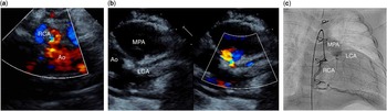
At 4 months of age, a routine echocardiogram showed prominent right coronary artery size and flow (Fig 2a). The left coronary artery originated from the pulmonary artery and had retrograde flow (Fig 2b). Speckling of colour flow in the right ventricular wall and ventricular septum suggesting collateral flow was present. There was no mitral regurgitation, endocardial fibroelastosis, or left ventricular dysfunction. The electrocardiogram did not show ischaemic changes. Subsequent cardiac catheterisation confirmed the echocardiographic findings (Fig 2c). The patient then underwent successful repair of the anomalous coronary artery. The postoperative course was complicated by a prolonged ventilator weaning time and aspiration requiring gastrostomy tube placement. He was discharged home 6 weeks after surgery, and he continues to do well with normal biventricular function and unobstructed pulmonary venous return on echocardiogram.
Echocardiography images demonstrating ( a ) the dilated right coronary artery (RCA) with prominent flow and ( b ) left coronary artery (LCA) originating from the main pulmonary artery (MPA) with retrograde flow. ( c ) Angiogram demonstrating dilated RCA with collateral flow to the LCA, which ultimately fills the MPA. Ao=aorta.

Discussion
To our knowledge, this is the first description of an infant with both anomalous left coronary artery from the pulmonary artery and total anomalous pulmonary venous return. This case emphasises several key points regarding the physiology, diagnosis, and presentation of anomalous left coronary artery from the pulmonary artery.
In anomalous left coronary artery from the pulmonary artery, myocardial ischaemia occurs secondary to the fall in pulmonary vascular resistance after birth. Initially, the left coronary artery vasculature dilates, which decreases the resistance and accommodates the lower perfusion pressure. Eventually, myocardial oxygen demand is not met. Collaterals develop between the right and the left coronary system, which leads to coronary steal. Oxygenated blood from the aorta enters the right coronary artery, bypasses the high-resistance vessels of the myocardium, and ultimately flows into the low-resistance pulmonary artery.Reference Lim and Matherne 7 In the presence of a lesion that causes elevated pulmonary artery pressures, such as a patent ductus arteriosus or ventricular septal defect, there is less coronary steal and adequate coronary perfusion and oxygen delivery to the myocardium. If the anomalous left coronary artery is unrecognised before repair of the associated lesion, left ventricular dysfunction can develop postoperatively leading to significant morbidity and mortality.Reference Callaghan, O’Hare and Casey 4 , Reference Ortiz, de Leval and Somerville 5
In our case, the patient’s elevated pulmonary artery pressures secondary to the total anomalous pulmonary venous return made it difficult to diagnose the anomalous left coronary artery by echocardiography. When retrograde flow is not present in the coronary artery, it can often appear as having a normal origin with normal flow. In our case, the left coronary artery appeared to have a normal origin by two-dimensional imaging, but was not well interrogated with color Doppler (Fig 1c). In the presence of a shunt lesion, this should raise suspicion for a coronary anomaly.
Although preoperative diagnosis may be difficult, coronary anomalies have been recognised in the operating room. Shetty et alReference Shetty, Thareen, Ramaiah, Narayan, Das and Chandrashekariah 8 described a case of a single coronary artery from the pulmonary artery with a ventricular septal defect that was recognised because of the inability to arrest the heart with administration of cardioplegia. Our patient required deep hypothermic circulatory arrest for the initial surgery, and thus the ineffectiveness of the cardioplegia was not recognised.
Finally, our patient was quite irritable after the initial surgery and presented with unexplained symptoms requiring admission to the hospital. These were attributed to colic and constipation, but in retrospect may have been secondary to angina.Reference Bland, White and Garland 9 The patient had follow-up imaging, but the coronary arteries were not adequately assessed. He did not have left ventricular dysfunction, echo-bright papillary muscles, mitral regurgitation, or evidence of ischaemia on electrocardiogram, which likely would have made us more suspicious of a coronary anomaly.
Anomalous left coronary artery from the pulmonary artery is a must-recognise entity. Although it is rare and can be difficult to diagnose, the presence of a left-to-right shunt lesion elevating pulmonary artery pressures and or resistance can make it even more challenging to identify. Therefore, it is crucial to have adequate coronary imaging with two-dimensional imaging and color Doppler, and to obtain serial imaging of the coronary arteries, especially in patients with unexplained irritability after cardiac surgery.
Acknowledgements
None.
Financial Support
This research received no specific grant from any funding agency or from commercial or not-for-profit sectors.
Conflicts of Interest
None.