Recent advances in CT technology allow imagers to perform high-quality examinations while maximising speed of acquisition and minimising radiation dose, features that are particularly important in young patients who may have greater sensitivity to ionising radiation. In this case, we describe the use of ultra-low-dose dual-source cardiac for the evaluation of a neonate with CHD. The use of rapid, high-pitch, dual-source technique limits motion artefact, providing a high level of anatomic detail without the need for sedation or breathholding, with the added benefit of a very low radiation dose. Several additional dose-reduction strategies were utilised, resulting in an effective dose lower than that expected from diagnostic cardiac catheterisation. Given these strengths, low-dose, high-pitch cardiac CT should be considered as an alternative to MRI or diagnostic cardiac catheterisation in neonates with complex CHD that require definitive anatomic evaluation.
Case report
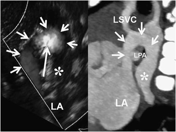
A 4-day-old full-term infant with prenatally diagnosed complex CHD had an initial postnatal echocardiogram with findings suspicious of supracardiac anomalous pulmonary venous connection. The pulmonary veins appeared to enter an ascending vein draining to the left atrium with elevated flow velocities on Doppler echocardiography concerning for stenosis (Fig 1). Echocardiogram was unclear regarding involvement of all veins, and whether the ascending vein passed between the left bronchus and left pulmonary artery, which would have argued for earlier intervention. Cardiac CT angiography was requested to completely characterise the pulmonary venous anatomy. Contrast-enhanced CT angiogram of the chest was performed utilising the following dose-reduction strategies: 70 kVp tube voltage, iterative reconstruction with reduced tube current (34 mAs, SAFIRE I30f 3; Siemens Healthcare, Malvern, Pennsylvania, United States of America), and high-pitch technique (pitch=3.0). The examination was conducted without sedation, and the infant tightly swaddled on the CT table with a technologist in the room for safety. Intravenous contrast was hand injected as fast as possible at a dose of 2 ml/kg. Real-time bolus tracking of contrast arrival in the ascending aorta was performed immediately following injection for acquisition timing. No electrocardiography-gating was used because of the heart rate, which was >140 beats/minute at the time of examination. Dose length product was 7 mGy cm with an estimated exam effective dose of 0.27 mSv when using a conversion factor corrected for patient age (k=0.039 mSv mGy−1 cm−1).Reference Dougeni, Faulkner and Panayiotakis 1
Sagittal image obtained by Doppler echocardiogram (left) and sagittal maximum intensity projection image (right) from cardiac CT. The echocardiogram image has been rotated counter-clockwise to optimise comparison between the modalities. The venous confluence (asterisk) drains to a vertical vein (short arrows) that shows aliasing on colour Doppler (long arrow), suggesting possible stenosis. On CT, the draining vein (short arrows) courses around the left pulmonary artery (LPA) and left mainstem bronchus to drain into the left atrium (LA). A small, patent left-sided superior vena cava is identified draining into the vertical vein (LSVC). No stenosis of the venous pathway is identified.

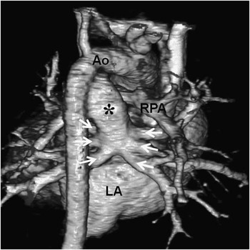
CT images showed a pulmonary venous confluence posterior to the left atrium that received three left-sided and three right-sided pulmonary veins (Fig 2). The confluence drained to a widely patent vertical vein that coursed over and around the left mainstem bronchus and left pulmonary artery (Fig 1), and drained into the superior aspect of the left atrium. A small left-sided superior caval vein, not identified at echocardiography, was seen draining into the vertical vein (Fig 1). Given the lack of obstruction of the anomalous pulmonary venous return and adequate oxygenation, the patient was discharged with close monitoring until the time of definitive surgical repair 4 months after the CT examination. CT findings were confirmed at the time of surgery, and the total anomalous pulmonary venous connection was repaired.
Coronal volume rendered image obtained from a posterior view in a 4-day-old female patient. Total anomalous pulmonary venous connection of the supracardiac type is demonstrated with six pulmonary veins (arrows) draining to a venous confluence (asterisk) separate from the left atrium (LA) that courses vertically over the left pulmonary artery (LPA). Image quality is excellent using low-dose, high-pitch technique (70 kVp, 34 mAs, pitch 3.0). Ao=aorta, RPA=right pulmonary artery.

Discussion
In the present case, ultra-low-dose CT angiography was used to characterise CHD in a neonate without the use of sedation. Echocardiography is the first-line imaging modality for non-invasive evaluation of prenatal and postnatal CHD; however, visualisation of posterior structures, such as the branch pulmonary arteries, aorta, and pulmonary veins, can be limited, particularly in patients with difficult imaging windows. Cardiac MRI is often the next test of choice for non-invasive imaging owing to its lack of ionising radiation and strength in flow assessment. The disadvantage is that lengthy examination time dictates need for sedation, which can be expensive, risky to the patient, and may limit availability.
Cardiac CT angiography is a rapid examination that provides high-resolution anatomic information. However, widespread utilisation in the neonatal population has been tempered by radiation exposure concerns. Recent technical advances in CT have provided cardiac imagers with multiple radiation dose-reduction techniques that, when combined, have an additive effect to drastically reduce dose, while maintaining the diagnostic quality. In this case, the estimated effective dose was 0.27 mSv, one-tenth of the estimated annual background radiation dose and significantly lower than the estimated 4.6 mSv median effective dose reported for paediatric diagnostic cardiac catheterisation.Reference Bacher, Bogaert, Lapere, De Wolf and Thierens 2 We utilised a reduced tube voltage of 70 kVp, an option that has recently become available. Decreasing kVp is a highly effective means for reducing dose,Reference Dougeni, Faulkner and Panayiotakis 1 and has the additional advantage of improved vascular enhancement owing to increased X-ray absorption of iodine. Significant reductions in tube current (mAs) can be accomplished by the use of iterative reconstruction. Iterative reconstruction generates images with decreased image noise compared with traditional filtered back projection methods.Reference Han, Grant, Garberich, Sedlmair, Lindberg and Lesser 3 Finally, high-pitch scans using dual-source CT mode utilises two X-ray tubes to accelerate image acquisition and reduce patient exposure. Scan times are <500 msecond, fast enough to eliminate respiratory motion artefacts and greatly reduce cardiac motion. This allows for diagnostic imaging without the need for sedation or controlled ventilation.Reference Lell, May and Deak 4 , Reference Han, Overman and Grant 5
In conclusion, ultra-low-dose high-pitch cardiac CT is a rapid and effective imaging technique that allows comprehensive non-invasive anatomic imaging in neonates with complex CHD without sedation and should be considered as an alternative to invasive catheterisation or MRI.
Acknowledgement
None.
Financial Support
This research received no specific grant from any funding agency, commercial, or not-for-profit sectors.
Conflicts of Interest
None.